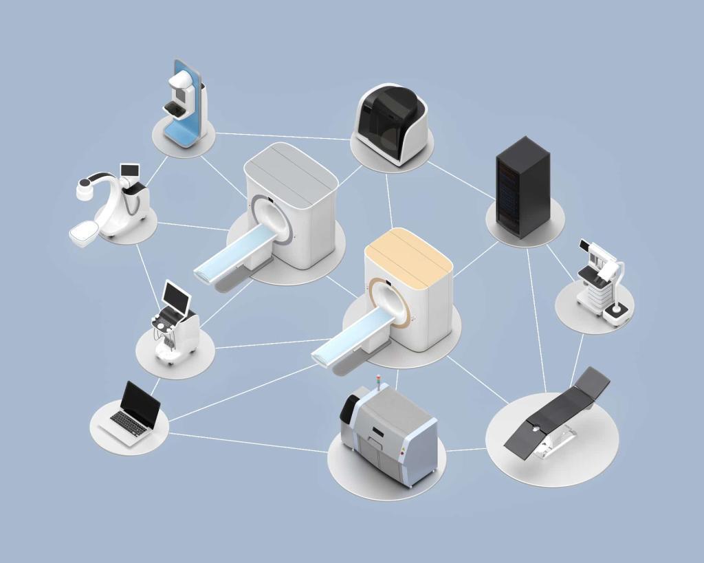
Medical imaging has always been central to modern healthcare, but in 2026, it is evolving into something far more integrated, intelligent, and widely accessible. Scanners are no longer confined to radiology departments, algorithms are supporting clinical decision-making in real time, and portable systems are extending diagnostic capability to community settings, remote regions, and even ambulances. At the same time, regulators and healthcare providers are placing greater emphasis on safety, transparency, and demonstrable patient value.
This article explores the key developments shaping medical imaging through 2026 and how they are changing clinical workflows, industry priorities, and patient experience.
Artificial intelligence becomes standard infrastructure
Artificial intelligence has moved from experimental trials and niche software pilots into routine clinical use. In 2026, many hospitals treat AI tools as core digital infrastructure within imaging departments. These systems assist radiologists with case prioritisation, automated measurements, structured reporting, and quality assurance, allowing specialists to focus more time on complex interpretation and communication with clinical teams.
In MRI, AI-based image reconstruction techniques accelerate scan acquisition while maintaining diagnostic quality, reducing appointment times and improving scanner throughput. In CT and X-ray, algorithmic triage identifies urgent findings such as intracranial haemorrhage, suspected pulmonary embolism, or pneumothorax, helping teams respond more rapidly in emergency settings.
Rather than displacing radiologists, AI increasingly supports them through digital augmentation. It pre-analyses images, proposes annotations, and highlights regions of interest, while the final judgement remains firmly in human hands. This collaborative model is now widely seen as the most realistic and clinically acceptable path forward.
From diagnosis to prognosis: imaging as a predictor of outcomes
A major conceptual shift underway in 2026 is the move from imaging as a purely diagnostic tool towards imaging as a prognostic and predictive resource. Radiomics and machine-learning approaches extract quantitative patterns from scans that are invisible to the naked eye but correlate with disease behaviour.
These techniques are already being explored in oncology to forecast treatment response, recurrence risk, and likely survival trajectories. In cardiovascular medicine, imaging biomarkers are used to assess which plaques are most likely to rupture and which patients are at risk of progression to heart failure. Instead of merely showing what disease looks like today, imaging is beginning to inform clinicians about how it may evolve tomorrow.
This shift aligns closely with personalised medicine. Prognostic imaging supports risk-stratified treatment planning, helping doctors match therapies to each patient’s biological profile. As the tools mature through 2026, more imaging reports are expected to include quantified metrics and predictive insights alongside traditional descriptive findings.
Multimodal and data-integrated diagnostics
Radiology is becoming increasingly interconnected with other clinical data sources. Rather than viewing imaging in isolation, clinicians in 2026 are beginning to work with multimodal decision-support systems that combine scans with laboratory results, genomic data, pathology reports, and electronic health records.
These platforms aim to provide a more comprehensive view of the patient’s condition. For example, an AI system may connect an imaging phenotype on MRI, a specific genetic mutation, and the results of a blood biomarker test, offering clinicians an integrated interpretation rather than separate fragments of information.
This development is also reshaping radiology training and identity. Radiologists are progressively seen not only as image readers but as central contributors to data-driven, multidisciplinary diagnostics. As a result, skills in informatics, data literacy, and collaboration across specialties are becoming increasingly important elements of professional practice.
Intelligence at the point of capture: AI on the scanner
A significant technical trend in 2026 is the growing use of “edge AI“, where algorithms run directly on imaging devices rather than relying solely on cloud processing. By embedding intelligence into scanners and portable systems, analysis can occur as images are acquired, enabling faster feedback and reducing dependence on network connectivity.
For example, real-time reconstruction and enhancement on MRI systems can present usable images more quickly, while embedded software on portable devices guides novice operators in probe positioning and scan acquisition. On-device processing also helps address data-protection concerns, as raw images can remain on the scanner instead of being transmitted externally for analysis.
This approach is particularly valuable in emergency medicine and remote healthcare environments, where rapid decision-making and operational resilience are critical.
Imaging beyond the hospital: mobility and point-of-care access
One of the most visible changes in 2026 is the expansion of imaging outside traditional radiology departments. Portable and point-of-care systems are increasingly used in emergency units, intensive care wards, GP surgeries, community clinics, and pre-hospital care.
Handheld ultrasound devices linked to smartphones or tablets are now widely used by clinicians across multiple specialities. They enable rapid bedside assessment, supporting tasks such as cardiac screening, trauma evaluation, obstetric monitoring, and guided procedures. AI-assisted overlays help less experienced users obtain usable images and interpret basic findings, broadening access without compromising safety.
This decentralisation is beginning to spread to higher-end modalities as well. Compact mobile CT systems are being trialled for stroke and head injury assessment in ambulances and rural health centres, enabling scanning earlier in the patient journey. The potential impact on emergency stroke care is particularly significant, as earlier imaging can support faster treatment decisions and better outcomes.
For health systems, the challenge in 2026 is less about acquiring portable equipment and more about integrating it into governance, training, reporting, and archiving frameworks in a coordinated and sustainable way.
Cloud platforms and enterprise imaging ecosystems
As imaging volumes increase and workflows become more distributed, healthcare providers are investing heavily in enterprise-level imaging infrastructure. Cloud-based platforms now host extensive archives of scans from multiple sites, modalities, and vendors, allowing clinicians to access images and reports from anywhere within the network.
These platforms also act as central hubs for AI deployment, image sharing, multidisciplinary review, and long-term data analysis. Industry consolidation and strategic partnerships are accelerating this shift, with major manufacturers and software companies aligning hardware, cloud architecture, and analytics into unified ecosystems.
For health organisations, the benefits include improved interoperability, more consistent reporting environments, and stronger foundations for innovation. However, the move to cloud-centred imaging also requires robust cybersecurity planning, careful cost management, and clear data-governance policies.
Value-based procurement and workforce-centred design
Economic pressure and rising scan demand mean that hospitals in 2026 are increasingly required to demonstrate measurable value from imaging investment. Procurement decisions are no longer driven solely by image resolution or technical specifications. Instead, purchasers focus on workflow automation, integration with clinical systems, impact on reporting times, and the ability to reduce repeat examinations.
Ergonomics and staff wellbeing are also gaining attention. Vendors are redesigning scanner interfaces, exam rooms, and control panels to reduce physical strain and cognitive burden on radiographers and radiologists. Automation is being used to streamline repetitive tasks such as protocol selection, dose calculation, and measurement generation.
These design priorities are particularly important at a time when imaging services face workforce shortages and growing caseloads. Technology that supports staff resilience and retention is now seen as a strategic necessity rather than a secondary benefit.
Stronger regulation and accountability for AI-driven imaging
With AI now deeply embedded in imaging workflows, regulatory oversight has become more rigorous. In 2026, developers and healthcare providers are expected to demonstrate transparency, robust evidence of safety and performance, and clear frameworks for human oversight.
Post-market monitoring, performance auditing, and incident reporting are increasingly formalised requirements. Hospitals introducing AI-enabled imaging tools are establishing governance committees, validation procedures, and documented protocols to ensure responsible adoption. There is greater recognition that algorithmic tools must be continually evaluated in real-world conditions, not only during pre-market testing.
Legal and ethical accountability is also evolving. Questions around liability—particularly when automated analysis influences clinical outcomes—are prompting health organisations to take a cautious, structured approach to deployment.
Evolving professional roles and patient expectations
Changes in professional culture and patient perception accompany the technological developments of 2026. Radiologists are becoming key interpreters of complex, data-rich diagnostic outputs and are expected to help colleagues understand both the strengths and limitations of AI tools. Many training programmes are adapting curricula to reflect this broader remit.
From the patient perspective, awareness of AI in healthcare is growing. Public surveys indicate a willingness to accept algorithmic support in diagnostics, provided clinicians remain clearly accountable, and systems are properly regulated. As imaging becomes more mobile and integrated into everyday clinical environments, patient contact with imaging professionals may also increase, reinforcing the importance of communication and reassurance.
Looking ahead: towards intelligent and connected imaging care
The overarching theme in medical imaging in 2026 is convergence. Hardware innovation, AI-driven analysis, portable access, and enterprise-level platforms are combining to create imaging systems that are more responsive, more predictive, and more closely interwoven with the broader care pathway.
A suspected stroke patient may be scanned before reaching the hospital, with images analysed in real time and reviewed simultaneously by specialists in different locations. A cancer follow-up scan may be automatically compared with previous studies, with quantitative metrics tracked over time and integrated into personalised treatment planning. A community GP may use a handheld ultrasound to resolve uncertainty during a consultation and avoid unnecessary referral.
These developments bring significant responsibility alongside opportunity. Success depends on thoughtful regulation, strong clinical leadership, sustained investment in workforce training, and careful attention to equity of access.
Handled well, the advances taking shape through 2026 have the potential to transform medical imaging from a predominantly diagnostic activity into a dynamic, intelligence-driven system that supports clinicians and patients across every stage of the healthcare journey.
Medical imaging in 2026 — Questions and Answers
What is the main direction of medical imaging in 2026?
Medical imaging in 2026 is becoming smarter, more connected, and more widely accessible. AI is now embedded in scanners and reporting systems, portable devices are taking imaging closer to the point of care, and imaging data is being integrated with other clinical information to support earlier diagnosis and more tailored treatment decisions. The focus is on improving clinical outcomes, speeding workflows, and extending access beyond large hospital centres.
How is artificial intelligence being used in imaging this year?
AI has moved firmly into day-to-day clinical practice. It assists radiologists by highlighting urgent findings, automatically generating measurements, and helping prioritise cases in busy departments. In MRI and CT, AI speeds up image reconstruction and reduces scanning time. In reporting workflows, it produces draft outputs that clinicians review and refine. Rather than replacing human expertise, AI serves as a supportive tool that helps specialists manage increasing workloads while maintaining accuracy.
Is imaging still mainly about diagnosis, or is its role changing?
Its role is expanding. Imaging is increasingly used not only to confirm the presence of a disease, but also to estimate how it may progress. Quantitative imaging and radiomics extract measurable features from scans that can indicate treatment response or likely outcomes. This supports personalised care by helping clinicians decide which therapies are most suitable for each patient, based on how their condition is expected to behave over time.
What does “multimodal diagnostics” mean in this context?
Multimodal diagnostics refers to the integration of imaging data with other forms of clinical information, such as laboratory tests, genomic findings, pathology results, and patient records. Instead of interpreting scans in isolation, radiologists and clinicians work with combined datasets that provide a fuller picture of the patient’s condition. This approach encourages closer collaboration across specialities and strengthens radiology’s role in multidisciplinary care.
How is AI being incorporated directly into imaging equipment?
Many imaging systems now include “on-device” or edge-based AI. Algorithms run inside the scanner or portable device itself, analysing data as it is captured. This allows faster feedback, supports real-time decision-making, and reduces reliance on external servers. It is especially helpful in urgent care, mobile imaging, and remote locations where network access may be limited. It also supports data protection practices by keeping sensitive images on local systems.
What changes are taking place in the way imaging is delivered?
Imaging is no longer confined to the main radiology suites. Portable and point-of-care systems are now widely used in emergency departments, intensive care units, GP settings, community clinics, and pre-hospital services. Handheld ultrasound devices have become common tools for bedside examination, and compact CT systems are beginning to support stroke assessment and trauma response outside traditional hospital environments. This shift brings imaging closer to the patient and shortens time to diagnosis.
How are cloud platforms influencing imaging services?
Cloud-based enterprise imaging platforms allow hospitals and clinics to store, share, and review images across multiple sites. Clinicians can access scans remotely, support virtual consultation, and collaborate more easily across departments. These platforms also provide central hubs for AI tools, audit processes, and long-term data analysis. However, they require careful planning around cybersecurity, governance, and system interoperability.
Are healthcare organisations changing how they choose imaging technology?
Yes. Procurement is increasingly based on value and workflow benefits rather than on hardware specifications alone. Hospitals look for systems that improve efficiency, reduce repeat scanning, integrate smoothly with existing IT infrastructure, and support staff wellbeing. Automation, ergonomic design, and usability are essential considerations, particularly in services facing staffing shortages and high demand.
What regulatory changes affect AI in imaging in 2026?
Regulation is becoming stricter and more structured. AI systems in imaging must demonstrate safety, transparency, and ongoing performance monitoring. Healthcare providers are expected to maintain governance frameworks for validation, oversight, and post-deployment review. This includes logging performance, handling incident reports, and ensuring clinicians remain accountable for final decisions. The overall aim is to promote innovation while maintaining patient safety and trust.
How are the roles of radiologists and imaging professionals evolving?
Radiologists are increasingly acting as data interpreters, clinical partners, and advisers on digital tools, rather than solely as image readers. They are involved in evaluating AI systems, guiding multidisciplinary care, and ensuring imaging insights are used effectively in treatment pathways. Training programmes are gradually incorporating data, communication, and collaborative skills to meet these new expectations.
What do these developments mean for patients?
Patients benefit from quicker access to imaging, faster decision-making in emergencies, and more personalised treatment planning. Portable imaging reduces travel and waiting times, while AI-supported analysis helps clinicians identify significant findings sooner. At the same time, there is continued emphasis on human oversight, transparency, and communication, so that technology strengthens care rather than replacing professional judgement.
What is the bigger picture for imaging in 2026?
Medical imaging is becoming an integrated part of the care pathway rather than a step that sits on its own. Scanners, software, and clinical systems are working together to provide earlier insight, broader access, and more informed treatment decisions. The challenge for health services is to balance innovation with responsible governance, investment in training, and equal access across regions and patient groups.
Handled thoughtfully, the direction taken in 2026 is helping imaging evolve from a primarily diagnostic activity into a connected, intelligent support system for clinicians and patients throughout the course of care.
Disclaimer
This article is intended for general information and educational purposes only. It does not constitute medical advice, clinical guidance, or a substitute for consultation with qualified healthcare professionals. The developments in medical imaging, artificial intelligence, and diagnostic technologies described here may vary across countries, healthcare providers, and clinical settings, and their use should always follow local regulations, professional standards, and institutional policies. Readers should not rely on this content to make decisions about diagnosis, treatment, or patient care. Open MedScience and the authors accept no responsibility for any loss, harm, or consequences arising from the interpretation or application of the information presented. Always seek advice from appropriately trained medical practitioners regarding individual health or clinical matters.
home » blog » medical technologies »