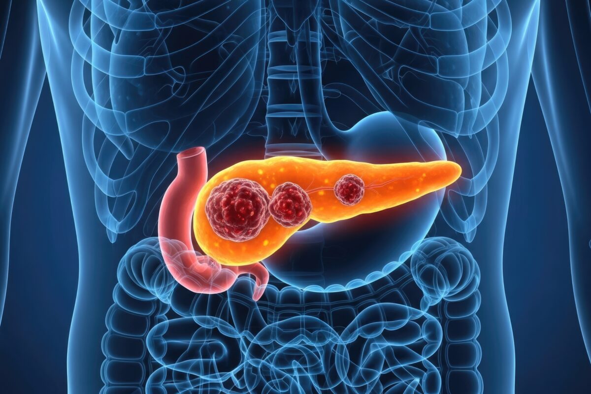
Pancreatic Cancer Research in 2025: Where Progress Is Finally Emerging
 By
By
Pancreatic cancer research in 2025 reveals crucial developments that may transform patient care and clinical practices.
Tumour growth imaging biomarkers play a crucial role in cancer diagnosis, prognosis, and treatment monitoring. These biomarkers help assess tumour development, response to therapy, and disease progression using various imaging techniques. Advances in medical imaging have led to the identification of several biomarkers that provide valuable insights into tumour biology.
Types of Imaging Biomarkers
Imaging biomarkers for tumour growth can be classified based on the imaging modality used. The most common techniques include positron emission tomography (PET), magnetic resonance imaging (MRI), computed tomography (CT), and ultrasound. Each modality provides different types of information about tumour characteristics.
Applications in Cancer Management
Imaging biomarkers are used in several aspects of cancer care, including early detection, treatment planning, and therapy response assessment. In precision oncology, biomarkers help stratify patients based on tumour characteristics, allowing for personalised treatment approaches. For instance, FDG-PET is used to monitor response in lymphoma patients, while ADC values from DWI-MRI predict treatment response in prostate cancer.
Future Perspectives
The integration of artificial intelligence (AI) with imaging biomarkers is expected to enhance tumour growth assessment. AI algorithms can analyse complex imaging data to identify patterns associated with tumour progression. Additionally, novel radiotracers and molecular imaging agents are being developed to provide deeper insights into tumour biology.
Imaging biomarkers continue to evolve, offering improved methods for detecting and monitoring tumours. Their role in guiding clinical decision-making is likely to expand, ultimately improving cancer outcomes.
home » Tumour Growth Imaging Biomarkers
Pancreatic cancer research in 2025 reveals crucial developments that may transform patient care and clinical practices.
Discover how Palmitoyl Tripeptide-1 enhances tissue remodelling and cellular processes in this informative article.