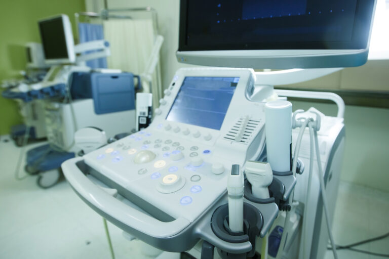
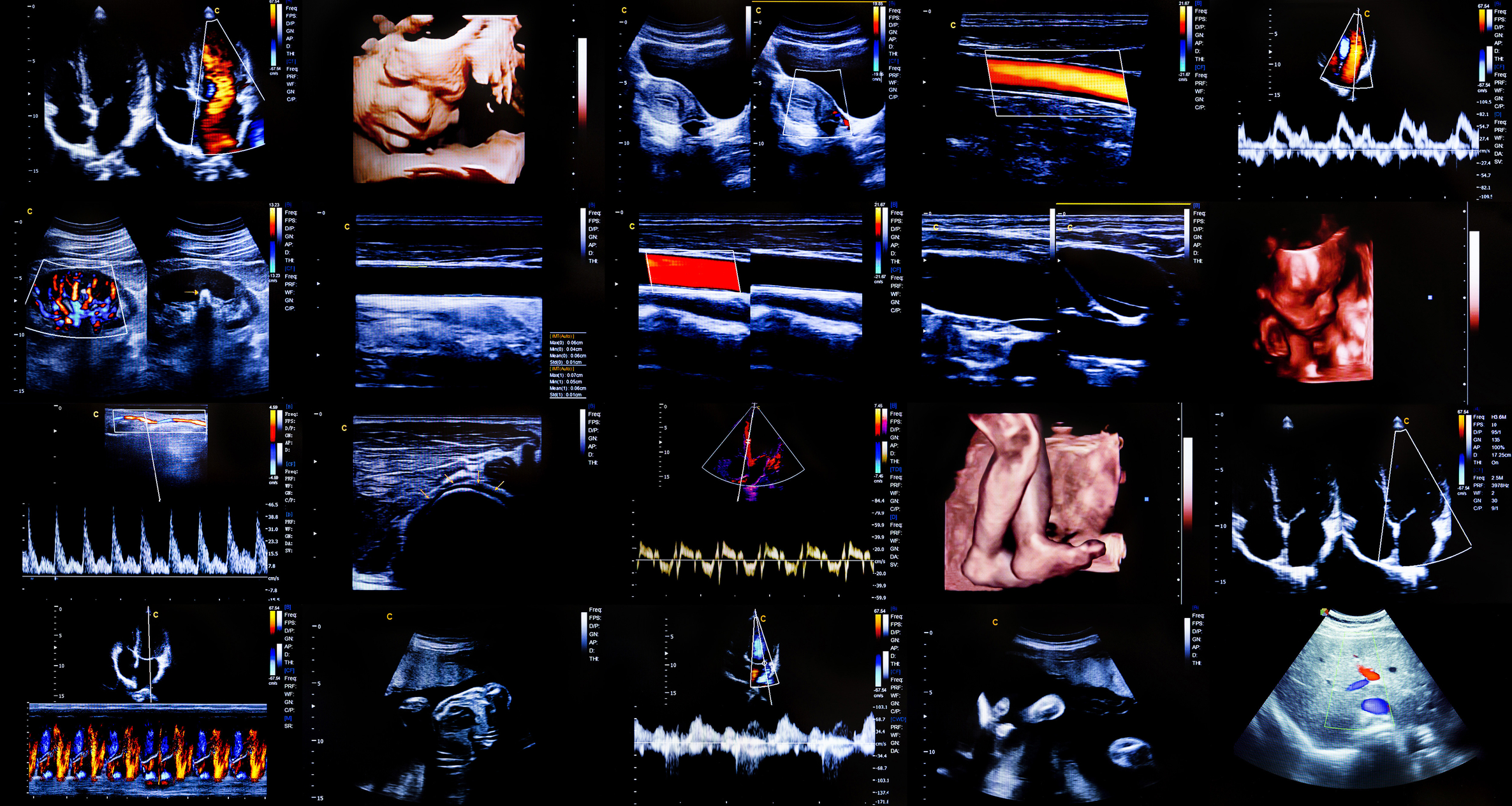
Ultra-Fast Ultrasound Imaging
Ultra-fast ultrasound imaging is an advanced technique that significantly enhances the speed and resolution of traditional ultrasound. Unlike conventional ultrasound, which operates at frame rates of around 50–100 frames per second (fps), ultra-fast ultrasound can achieve thousands of frames per second. This breakthrough allows for more detailed visualisation of rapid physiological processes, making it an invaluable tool in both research and clinical applications.
Principles of Ultra-Fast Ultrasound
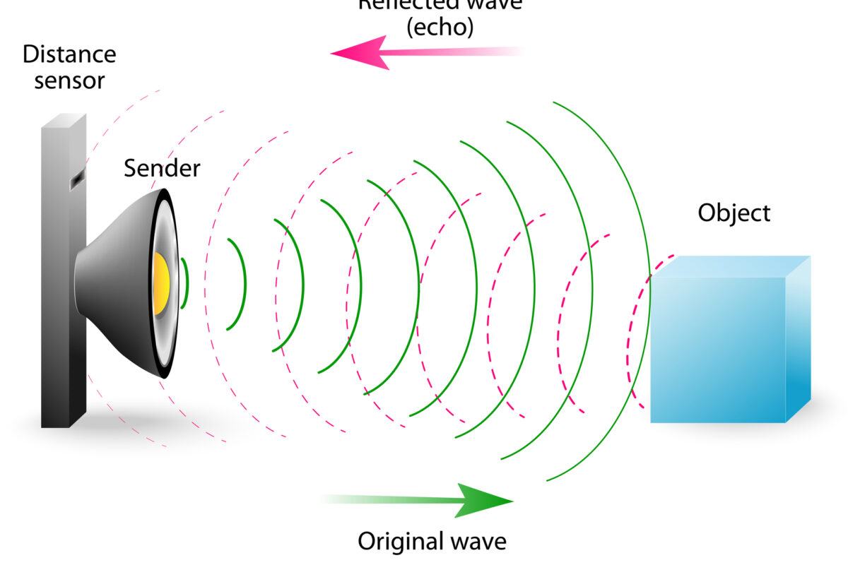
Traditional ultrasound imaging relies on sequential scanning of tissue using focused beams. This approach limits the frame rate because each pulse must be emitted, received, and processed before the next one is sent. Ultra-fast ultrasound overcomes this limitation by using plane waves or diverging waves, which illuminate the entire field of view with a single pulse. Echoes from these waves are then processed using sophisticated computational algorithms to reconstruct high-resolution images at an unprecedented speed.
Clinical Applications
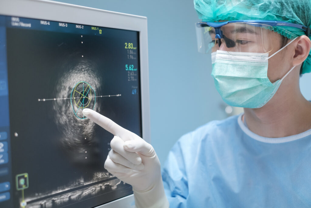
The ability to capture thousands of frames per second has opened new possibilities for medical imaging. Some key applications include:
Cardiovascular Imaging
Ultra-fast ultrasound enables precise assessment of heart function by capturing the movement of heart valves and myocardial tissues in real-time. It allows for accurate measurement of myocardial strain, which is essential for early detection of heart diseases. Additionally, it can improve the diagnosis of aortic stenosis and valve dysfunction.
Neurovascular Imaging
In neurology, ultra-fast ultrasound is used to image blood flow in the brain. The high frame rate allows for the detection of microvascular changes, which are crucial in diagnosing strokes and neurodegenerative diseases. Doppler techniques combined with ultra-fast ultrasound provide highly detailed maps of cerebral blood flow dynamics, offering a non-invasive alternative to traditional imaging methods such as MRI.
Elastography
Ultra-fast ultrasound is also used in shear wave elastography, a technique that measures tissue stiffness. This is particularly useful in oncology, where tumour stiffness can indicate malignancy. The high speed of ultra-fast ultrasound improves the accuracy of elastography, allowing for better differentiation between benign and malignant lesions.
Advantages Over Conventional Ultrasound
Ultra-fast ultrasound provides several benefits over traditional methods:
- Higher Temporal Resolution: The increased frame rate allows for the visualisation of rapid biological processes, such as muscle contractions and blood flow dynamics.
- Improved Sensitivity: Small-scale vascular structures and microcirculatory changes can be detected more easily.
- Reduced Motion Artefacts: Because images are acquired much faster, motion blur caused by breathing or heartbeats is minimised.
- Non-Invasive and Radiation-Free: Like conventional ultrasound, ultra-fast imaging is safe and does not expose patients to ionising radiation.
Challenges and Future Prospects
Despite its advantages, ultra-fast ultrasound imaging presents challenges, including the need for advanced computing power to process large datasets. Additionally, integrating this technology into portable and cost-effective clinical devices remains a key area of development.
As research continues, ultra-fast ultrasound is expected to become a standard tool in medical diagnostics, improving the early detection and monitoring of various diseases.
home » Ultra-Fast Ultrasound Imaging
 By
By