
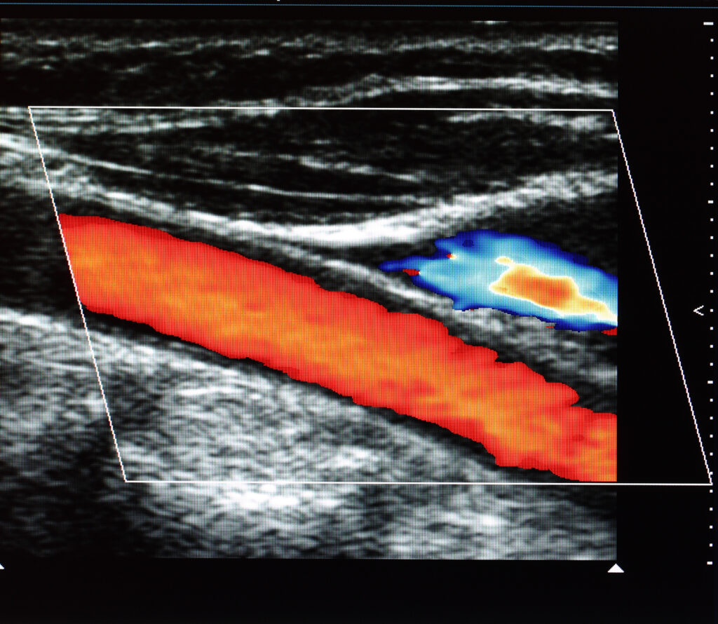
Artificial Intelligence Transforms the Future of Ultrasound Imaging
 By
By
Learn about the benefits of AI in ultrasound imaging, from automated analysis to enhanced decision support in diagnostics.
By
Learn about the benefits of AI in ultrasound imaging, from automated analysis to enhanced decision support in diagnostics.
 By
By
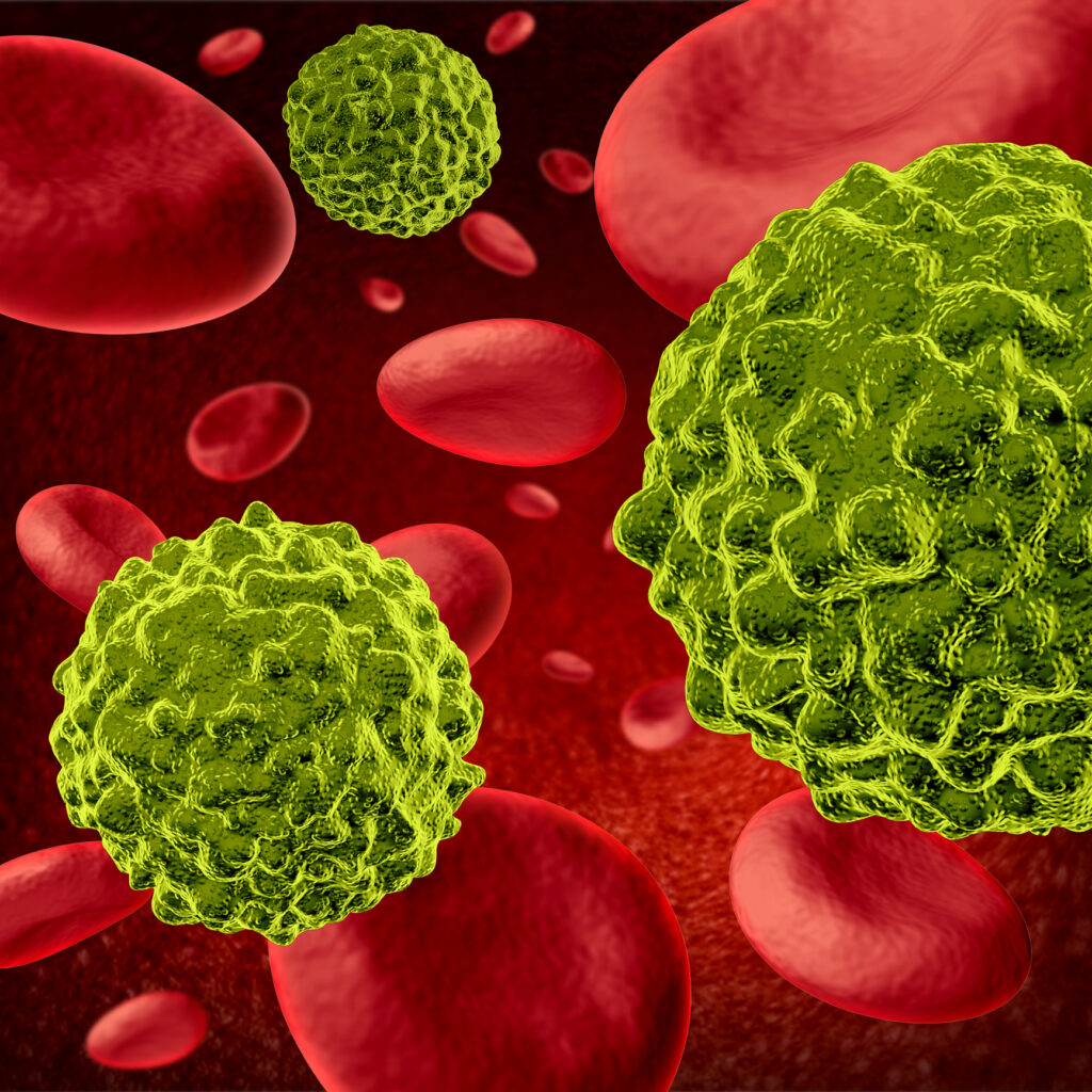
Yttrium-90 DOTA-FF-21101 targets P-Cadherin in solid tumours, delivering beta radiation for precise cancer treatment.
 By
By
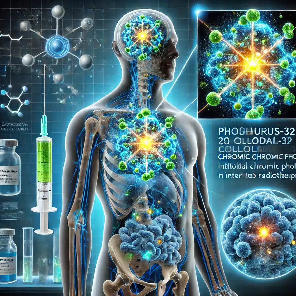
Phosphorus-32 Colloidal Chromic Phosphate delivers precise beta radiation for treating malignant effusions and cystic tumours.
 By
By
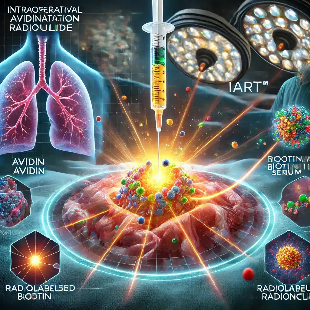
Intraoperative Avidination Radionuclide Treatment utilises avidin-biotin technology to deliver targeted tumour radiation therapy.
 By
By
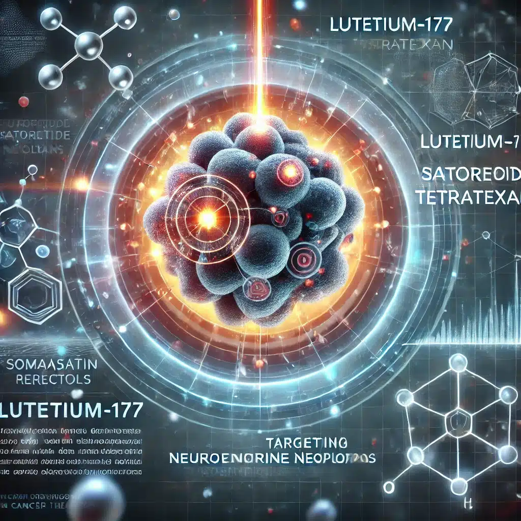
Lutetium-177 Satoreotide Tetratexan is a radiopharmaceutical targeting somatostatin receptors, providing advanced therapeutic options for neuroendocrine neoplasms effectively.
 By
By
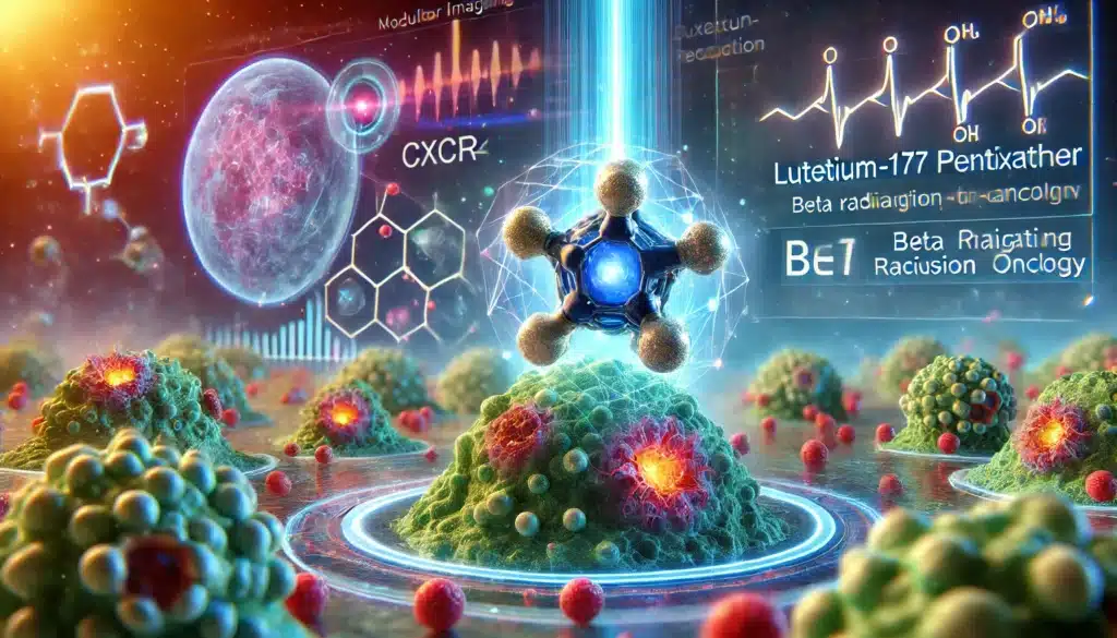
Lutetium-177 Pentixather targets CXCR4-expressing tumours, delivering precise beta radiation therapy to eliminate malignant cells effectively.
 By
By
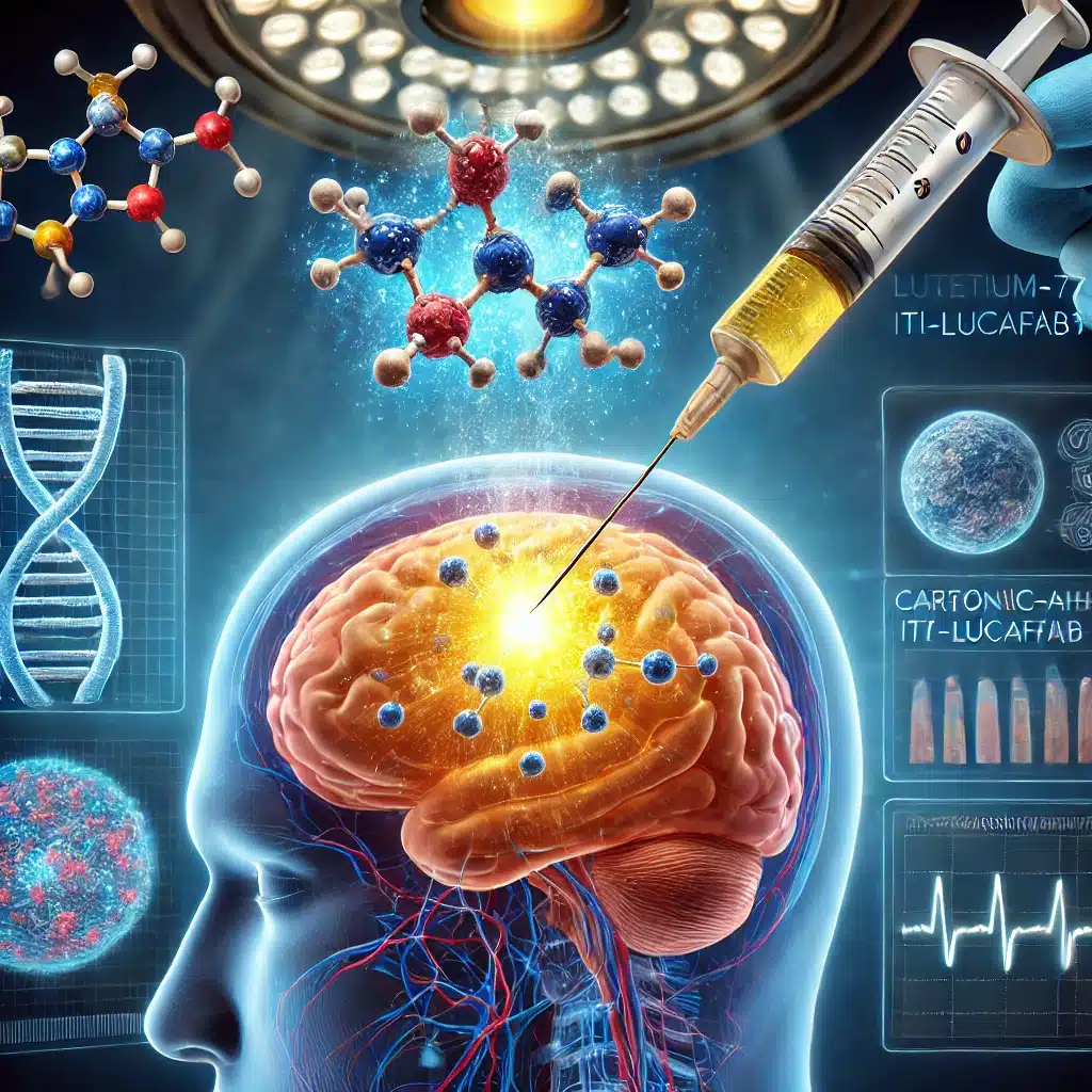
Targeting glioblastoma with Lutetium-177 ITM-31 offers precise intracavitary treatment, reducing recurrence by specifically addressing residual cancer cells post-surgery.
 By
By
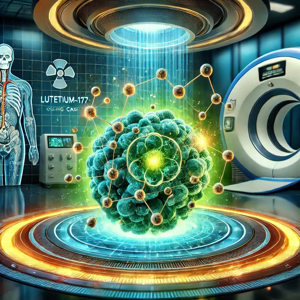
Lutetium-177 DPI-4452 pioneers precision radiotheranostics, revolutionising treatment for CAIX-expressing solid tumours through targeted imaging, therapy, and personalised oncology care advancements.
 By
By
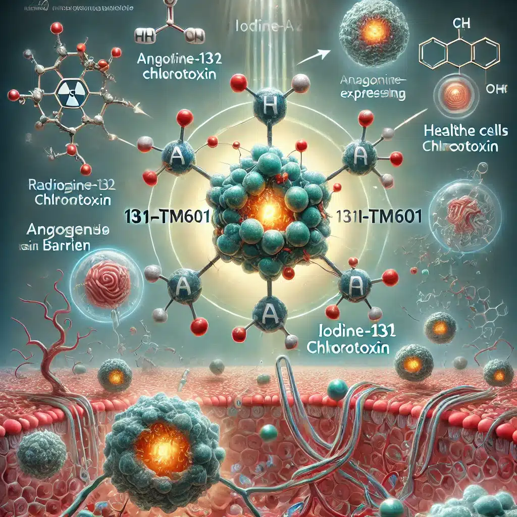
Iodine-131 TM601, a synthetic radiolabelled peptide, targets tumour cells expressing Annexin A2, delivering therapeutic radiation and exhibiting anti-angiogenic properties effectively.
 By
By
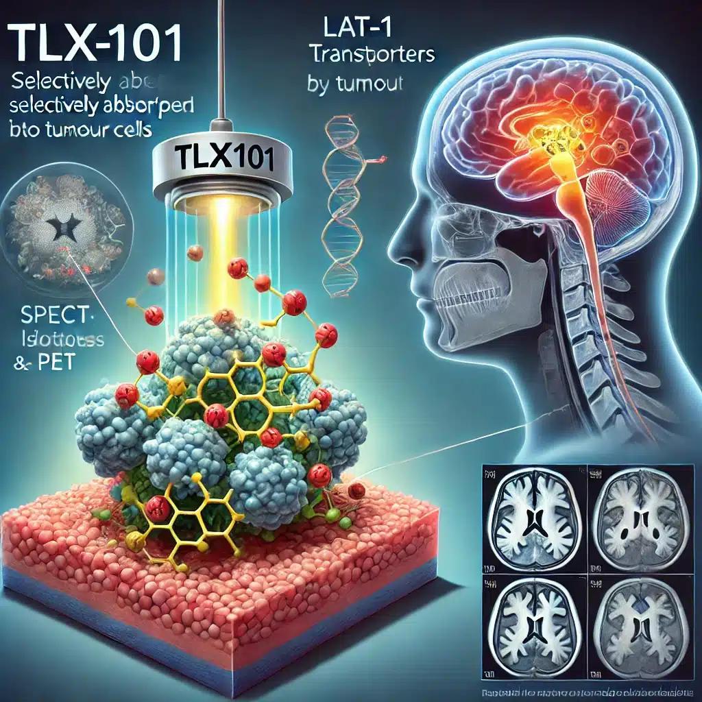
TLX101 is a revolutionary radiopharmaceutical targeting gliomas, offering dual imaging and therapy through tumour-selective uptake, enhancing treatment outcomes.
 By
By
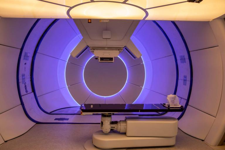
Proton therapy in cancer treatment offers precise targeting, reducing damage to healthy tissues and improving outcomes.
 By
By
Blastoma tumours require advanced imaging techniques like MRI, CT, PET, and ultrasound for accurate diagnosis and staging.
 By
By
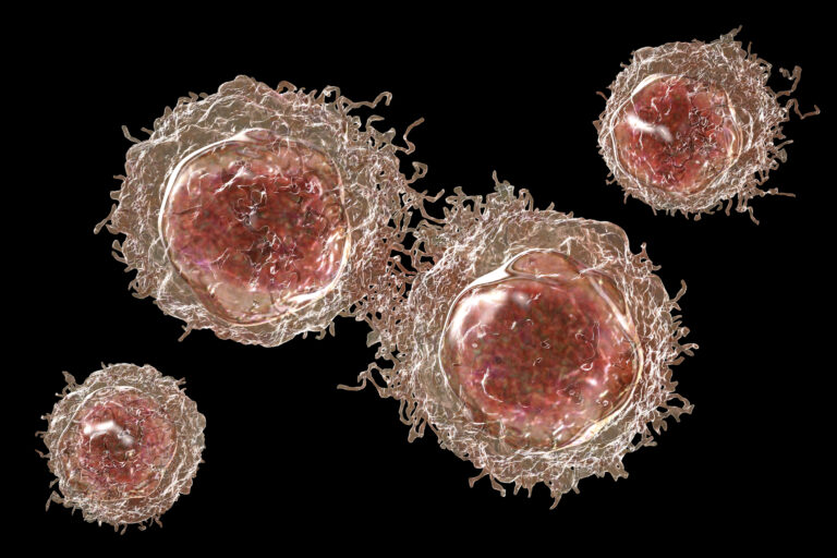
CAR T-cell therapy revolutionises cancer care, bringing hope where traditional treatments have been insufficient.
 By
By
Linear Energy Transfer is vital for understanding radiation’s biological effects and optimising cancer radiation therapy.
 By
By
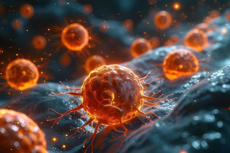
225Ac-FPI-1434 advances in clinical trials, targeting IGF-1R in chemo-resistant solid tumors with promising results.
 By
By
225Ac-DOTATOC advances GEP-NET treatment, offering targeted, effective therapy for patients resistant to traditional methods.
 By
By
Ultrasound Localization Microscopy offers unprecedented microvascular insights, transforming diagnosis and treatment through super-resolution imaging.
 By
By
Pancreatic cancer demands accurate prognosis, improved treatment strategies, and PET radiomic analysis for optimised post-resection outcomes.
 By
By
Lutetium-177 Dotatate selectively binds to SSTRs on NET cells, delivering targeted radiation, minimizing collateral damage, and reducing side effects.
By
Iodine-131 Iobenguane (I-131 MIBG), a radiopharmaceutical agent, enables early diagnosis and targeted treatment of neuroendocrine tumours, improving patient outcomes.
 By
By
Topics on PET imaging, automated radiosynthesis, breast cancer, prostate cancer therapy and ultrasound.