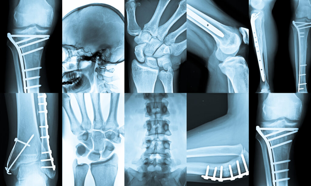
X-ray Phase Contrast Imaging
X-ray phase contrast imaging (XPCI) is an advanced imaging technique that enhances contrast by exploiting phase shifts in X-rays as they pass through different materials rather than relying solely on absorption differences. This approach provides superior visualisation of low-density materials, such as soft tissues, polymers, and biological specimens, which are often difficult to distinguish in conventional X-ray imaging.
Principles of XPCI
Traditional X-ray imaging relies on differences in attenuation, where denser materials absorb more X-rays, leading to contrast in the resulting image. However, many soft tissues have similar attenuation properties, making it challenging to differentiate them. XPCI, on the other hand, detects variations in the phase of the X-ray wavefront as it travels through a sample. This phase shift occurs due to differences in the refractive index of the material, which is a function of its electron density. By capturing these phase variations, XPCI provides a much higher level of detail, particularly for structures with low inherent contrast.
Several methods are used to extract phase information from an X-ray beam, with the most common techniques including propagation-based imaging, grating interferometry, analyser-based imaging, and edge-illumination phase contrast. Each technique has its advantages and is suited to different experimental setups and imaging needs.
XPCI Techniques
- Propagation-Based Imaging (PBI): Also known as in-line phase contrast, this method relies on free-space propagation of the X-ray beam after passing through the object. Phase differences lead to interference effects, which enhance edge contrast in the image. PBI is simple to implement but requires a high degree of spatial coherence in the X-ray source.
- Grating-Based Interferometry: This method uses a series of X-ray diffraction gratings to encode phase shifts and retrieve phase information. It is particularly useful for quantitative phase imaging and can be implemented with conventional X-ray sources.
- Analyser-Based Imaging (ABI): This technique employs a crystal analyser to detect changes in X-ray refraction. ABI is highly sensitive to phase variations and is often used in medical and material science applications.
- Edge-Illumination Phase Contrast: This method exploits the differential refraction of X-rays at material interfaces to enhance contrast. It is compatible with polychromatic X-ray sources and does not require highly coherent radiation, making it a practical choice for medical imaging.
Applications of XPCI
XPCI has significant potential in various fields, including medical diagnostics, materials science, and biological imaging. In medicine, it offers improved visualisation of soft tissues, aiding in the early detection of diseases such as cancer and lung conditions. In materials science, it provides detailed imaging of composite structures and nanomaterials.
With ongoing advancements in X-ray source technology and detector sensitivity, XPCI is becoming more accessible, bringing it closer to routine clinical and industrial applications.
home » X-ray Phase Contrast Imaging

 By
By