Breast Imaging
Breast imaging is a specialised area in diagnostic radiology that focuses on examining and assessing breast tissue to detect abnormalities, including breast cancer. The field encompasses a variety of imaging techniques and procedures, each with unique advantages and applications. These methods have significantly improved breast cancer’s early detection and treatment, saving countless lives.
Screening mammography is a preventive measure that employs low-dose X-rays to identify breast cancer before any symptoms become evident. By detecting cancerous changes early, this technique enables prompt intervention and treatment, thereby increasing the chances of a successful outcome. Women with a higher risk of breast cancer, such as those with a family history, may begin screening mammography earlier than those with average risk.
Diagnostic mammography, on the other hand, is employed when symptoms or abnormalities are already present. This method, which uses X-rays, examines the breast tissue to confirm any changes and determine the treatability of cancer. Diagnostic mammography is crucial in guiding subsequent medical decisions and treatment.
Breast tomosynthesis, also known as 3D mammography, is an advanced technique based on digital mammography. It captures multiple X-ray images from different angles and generates a three-dimensional reconstruction of the breast tissue. This technology provides radiologists with more detailed information and helps reduce false-positive findings compared to traditional mammography.
Ultrasound imaging is another essential tool in breast imaging. It uses high-frequency sound waves to differentiate between cysts, which are fluid-filled sacs, and solid lumps, which may be cancerous. Unlike mammography, ultrasound does not involve radiation exposure and is often used with other imaging modalities.
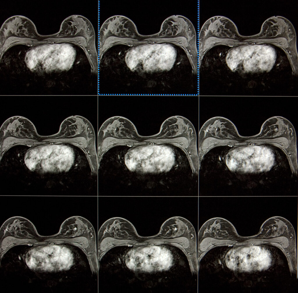
Breast magnetic resonance imaging (MRI) is a highly sensitive technique to identify early-stage breast cancer, especially in women with dense breast tissue or a high risk of developing the disease. It can also determine the extent of cancer following a diagnosis.
In addition to these primary imaging techniques, several other specialised procedures can aid in diagnosing and treating breast cancer. Breast hook wire localisation helps surgeons locate and remove small lesions during surgery. Breast fine needle aspiration (FNA) involves extracting cells from a suspicious area using a thin needle for further analysis. Breast core biopsy and vacuum-assisted core biopsy obtain tissue samples using a larger needle or vacuum device, allowing for a more accurate diagnosis.
home » breast imaging

 By
By