Heart imaging has undergone remarkable advances over the past few years. Advances in hardware, software, computing power and data science are changing how clinicians diagnose, monitor and treat cardiovascular disease. These innovations are improving accuracy, reducing invasive procedures and supporting more personalised approaches to patient care. This article explores the most important developments now influencing practice, covering CT, MRI, echocardiography, image analysis and the growing role of artificial intelligence.
Photon-Counting CT and High-Resolution Coronary Imaging
One of the most talked-about developments in cardiac imaging is the introduction of photon-counting CT (PCCT). Traditional CT scanners measure the total energy of X-rays that pass through the body, whereas PCCT registers each photon individually. This approach offers substantially better spatial resolution, improved contrast and reduced image noise. In cardiac applications, this means a clearer view of coronary arteries, plaque features and stents.
PCCT scanners also help tackle a persistent challenge in coronary CT: heavy calcification. In patients with extensive calcium deposits, standard CT can produce “blooming” artefacts, making it harder to assess the degree of narrowing. PCCT reduces this artefact, enabling a more confident assessment. This is particularly beneficial for older patients or those with long-standing vascular disease, for whom diagnostic uncertainty has traditionally been higher.
In addition, PCCT opens the door to multi-energy imaging. By separating X-rays into different energy levels, the scanner can distinguish tissues and materials that previously appeared similar. This will help with plaque characterisation, the identification of vulnerable lesions, and the improved evaluation of stents. As scanner availability increases, cardiac CT is likely to move even further towards the centre of coronary assessment.
A related development is the routine use of CT-derived fractional flow reserve (FFRCT). This technique uses computational fluid dynamics to predict the likelihood that a coronary stenosis is causing significant blood-flow limitation. Recent work within the NHS has shown that FFRCT can reduce the number of invasive coronary angiograms required. By improving triage, it also shortens waiting lists and lowers costs.
The Expanding Capabilities of Cardiac MRI
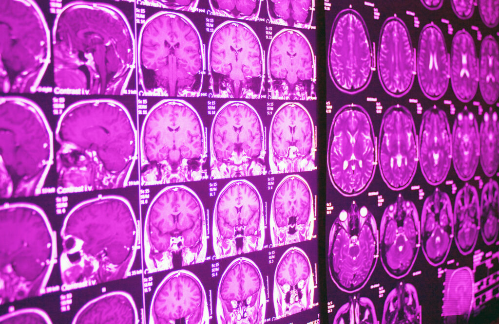
Cardiac magnetic resonance (CMR) remains the most versatile and informative modality for functional assessment and tissue characterisation. Over the past few years, CMR has grown even more capable with the refinement of mapping techniques, free-breathing protocols and compressed-sensing approaches that speed up data acquisition.
Quantitative Mapping
T1- and T2-mapping have become central to the evaluation of cardiomyopathies and myocarditis. These maps provide absolute values for tissue properties, helping clinicians detect diffuse disease that might be missed on standard imaging. Extracellular volume (ECV) mapping goes further, estimating the proportion of the myocardium composed of extracellular matrix. Elevated ECV is a marker of fibrosis, infiltration or oedema, offering a sensitive measure of early disease.
Real-Time and Free-Breathing Imaging
A significant barrier for many patients during MRI is the need for repeated breath-holds. New real-time imaging sequences that acquire data continuously without breath-holding are changing this. These sequences are especially helpful for patients with heart failure, arrhythmias or limited ability to follow complex instructions. They also improve image consistency, leading to more reliable quantification.
Magnetic Resonance Fingerprinting
Magnetic resonance fingerprinting (MRF) is an emerging technique that aims to simultaneously extract multiple tissue parameters. Instead of acquiring dedicated sequences for T1, T2 and other maps, MRF uses a single acquisition with varying imaging properties. Advanced reconstruction algorithms then match these “fingerprints” to a large database to determine tissue characteristics. While still in early clinical evaluation, MRF has the potential to significantly shorten scan times.
Growing Impact in Cardiomyopathies
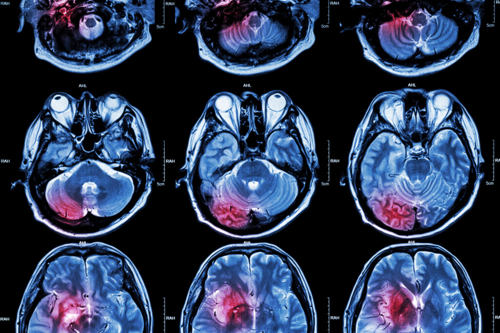
CMR is now central to the evaluation of hypertrophic, dilated and arrhythmogenic cardiomyopathies. Its ability to visualise scars, fibrosis, and structural abnormalities supports diagnosis, risk assessment, and family screening. Increasingly, CMR findings are being integrated with genetic information to guide patient management. This combination helps identify individuals at higher risk of arrhythmias, supporting decisions about implantable cardioverter-defibrillators.
Echocardiography: From 2D Images to 3D Quantification and Advanced Strain
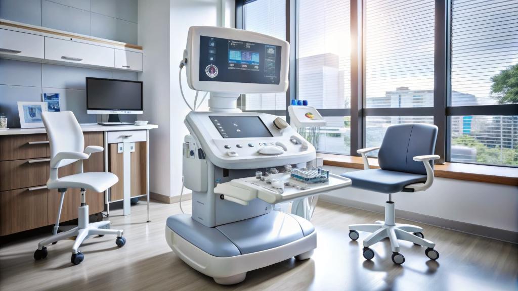
Echocardiography remains the most widely used cardiac imaging technique thanks to its portability, safety and speed. Recent advances, however, have shifted it from a predominantly qualitative tool to a more quantitative and reproducible one.
Three-Dimensional Echocardiography
3D echo has become increasingly robust, offering a more accurate assessment of ventricular volumes and ejection fraction. This is particularly valuable for right-ventricular evaluation, where 2D measurements often struggle due to the right ventricle’s complex shape. 3D techniques also provide improved views of valve anatomy, which aid planning for structural interventions, such as transcatheter mitral and tricuspid procedures.
Large reference datasets for 3D left-atrial and right-ventricular values have recently been established. These reference values will support better interpretation of individual scans across all patient groups.
Strain Imaging and Early Detection of Dysfunction
Speckle-tracking echocardiography, especially global longitudinal strain (GLS), has become mainstream. GLS is more sensitive than ejection fraction for detecting early myocardial impairment. It is widely used for monitoring chemotherapy, evaluating valvular disease, and evaluating cardiomyopathy. Advances in automation have made GLS quicker to obtain, improving its clinical uptake.
AI and Machine Learning in Cardiac Imaging
One of the most influential trends reshaping the field is the integration of artificial intelligence across imaging modalities. AI now contributes to nearly every stage of the imaging pathway: acquisition, reconstruction, segmentation, quantification and diagnosis.
Workflow Acceleration and Automation
Deep-learning reconstruction algorithms allow lower-dose CT scans with maintained image quality. In MRI, AI speeds up undersampled acquisitions, cutting scan times and improving patient comfort. Automated segmentation tools now assist with chamber volume calculations, valve assessment and tissue quantification, reducing clinician workload and improving consistency.
Diagnostic Support
AI systems trained on echo, CT or CMR data can highlight abnormalities, flag measurements that fall outside expected ranges and even propose likely diagnoses. Of particular interest is the use of AI to analyse standard tests, such as ECGs, to predict structural heart disease. Studies have shown that machine-learning models can identify individuals who may benefit from further cardiac imaging, improving early detection.
Tissue Characterisation and Scar Segmentation
In CMR, late gadolinium enhancement (LGE) is the standard method for detecting fibrosis or scar tissue. Traditionally, segmentation of LGE areas has been manual and time-consuming. AI-based segmentation algorithms now provide automated, reproducible contouring, enabling more reliable quantification. This has significant implications for prognostic scoring in cardiomyopathies and post-infarct patients.
Plaque Analysis and Risk Prediction
In coronary CT, AI is advancing plaque characterisation by classifying plaque composition, identifying high-risk features and supporting functional assessment. Combining anatomical and functional information from CT scans with machine-learning models enables more refined risk prediction. This could eventually support personalised decisions about preventive therapy.
Developments in Structural Heart Disease Imaging
The growth of transcatheter valve interventions has pushed imaging to the forefront of pre-procedural planning. High-resolution CT, 3D echo and CMR all contribute unique information.
Aortic Valve Disease
CT is now the standard for planning transcatheter aortic valve implantation (TAVI). It provides precise measurements of annulus size, valve morphology, vascular access and calcium distribution. As spatial resolution improves, assessment becomes more accurate, reducing the risk of complications.
Mitral and Tricuspid Interventions
3D transoesophageal echocardiography offers exceptionally detailed views of the mitral and tricuspid valves. This supports the selection of appropriate devices for transcatheter edge-to-edge repair, valve replacement and annuloplasty. CT provides complementary information on leaflet anatomy, subvalvular structures, and landing zones for devices.
Post-Procedural Assessment
CMR is increasingly used to evaluate regurgitant volumes and ventricular remodelling after valvular intervention. Its accuracy in flow quantification helps determine whether further treatment is required.
Cardiac Imaging in Ischaemic Heart Disease
Coronary disease remains a major cause of global morbidity and mortality. Imaging innovations are refining its diagnosis and helping avoid unnecessary invasive procedures.
Perfusion Imaging
CT perfusion and stress CMR are becoming more widely available. Both modalities assess myocardial blood flow under stress, improving identification of flow-limiting lesions. Combining perfusion with anatomical CT or CMR data offers a comprehensive evaluation of both structure and function in a single visit.
Reducing Invasive Procedures
The integration of FFRCT into the clinical pathway is reducing the need for invasive coronary angiography. In selected patients, CT alone can now guide management with high accuracy. This improves the patient experience, reduces procedural risk, and conserves healthcare resources.
Looking Ahead: Integration and Personalised Care
While individual innovations are important, the future of heart imaging lies in integration. Bringing together CT, MRI, echocardiography, ECG data, blood biomarkers, and genetic information will create a more complete picture of cardiovascular health. Advances in cloud computing, interoperability and AI will make this level of data fusion increasingly achievable.
Standardisation is another major priority. As new techniques become mainstream, consistent acquisition protocols, reference values, and reporting methods will be essential. This will allow clinicians to compare results across centres and time points with greater confidence.
Finally, accessibility remains a challenge. Some of the newest technologies, such as PCCT and advanced MRI, are currently available only at specialist centres. Wider adoption will depend on cost, training and local infrastructure.
Disclaimer
This article is provided for general information and educational purposes only. It is not intended to offer medical advice, establish clinical guidance, or replace consultation with qualified healthcare professionals. The technologies, methods and research developments described are subject to ongoing evaluation, regulatory review and variation between healthcare systems. Clinical decisions should always be based on a full assessment by appropriately trained clinicians, supported by locally approved protocols and the most current evidence. Open MedScience does not guarantee the completeness, accuracy or continued relevance of the content and accepts no liability for any loss or harm arising from reliance on the information presented. Readers should seek professional medical advice for any questions or concerns relating to diagnosis, treatment or patient care.
You are here: home » diagnostic medical imaging blog »


