Modern medicine owes much of its power to the ability to see inside the human body. From identifying fractures and tumours to tracking disease progression and guiding surgery, medical imaging has redefined both diagnosis and treatment. Yet this extraordinary capability began not with advanced computers or artificial intelligence, but with the curiosity and persistence of a few determined scientists who sought to understand the unseen.
The story of medical imaging spans more than a century and bridges physics, chemistry, biology, engineering, and computing. It is a story of breakthroughs born of observation, accidents that became revolutions, and the human drive to see what was once invisible. The pioneers behind these discoveries changed not only medicine but the way humanity perceives itself.
Wilhelm Conrad Röntgen: The Spark That Started It All
The dawn of medical imaging began almost by chance. In November 1895, German physicist Wilhelm Conrad Röntgen was experimenting with cathode rays — streams of electrons in vacuum tubes — when he noticed that a fluorescent screen nearby glowed, even though the tube was shielded. Whatever was passing through the covering was invisible yet capable of penetrating matter. Röntgen called them “X-rays” to signify their unknown nature.
His curiosity led him to place various objects between the tube and a photographic plate. When he asked his wife, Anna Bertha, to rest her hand in the path of the rays, the resulting image showed the bones of her hand and the faint circle of her wedding ring. It was both eerie and astonishing. The image—the first X-ray photograph—revealed the hidden architecture of the human body.
Within months, hospitals across Europe and America were using X-rays to locate bullets, fractures, and swallowed objects. For the first time in history, doctors could peer beneath the skin without making an incision. In 1901, Röntgen received the first Nobel Prize in Physics for his discovery, and medical imaging was born.
Marie and Pierre Curie: Harnessing Invisible Energy
The next great leap came from another remarkable scientific partnership — Marie and Pierre Curie. While investigating the mysterious emissions from uranium salts, the Curies discovered two new elements: polonium and radium. Their pioneering research introduced the concept of radioactivity and revealed that certain materials emitted powerful, penetrating rays capable of exposing photographic film and affecting living tissue.
The Curies’ discoveries transformed both science and medicine. Radium soon became a key source for radiographic imaging and radiotherapy, while their research laid the groundwork for nuclear medicine decades later. Yet Marie Curie’s contribution extended beyond theory. During the First World War, she developed mobile X-ray units — known as “Little Curies” — and trained nurses and doctors to use them on the front lines. These vehicles allowed field medics to locate bullets and fractures quickly, saving thousands of lives.
Marie Curie remains the only person ever to win Nobel Prizes in two different sciences — Physics and Chemistry. Her work combined intellectual brilliance with compassion, embodying the spirit of science in the service of humanity.
Allan MacLeod Cormack and Godfrey Hounsfield: Seeing in Slices
By the mid-20th century, X-rays could show the body in two dimensions, but overlapping structures often obscured vital details. The next breakthrough came from the combination of mathematics, physics, and emerging computing technology.
Allan MacLeod Cormack, a South African physicist, developed mathematical methods to reconstruct cross-sectional images from X-ray data. Around the same time, British engineer Godfrey Hounsfield, working at EMI Laboratories in London, independently conceived the idea of scanning the body from multiple angles and using a computer to piece the data together.
The result was computed tomography (CT), a technique that produces detailed “slices” of the body by measuring the absorption of X-rays at thousands of points. The first clinical CT scanner, installed at Atkinson Morley’s Hospital in 1971, produced astonishingly clear images of the brain, revealing strokes, tumours, and haemorrhages that were invisible on conventional X-rays.
CT scanning revolutionised diagnostic medicine almost overnight. It provided doctors with a non-invasive window into the brain, chest, and abdomen, transforming the speed and precision of diagnosis. In 1979, Cormack and Hounsfield shared the Nobel Prize in Physiology or Medicine for their invention. The world of radiology had moved from flat images to three-dimensional visualisation.
Raymond Damadian: Listening to the Body’s Magnetic Pulse
While CT was revolutionising radiology, another visionary was exploring a radically different idea — imaging with magnetism rather than radiation. In the early 1970s, American physician and biophysicist Raymond Damadian discovered that cancerous tissues could be distinguished from healthy ones using nuclear magnetic resonance (NMR). He proposed harnessing this property to create images of the human body.
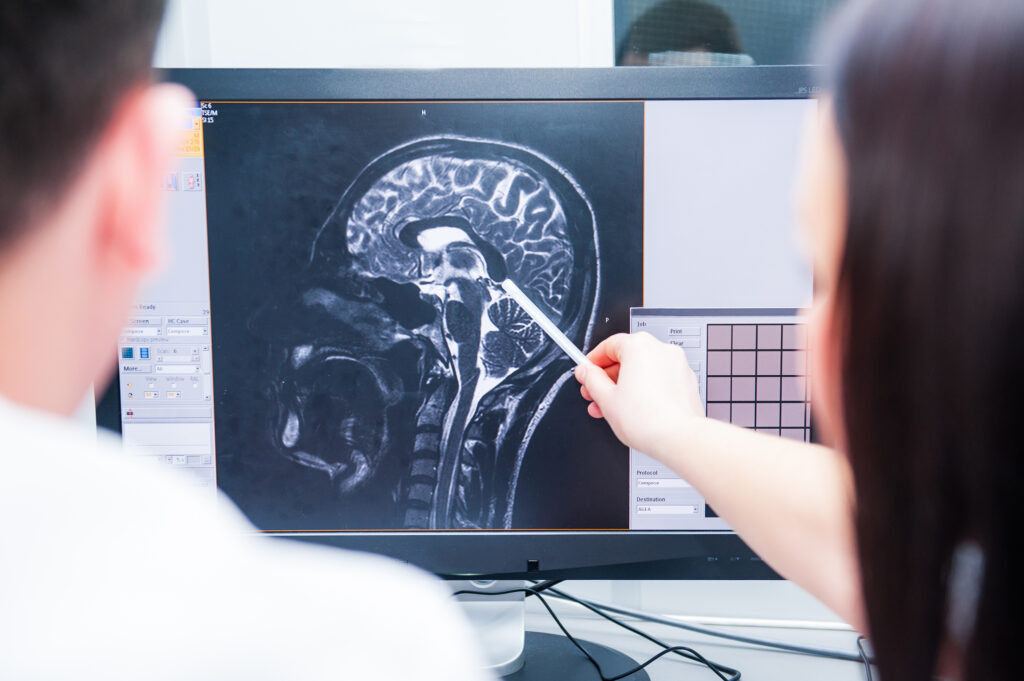
Damadian’s team built the first whole-body magnetic resonance scanner, which successfully produced an image in 1977. Magnetic resonance imaging (MRI) relies on magnetic fields and radio waves to detect the response of hydrogen nuclei within water and fat molecules in the body. The result was a detailed, radiation-free picture of internal anatomy — particularly soft tissues that X-rays struggled to capture.
MRI transformed clinical diagnostics. It became the gold standard for neurological, musculoskeletal, and cardiovascular imaging. Its ability to visualise the brain, heart, and joints in exquisite detail has made it indispensable to modern medicine. Unlike CT, MRI can differentiate between subtle variations in soft tissue, providing insights that often determine life-saving treatment decisions.
Ian Donald: The Sound of a Beating Heart
The story of imaging would be incomplete without recognising the contribution of Ian Donald, the Scottish obstetrician who pioneered medical ultrasound. In the 1950s, Donald borrowed sonar equipment — originally used to detect submarines — and adapted it for medical purposes. By transmitting high-frequency sound waves through tissue and measuring their echoes, he was able to create images of internal organs.
Donald’s experiments led to the first practical ultrasound scanner, which soon became the cornerstone of obstetric care. For the first time, clinicians could observe the developing foetus safely and in real time, without exposure to radiation. Parents could see their unborn child, hear its heartbeat, and witness early life unfold before their eyes.
Ultrasound’s influence extended far beyond pregnancy care. It became essential in cardiology, oncology, and emergency medicine, offering a quick, safe, and inexpensive method to view the body in motion. Donald’s ingenuity transformed sound into sight, proving once again that the tools of one discipline could illuminate another.
Paul Lauterbur and Peter Mansfield: Perfecting MRI
Although Raymond Damadian’s work demonstrated MRI’s potential, it was the contributions of Paul Lauterbur and Peter Mansfield that turned it into a practical medical tool.
In 1973, American chemist Paul Lauterbur introduced the use of magnetic field gradients, enabling spatial information to be encoded and reconstructed into images. Two years later, British physicist Peter Mansfield developed mathematical techniques that dramatically improved imaging speed and resolution, making MRI suitable for routine clinical use.
Their work converted Damadian’s theoretical concept into a viable diagnostic method. MRI scanners spread rapidly across hospitals during the 1980s, offering doctors an unprecedented view of the human body. For their achievements, Lauterbur and Mansfield received the 2003 Nobel Prize in Physiology or Medicine.
Together, these scientists reshaped modern radiology, demonstrating how collaborative innovation could bridge theory and application.
Rosalind Franklin and Marie Curie: Women Who Illuminated the Invisible
Women have played vital roles in the development of imaging, though their contributions were not always recognised in their lifetimes. Rosalind Franklin, a British chemist and X-ray crystallographer, produced the now-famous “Photograph 51,” which revealed the helical structure of DNA. Her precision and technical mastery with X-ray diffraction provided the missing evidence that enabled Watson and Crick to model the DNA double helix — one of the most significant discoveries in science.
Marie Curie’s influence, of course, remains immense. Beyond her discoveries, her insistence on scientific integrity and her humanitarian application of research set a precedent for generations of scientists. Their shared legacy underscores that the progress of medical imaging has always depended on intellect, perseverance, and courage—not gender or status.
From Film Plates to AI Pixels: The Continuing Revolution
Medical imaging has evolved far beyond its early mechanical roots. The 20th century brought film-based radiography, fluoroscopy, CT, MRI, ultrasound, and PET imaging. The 21st century is defined by digital transformation and integration. Hybrid systems like PET-CT and PET-MRI combine functional and anatomical imaging, allowing doctors to map metabolism, blood flow, and cellular activity alongside structural detail.
Artificial intelligence is now the newest frontier. Deep learning algorithms can analyse vast volumes of imaging data, detect subtle abnormalities, and assist radiologists in making faster, more accurate diagnoses. These systems are not replacing clinicians but enhancing their capabilities — reducing errors, improving workflow, and personalising treatment planning.
In parallel, molecular imaging is allowing scientists to visualise disease at the biochemical level, revealing the earliest changes in cancer, neurodegeneration, and cardiovascular conditions. Imaging is no longer limited to anatomy; it is now exploring metabolism, genetics, and cellular function.
Looking Ahead: Vision, Innovation, and Humanity
The journey from Röntgen’s flickering screen to today’s AI-powered diagnostics represents one of the most extraordinary transformations in human knowledge. Yet, at its core, the story of medical imaging remains one of curiosity, persistence, and collaboration.
Each pioneer — from Röntgen and the Curies to Hounsfield, Damadian, Donald, and Franklin — shared a willingness to look beyond the visible. Their discoveries often began as side experiments, unexpected observations, or ideas dismissed by their peers. But through determination and imagination, they changed how medicine understands the body and, ultimately, how humanity understands itself.
As the next generation of scientists and clinicians continues to refine imaging technologies, new questions arise. How can we make advanced imaging accessible globally? How can AI assist diagnosis ethically? How do we balance precision with compassion in an increasingly digital world? The pioneers of the past offer an answer: innovation must always serve humanity.
Conclusion
The history of medical imaging is a testament to human ingenuity. From Röntgen’s first X-ray image to the breathtaking clarity of modern MRI and PET scans, each discovery has built upon the last, extending our capacity to diagnose, heal, and understand. These pioneers transformed invisible phenomena into tools that save lives daily.
Today, as we enter an era defined by artificial intelligence, quantum detection, and molecular visualisation, their spirit endures. They remind us that every great leap in science begins with the courage to look deeper — to illuminate what lies beyond sight.
Disclaimer
This article, “The Pioneers of Medical Imaging: The People Who Changed the Way We See Ourselves”, is published by Open MedScience for educational and informational purposes only. It is not intended to provide medical or professional advice. While efforts have been made to ensure accuracy, Open MedScience accepts no responsibility for any errors or omissions, or for any outcomes resulting from the use of this information.
home » blog » education »