Summary: Ultrasound has become an invaluable tool in the detection and management of cancer, offering a non-invasive, cost-effective, and widely accessible diagnostic option. With the ability to distinguish solid from cystic lesions and identify tumour characteristics in real time, ultrasound plays a pivotal role across a range of cancers, particularly breast, thyroid, liver, and prostate cancers. This article explores the fundamentals of ultrasound technology, its applications in oncology, limitations, advancements, and the future potential of this imaging technique in enhancing early cancer detection.
Introduction: Understanding Ultrasound in Medical Imaging
Ultrasound imaging, also known as sonography, is a diagnostic technique that uses high-frequency sound waves to create images of structures within the body. In the field of oncology, ultrasound provides critical insights into tumour detection and assessment, assisting in staging, biopsy guidance, and treatment monitoring. Due to its high sensitivity in detecting structural abnormalities, ultrasound is particularly beneficial in screening programmes for high-risk populations and provides a crucial tool in early cancer detection.
The Science Behind Ultrasound Technology
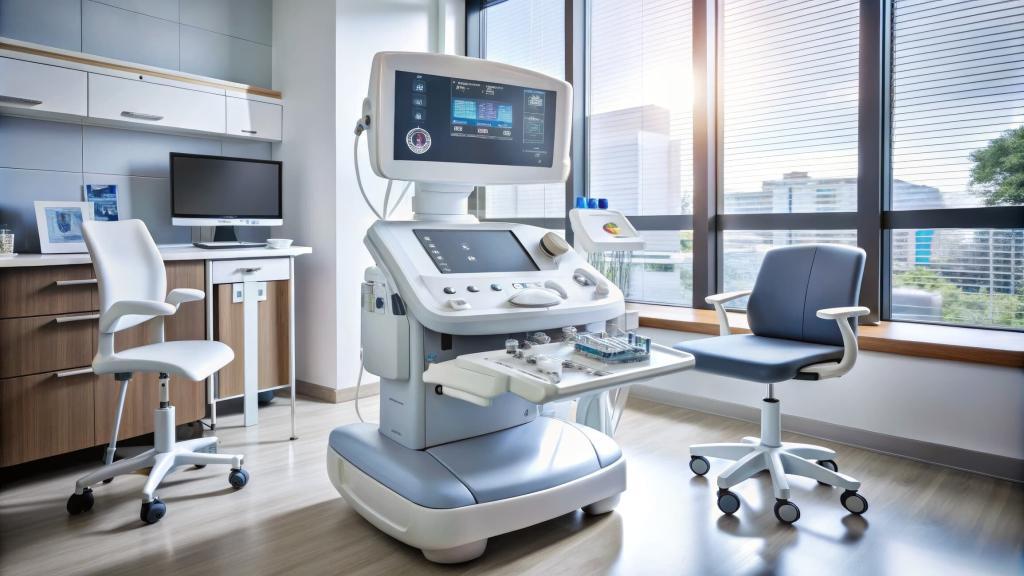
Ultrasound technology operates by transmitting sound waves through body tissues. These waves, which are beyond the range of human hearing, bounce off tissues with different densities, producing echoes. A transducer then captures these echoes and translates them into visual images on a screen. The technique is entirely safe, as it uses no ionising radiation, making it suitable for repeated examinations and particularly valuable for tracking tumour changes over time.
Key advantages of ultrasound include:
- Real-time Imaging: Enables immediate observation and evaluation.
- No Radiation Exposure: Suitable for pregnant individuals and frequent monitoring.
- Cost-Effective and Accessible: Widely available across clinical settings and affordable compared to other imaging modalities.
However, ultrasound has limitations, notably its dependence on operator skill and difficulties in imaging areas with dense or thick tissue structures. Despite these constraints, it is a powerful initial diagnostic tool, especially in areas where other imaging modalities, like MRI or CT scans, may be less accessible or practical.
Applications of Ultrasound in Cancer Detection
Breast Cancer Detection
Ultrasound is a primary diagnostic tool in breast cancer screening, especially in younger women with denser breast tissue, where mammograms may be less effective. The technique enables clinicians to distinguish between solid masses and cystic lesions, often detected as lumps, and to assess tumour characteristics such as size, shape, and vascularity.
Ultrasound is also instrumental in guiding biopsies to ensure accurate sampling from suspicious areas. Studies have shown that in combination with mammography, ultrasound significantly improves the accuracy of breast cancer detection, particularly in cases where mammography alone might miss smaller or more challenging lesions.
Thyroid Cancer Detection
In recent years, ultrasound has become the preferred method for detecting and evaluating thyroid nodules, which are prevalent but often benign. Ultrasound helps assess the nodule’s size, echotexture, and vascularity and can identify suspicious features such as microcalcifications or irregular borders indicative of malignancy.
High-resolution ultrasound is also used to guide fine-needle aspiration biopsies of thyroid nodules, improving diagnostic accuracy. Notably, the American Thyroid Association recommends ultrasound as a first-line imaging modality in the detection and evaluation of thyroid nodules.
Liver Cancer Detection
Liver cancer, including hepatocellular carcinoma (HCC), is often screened in high-risk populations, such as individuals with chronic hepatitis or cirrhosis, through ultrasound. Ultrasound is non-invasive and can detect early tumour formations, which is critical in liver cancer due to its aggressive nature.
Doppler ultrasound further enhances liver cancer detection by assessing blood flow within the liver and tumour, identifying characteristic vascular patterns of malignancy. It can also monitor for liver metastases in patients with other primary cancers. Despite its limitations in detecting smaller lesions, ultrasound remains a mainstay in liver cancer surveillance, often paired with alpha-fetoprotein blood tests in high-risk groups.
Prostate Cancer Detection
Transrectal ultrasound (TRUS) is widely used in prostate cancer diagnostics, providing valuable images for assessing prostate volume, structure, and irregularities. TRUS is frequently employed in guiding biopsies to sample prostate tissue accurately and has become integral in the diagnostic pathway for prostate cancer.
While MRI has become more prominent in detecting and staging prostate cancer, ultrasound remains crucial in initial screenings, especially in resource-limited settings. Research is ongoing to improve ultrasound accuracy for prostate cancer, such as contrast-enhanced and elastography ultrasound, which can better differentiate malignant from benign tissues.
Gynaecological Cancers
Ultrasound is a primary diagnostic tool in detecting and evaluating ovarian and endometrial cancers. Transvaginal ultrasound (TVUS) allows for high-resolution imaging of the ovaries, uterus, and surrounding tissues, which aids in identifying masses, cysts, or other abnormalities.
In ovarian cancer, ultrasound can help detect abnormal growths, distinguish benign from malignant lesions, and provide information on tumour size and spread. Doppler ultrasound also helps assess blood flow to suspicious areas, enhancing diagnostic accuracy. As gynaecological cancers are often asymptomatic in early stages, ultrasound provides a non-invasive screening tool, especially for high-risk patients.
Limitations of Ultrasound in Cancer Detection
While ultrasound is a versatile and invaluable imaging tool, it has some limitations that must be acknowledged. The image quality can be affected by the skill and experience of the operator, as well as by patient factors such as body habitus. Ultrasound is also less effective at visualising deep-seated or obscured organs, as sound waves cannot penetrate bone or dense tissues well.
Moreover, ultrasound’s sensitivity in detecting very small tumours can be lower compared to CT or MRI scans. This limitation is particularly notable in areas like the lung, where ultrasound is generally not used due to interference from air-filled structures. Additionally, while ultrasound can suggest the presence of malignancy, it cannot provide definitive diagnoses, which require histological analysis through biopsy.
Technological Advancements in Ultrasound for Cancer Detection
Recent advancements in ultrasound technology have significantly improved its capabilities in oncology. These include:
Doppler Ultrasound
Doppler ultrasound assesses blood flow, which is crucial in differentiating benign from malignant tumours, as cancers often exhibit characteristic vascular patterns. Colour Doppler and power Doppler have enhanced the accuracy of ultrasound in assessing tumour vascularity, especially in liver and thyroid cancers.
Elastography
Ultrasound elastography is a newer technique that evaluates tissue stiffness, offering additional diagnostic information for cancer detection. Since malignant tumours tend to be stiffer than benign lesions, elastography is particularly useful in detecting breast, liver, and thyroid cancers. Studies have shown that elastography improves the specificity of ultrasound, reducing unnecessary biopsies.
Contrast-Enhanced Ultrasound (CEUS)
Contrast-enhanced ultrasound involves the injection of microbubble contrast agents, which improve the visualisation of blood flow and vascular patterns within tumours. CEUS has shown promise in liver cancer detection, allowing for a more detailed assessment of tumour vascularity without the need for radiation or contrast agents used in CT and MRI. CEUS is becoming more widely adopted for its ability to enhance ultrasound’s sensitivity in detecting small and less conspicuous tumours.
Artificial Intelligence (AI) Integration
AI is increasingly being integrated into ultrasound systems to enhance cancer detection. Algorithms can assist in identifying and characterising suspicious lesions, reducing operator dependency and improving diagnostic accuracy. AI-driven ultrasound is being explored in breast, thyroid, and prostate cancer screenings, where early studies show potential for enhancing diagnostic precision and efficiency.
Future Directions: Ultrasound’s Expanding Role in Oncology
Ultrasound’s role in cancer detection continues to expand, with ongoing research focused on improving image quality, diagnostic accuracy, and integration with other modalities. Innovations such as portable and handheld ultrasound devices are making it easier to use this technology in remote or low-resource settings, where access to other imaging modalities may be limited.
Future developments in ultrasound-guided therapies are also promising, with high-intensity focused ultrasound (HIFU) showing potential in treating certain tumours non-invasively. The use of ultrasound in combination with other imaging modalities, like MRI, is being explored to create more comprehensive and accurate diagnostic pathways, particularly in challenging cases like prostate and liver cancers.
Conclusion
Ultrasound has firmly established itself as a vital tool in the detection and management of various cancers. Its non-invasive, cost-effective, and widely accessible nature makes it suitable for early detection, biopsy guidance, and ongoing tumour monitoring. Despite certain limitations, advancements in ultrasound technology and AI integration are enhancing its accuracy and expanding its applications in oncology.
As research continues, ultrasound is expected to play an even more significant role in cancer diagnostics, particularly in underserved areas. With its potential for integration into multimodal diagnostic approaches and use in ultrasound-guided therapies, ultrasound holds a promising future in improving early cancer detection, patient outcomes, and overall cancer care.
Disclaimer
The information presented in this article, Ultrasound Unveiled: Transforming Cancer Detection and Diagnosis, is intended for general informational and educational purposes only. It is not a substitute for professional medical advice, diagnosis, or treatment. The content reflects current knowledge and understanding at the time of publication (9 November 2024) and may be subject to change as further research and technological advancements emerge.
Readers are advised to consult qualified healthcare professionals for any concerns or questions regarding medical conditions or diagnostic procedures. Open Medscience does not accept any liability for decisions made based on the information provided in this article. The views expressed are those of the authors and contributors and do not necessarily reflect the official policy or position of Open Medscience or its affiliates.
home » blog » medical imaging modalities »


