Summary: Artificial intelligence is transforming medical imaging by automating repetitive tasks, including scan triage, measurements, and quality checks. While AI in radiology now matches or outperforms humans in narrow, well-defined tasks, it lacks the broader contextual reasoning, adaptability, and accountability of trained professionals. Current AI tools are assistive rather than autonomous, operating under strict regulatory oversight. The near future will see AI deeply embedded in imaging workflows, streamlining processes and improving consistency, but not replacing human expertise. The long-term outlook is one of collaboration, with AI acting as a powerful partner to deliver faster, safer, and more consistent care.
Keywords: Artificial intelligence in radiology, AI medical imaging, Radiology automation, Imaging workflow AI, AI in healthcare UK, Medical imaging future.
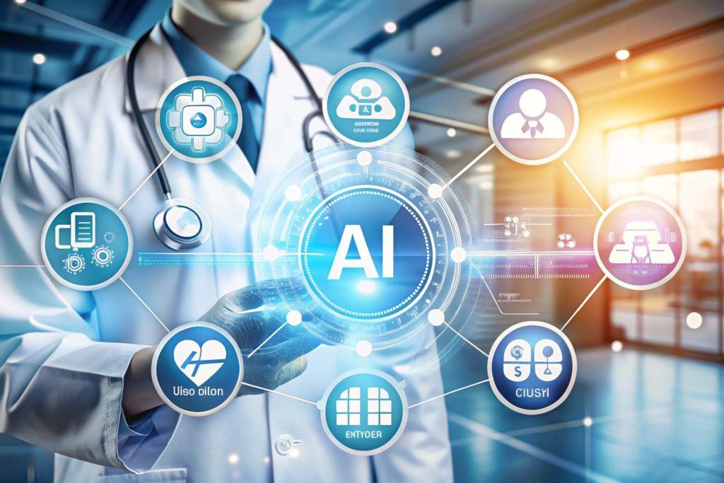
The Future of AI in Healthcare UK
The question of whether artificial intelligence will one day replace radiologists and reporting radiographers has moved beyond academic speculation and into the arenas of hospital boardrooms, healthcare policy debates, and public discussion. To answer it meaningfully, one must first clarify what “take over” actually entails in the context of medical imaging, then examine the genuine capabilities of AI systems as they are currently deployed, and finally consider the realities of day-to-day clinical practice.
Automation is already reshaping parts of the radiology workflow, particularly in repetitive technical processes. Yet the delivery of patient care continues to demand the clinical judgement, nuanced interpretation, and human accountability of trained professionals. The current wave of AI innovation in imaging is impressive in its ability to execute narrow tasks with speed and consistency; however, its role today is assistive rather than autonomous. This article examines the current state of AI, its limitations, and how it is likely to integrate into the future of radiology, without displacing the human expertise that underpins safe and effective care.
Understanding the Medical Imaging Workflow
Medical imaging is not a single act of image interpretation; it is a complex chain of interlinked steps that begin long before a scan is reviewed on a reporting screen. It begins with selecting the appropriate imaging test for a given clinical indication, ensuring that the requested examination is both necessary and safe. The next stage involves acquiring the image itself, which must be performed to a high and consistent standard to ensure diagnostic reliability. Once images are produced, the professional must identify relevant abnormalities, interpret them in light of the patient’s medical history, physical examination findings, and any prior imaging, and then synthesise these elements into a coherent conclusion. Finally, this information must be communicated clearly to the referring clinician, often with recommendations for further investigation or treatment.
AI systems excel at specific, well-defined pattern recognition challenges within this chain, such as segmenting structures, detecting certain abnormalities, or carrying out measurements with millimetre-level precision. However, clinical practice is rarely as clean or predictable as the training environment of a neural network. Patients present with incomplete histories, rare conditions, or unusual combinations of symptoms and signs. Motion artefacts or equipment limitations may compromise image quality. Contextual decision-making, weighing one finding against the totality of a patient’s situation, remains an area where human reasoning surpasses any algorithm currently in use. This is why AI has been integrated so far as an assistive layer rather than a replacement for the professionals guiding the process from start to finish.
The Current State of Artificial Intelligence in Radiology
In certain narrow applications, AI has achieved performance levels that are comparable to or exceed the average human reader. In breast screening, for example, algorithms can flag areas of concern on mammograms with a high degree of sensitivity and specificity. For chest radiographs, AI can identify urgent pathologies such as pneumothorax or severe pneumonia and help prioritise these cases for rapid review. In the emergency setting, head CT scans for stroke can be triaged automatically, enabling faster access to life-saving interventions. In orthopaedic trauma, AI can help detect subtle fractures that might otherwise be missed in a busy emergency department. Oncology workflows have also benefited from automated tumour measurement and tracking, ensuring consistency in follow-up studies.
The strength of these systems lies in their ability to perform with remarkable reliability when the input images are of high quality and closely resemble the data on which the model was trained. But three significant limitations continue to prevent AI from functioning as a wholesale replacement for human radiology expertise. The first challenge is generalisation: models trained on data from one environment may underperform when deployed in a different hospital, with different equipment, patient demographics, or protocols. The second is the tendency for narrow systems to overlook findings outside their programmed remit. An algorithm optimised for lung nodule detection, for example, may miss a vital fracture visible in the same image. The third is the absence of a broader clinical context. AI may excel at measuring the size of a lesion. Still, it is the clinician who must determine whether that measurement represents a clinically significant change when considered in relation to the patient’s overall presentation.
Evidence and Regulation for AI in Medical Imaging
Moving from research prototypes to clinically deployed systems requires far more than a high retrospective accuracy score. Prospective clinical trials and real-world monitoring are crucial for confirming that an AI tool not only performs well in controlled conditions but also delivers tangible benefits in everyday practice. Regulatory bodies in the UK and EU currently approve AI systems in medical imaging as assistive devices, meaning they are intended to support, not replace, professional judgement. These approvals come with stringent requirements for human oversight, ensuring that the accountability for a diagnosis remains with a qualified clinician. This regulatory stance reflects both a precautionary approach to patient safety and an acknowledgement of the limitations of current AI technology.
Where Radiology Automation Fits into the Imaging Workflow
AI has already become embedded in various points across the imaging pathway. At the preparatory stage, systems can help determine the most appropriate scanning protocol for a patient’s clinical indication and can carry out automated safety checks, for example, by cross-referencing a patient’s history for contraindications to contrast agents. Scheduling algorithms can also distribute workload more efficiently, reducing bottlenecks.
During image acquisition, AI can assist by automatically positioning the patient and applying motion correction to reduce blurring. Real-time quality control tools can flag when an image is suboptimal, allowing immediate re-acquisition and reducing the need for patients to be recalled. Ultrasound systems can guide operators to capture standardised views, making results more reproducible between operators and sites.
In interpretation, AI can act as a second set of eyes, highlighting urgent cases for priority review and automatically segmenting and measuring structures to save time. Some systems generate draft reports based on detected features, which the reporting professional can then refine.
Ultimately, AI plays a role in communicating findings. Automated structuring of reports and integration with speech recognition systems can make documentation faster and more uniform. Certain platforms can also link imaging findings directly to care pathways, ensuring that results are acted upon promptly and in accordance with established guidelines.
Challenges in Imaging Workflow AI
Alongside these advances, several challenges must be addressed if AI is to be trusted as a core part of the imaging process. Data security and patient privacy are crucial, especially when AI relies on large volumes of clinical data for training and validation. Emerging solutions such as federated learning, which keeps patient data within the originating institution, and synthetic data generation, which creates artificial but statistically representative datasets, are helping to address these concerns.
Another pressing issue is fairness. AI systems trained on datasets that under-represent certain patient groups may perform less accurately for those populations, risking disparities in care. Ongoing auditing and retraining are essential to ensure equitable performance across diverse patient cohorts.
The integration of AI into imaging workflows is also changing the roles of human professionals. Rather than making them redundant, automation is shifting the skill set required. There is an increasing need for radiologists and radiographers who can oversee AI systems, verify their outputs, and communicate nuanced findings to patients and referring clinicians. Expertise in integrated diagnostics—combining imaging with pathology, genomics, and other data sources—is likely to become more valuable in the coming years.
The Next Frontier: Foundation and Multimodal AI in Medical Imaging
The latest wave of AI development is shifting towards foundation models and multimodal systems that can interpret medical images in conjunction with other clinical information. These models may be able to generate first-pass reports for straightforward cases, track disease progression in oncology with high precision, or guide real-time ultrasound scanning while alerting the operator to potential anomalies. The role of these tools will not be to act as independent diagnosticians but to function as collaborative partners, streamlining the mechanical aspects of image interpretation so that human professionals can focus on higher-level clinical reasoning and patient care.
Will Fully Autonomous AI Replace Professionals?
In certain narrowly defined settings, fully autonomous AI may eventually become a practical reality. Screening programmes with clearly specified image types and target pathologies, such as diabetic retinopathy or specific cancer screening initiatives, are possible candidates. Even then, the deployment of such systems will likely include a safety net of human oversight, both to safeguard against errors and to meet legal and ethical requirements for accountability. The practice of medicine evolves cautiously, and the introduction of new technology is subject to thorough validation and governance before it becomes embedded in routine care.
The Patient View: AI in Healthcare UK
From a patient’s perspective, the integration of AI into medical imaging has the potential to deliver meaningful benefits. Faster triage in stroke can mean earlier treatment and better outcomes. Automated detection of subtle fractures can prevent missed diagnoses that might otherwise lead to long-term disability. Consistent measurement of tumours over the course of cancer treatment can improve the accuracy of assessing therapeutic response. Expanding access to standardised ultrasound examinations can make high-quality imaging available in more settings, including those with limited specialist expertise. These improvements, however, rely on careful integration of AI into the workflow and ongoing performance monitoring to ensure that the promised benefits are realised in practice.
Conclusion
If “take over” is understood to mean the complete replacement of radiology professionals, then AI is unlikely to achieve this in the foreseeable future. If, however, the phrase is taken to describe the transformation of imaging workflows through the automation of routine tasks and the augmentation of human capability, then the process is already well underway. The most apt analogy can be found in the aviation industry, where autopilot systems handle the predictable and repetitive elements of flight. At the same time, trained pilots remain in control, ready to respond to any unexpected events.
With rigorous testing, robust governance, and thoughtful integration, AI can help to deliver imaging services that are faster, safer, and more consistent, all without eroding the central role of human expertise. In the years to come, the most effective medical imaging departments will be those that embrace AI not as a competitor but as a powerful partner—one that enhances the skill of the professionals who interpret the images and make the decisions that matter most to patients.
Disclaimer
The information provided in this article is for general educational and informational purposes only and should not be considered as medical advice, diagnosis, or treatment. While every effort has been made to ensure the accuracy and reliability of the content at the time of publication, advances in research, technology, and regulation may result in changes over time. Artificial intelligence systems in medical imaging remain subject to clinical oversight, regulatory approval, and professional judgement. Readers should not rely solely on AI-related information when making decisions about medical care. Always consult a qualified healthcare professional for advice regarding any medical condition, imaging result, or treatment plan. Open MedScience and the author disclaim any liability for actions taken or not taken based on the content of this article.