Summary: Every year, the onset of January symbolises both a literal and figurative fresh start, encouraging reflection on achievements of the past while inspiring hopes for the future. In the medical world, the new year’s dawn has often witnessed the unveiling, introduction, or crucial refinement of imaging breakthroughs. These pioneering inventions have consistently transformed healthcare from the early discoveries of X-ray technology announced in January 1896 to subsequent developments in computed tomography (CT), magnetic resonance imaging (MRI), ultrasound, and beyond. By enabling precise diagnoses and guiding sophisticated treatments, medical imaging technologies have reshaped patient care around the globe. This article explores how historic milestones, many of which took centre stage on or around New Year’s Day, have led to modern imaging marvels that continue to save lives and catalyse scientific progress.
Keywords: Medical Imaging; X-rays; MRI; Ultrasound; CT Scan; Technological Innovations.
Introduction: A Festive Catalyst for Innovation
The transition from one year to the next is often a period of celebration, hope, and renewed commitment. For centuries, science and medicine practitioners have used the New Year as an opportunity to pause and reflect on their progress, formalise new goals, and launch promising projects. In the area of healthcare, this date has occasionally served as a historical landmark for announcing discoveries or achievements, allowing researchers and clinicians to unite their sense of purpose with the festive, forward-looking spirit of the calendar’s turning point.
Throughout medical history, imaging stands out as one of the most transformative fields, offering doctors the invaluable ability to peer into the human body without making a single incision. Nearly all of these technologies—ranging from the earliest, grainy X-ray films to today’s sophisticated PET and functional MRI scans—emerged with a certain element of drama, often capturing global attention when first showcased. It is fascinating to note that the first practical announcements of several medical imaging innovations arrived around New Year’s Day, reinforcing the sense that a fresh era was beginning, not just on the calendar but also in the medical domain.
Over time, as these imaging devices migrated from research laboratories to hospitals, their broader deployment radically shifted how physicians diagnose, treat, and follow up with their patients. The result is a world where the interior of the human body can be examined in real-time, abnormalities detected at ever-earlier stages, and personalised treatments devised with higher precision than ever before.
X-rays and the New Year Revelation
Modern medical imaging began with the discovery of X-rays by Wilhelm Conrad Röntgen in late 1895. Although Röntgen’s initial experiments took place before the New Year, it was January 1896 that truly brought X-rays into the public spotlight. Newspapers around the world—some as early as the first days of January—reported on his astonishing success in creating images of the internal structures of the human body. This revolutionary feat was quickly hailed as “the new photography,” stirring equal measures of excitement and curiosity.
From Laboratory Marvel to Public Demonstration
During those early days, the concept of invisible rays passing through flesh and bone seemed almost magical to those who had never encountered it. The first images produced—famously Röntgen’s wife’s hand, complete with clear outlines of her bones and a ring—sparked intense interest. By January 1896, scientists worldwide began replicating Röntgen’s experiment. On 2 January 1896, for instance, the earliest known hospital-based X-ray procedure in the United Kingdom took place in Glasgow, marking this new tool’s first real-world medical application. While the date was technically 2 January, the synergy with New Year’s festivities amplified the sense that medicine was entering a new era.
Impact on Early Healthcare
X-ray imaging revolutionised diagnosis. Doctors finally had a non-invasive way to detect bone fractures, locate foreign objects (like bullets or metal fragments), and later, with improved contrast agents, examine soft tissue. The swift acceptance of this novel technology across multiple medical fields meant that by the early 20th century, X-ray machines had become a common sight in hospitals around the globe. The discovery was so significant that Röntgen received the very first Nobel Prize in Physics in 1901.
Transformational Progress: Ultrasound and CT Scanning
Following the excitement surrounding X-rays, the early to mid-20th century witnessed several leaps in imaging technology, but two stand out as particularly significant for their eventual introduction around the start of a new year: ultrasound and computed tomography (CT).
Ultrasound: From Sonar to Sonomedicine
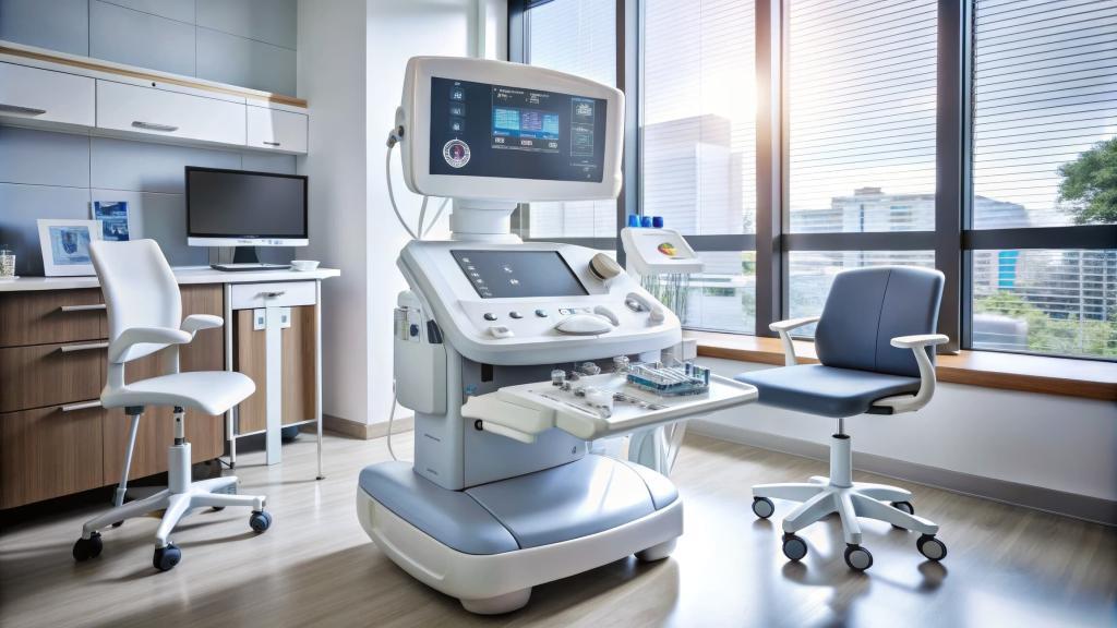
Initially developed from underwater sonar technology used in the Second World War, ultrasound made its way into medical examinations in the late 1940s and 1950s. By the 1960s, portable and more refined devices were being tested in hospitals. Early on, these instruments were used in obstetrics to monitor foetal development. However, many improvements—in both the software that interpreted the echo data and the probes that emitted ultrasonic waves—were introduced and publicly demonstrated at major medical conferences held in early January. This timing added an extra dimension of excitement, as the new year would bring a new generation of ultrasound machines capable of sharper images and real-time motion.
As the decades wore on, ultrasound’s use expanded to cardiology (echocardiography), vascular imaging, and soft-tissue examinations. Its safety profile, cost-effectiveness, and non-invasive nature made ultrasound an indispensable tool worldwide, especially in resource-limited settings where more advanced machines were not readily available.
CT Scanning: Slicing Through the Body—Virtually
Computed tomography, or CT, often attributes its invention to British engineer Sir Godfrey Hounsfield. Though the first human scans took place in the early 1970s, the official unveiling of this ground-breaking technology coincided with January announcements. Early in 1972, the public began hearing about a device that could compile multiple X-ray images into a three-dimensional slice of the body, enabling doctors to see internal structures with remarkable clarity.
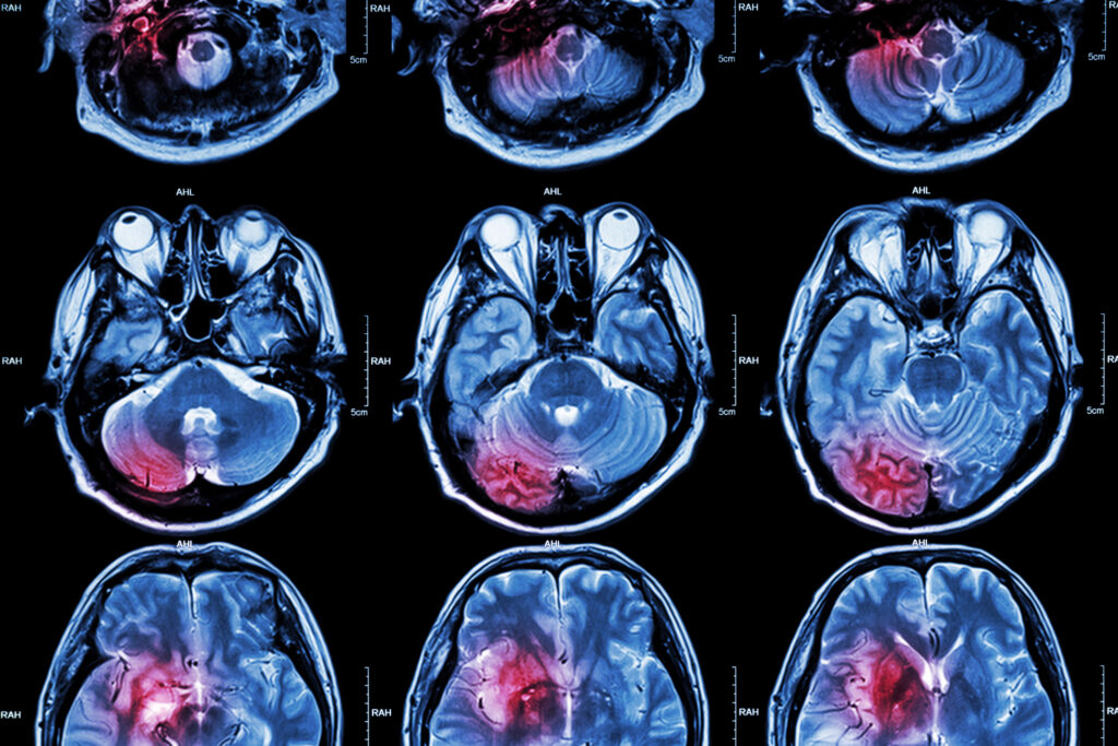
When CT scanners reached clinics, they completely redefined diagnostic precision. Brain scans were among the first to benefit, offering unprecedented views of tumours, lesions, and other abnormalities. Over time, refinements in the scanning process, coupled with faster computers, allowed entire body sections to be scanned within seconds. This not only improved patient comfort but also enhanced diagnostic accuracy.
Magnetic Resonance Imaging: A Vision for the Future
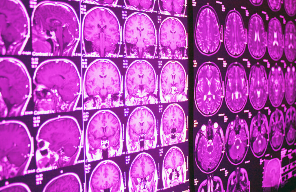
While X-rays rely on radiation and ultrasound on sound waves, magnetic resonance imaging (MRI) capitalises on the natural magnetic properties of the human body. By the late 1970s, researchers, including Paul Lauterbur and Sir Peter Mansfield, had perfected the concept of MRI to produce highly detailed images of soft tissue. The early 1980s saw increased clinical trials and commercial availability, with pivotal research milestones often reported during January conferences.
Mechanics and Early Demonstrations
MRI operates by aligning hydrogen nuclei in the body using a powerful magnetic field, then disturbing that alignment with radiofrequency pulses. As these nuclei return to their original orientation, they emit signals that a scanner detects and reconstructs into detailed images. The scanning process, which was initially quite lengthy, improved over the years to become faster and more comfortable.
Applications and Advantages
The biggest advantage of MRI, announced with gusto when it first entered the mainstream, is its ability to detail soft tissues—muscles, ligaments, the spinal cord, and even subtle changes in brain matter—better than any other imaging modality. Early January press releases and scientific gatherings made the revolutionary potential of MRI known to hospitals and clinicians, stirring significant media interest. To this day, MRI remains the gold standard for diagnosing a wide range of conditions, from spinal disc injuries to brain tumours and neurological diseases such as multiple sclerosis.
Nuclear Medicine and PET Scans: Pushing Boundaries
Where X-rays, ultrasound, and MRI concentrate on structural imaging, nuclear medicine images metabolic and molecular processes within the body. One of the most prominent forms of nuclear medicine imaging is the positron emission tomography (PET) scan. In essence, a radioactive tracer is injected into the patient, and as it travels through the body, a PET scanner detects positron emissions that highlight metabolic activity.
Historic Milestones in Early January
While the concept of nuclear medicine began to solidify in the mid-20th century, PET scanning’s notable expansions into oncology and neurology often gained attention during January symposiums. These gatherings introduced innovations such as more refined tracers or advanced computational techniques to interpret the data. The proximity of these announcements to the start of a new year underscored the sense that diagnostic medicine was stepping into unexplored territory, with the capability to monitor cancers at the cellular level or examine brain function in living patients.
Transformative Impact on Diagnosis and Treatment
The synergy of PET with CT or MRI (as in PET-CT or PET-MRI) has proven revolutionary. By merging functional and structural information, clinicians can accurately pinpoint abnormal areas. This integrated approach is crucial for cancer detection, staging, and monitoring response to therapy. Early-year public announcements often showcased real patient case studies, demonstrating how these scanning methods enabled earlier intervention and more effective treatments.
Modern Realities: AI Integration and 3D Imaging
Medical imaging did not pause once these major modalities were established. On the contrary, contemporary researchers continue to use the new year as a launchpad for unveiling the latest enhancements, particularly those powered by artificial intelligence (AI).
AI-Assisted Diagnosis
Today’s radiologists increasingly use AI algorithms to sift through large volumes of images, highlighting suspicious lesions or anomalies that might warrant further investigation. Early-stage prototypes of AI-assisted systems were sometimes introduced at January tech forums or medical gatherings, emphasising how an algorithm could handle repetitive tasks—like scanning thousands of mammograms—while the clinician focused on cases requiring personalised attention.
3D and 4D Imaging
An additional development that often gains attention around January is 3D and 4D imaging techniques. These are vital not only for diagnosing complex conditions but also for planning surgeries. In 3D reconstructions, doctors can rotate or slice through an image in multiple dimensions, revealing angles or perspectives that 2D scans might miss. Going a step further, 4D imaging adds the time component, capturing dynamic processes such as the beating heart in real-time. Many of these technologies have been spotlighted at the onset of the year, underlining their “future is now” appeal.
Looking Ahead: Evolving Frontiers and the Next New Year
In tracing the development of medical imaging breakthroughs, it becomes clear that scientific exploration often aligns neatly with the collective renewal that comes each January. The new year’s excitement provides a powerful backdrop for unveiling the next wave of technological marvels, stirring hope and possibility within the medical community and the wider public.
Future Modalities
As we look to the future, several advanced concepts are in development, waiting for their moment to shine—potentially at a forthcoming January conference or via a ground-breaking New Year’s announcement. Among the most eagerly anticipated are quantum-enabled imaging devices, which may offer previously unimaginable levels of detail. Researchers are also working on hyperpolarised MRI, which boosts signal strength in the body’s molecules, and advanced optical imaging methods capable of gleaning real-time molecular data.
Personalised Medicine and Beyond
One of the most profound ways imaging will evolve is by integrating directly into personalised medicine strategies. As genome sequencing becomes more accessible, imaging could link with genetic information to craft tailor-made treatment protocols. For instance, a patient with a known genetic predisposition to a particular cancer might undergo more frequent and finely tuned scans, enabling earlier detection than generic screening guidelines. Announcements about these combined strategies often surface in the year’s opening weeks, reflecting the widespread determination to push the boundaries of modern healthcare.
Ethical and Practical Considerations
Rapid advances in medical imaging raise questions about cost, accessibility, and data protection. High-end imaging machines remain expensive, and not every region or country can secure them easily. Additionally, AI-driven tools create large volumes of patient data that require responsible handling to maintain confidentiality. Policymakers, healthcare systems, and technology providers thus face the perennial challenge of balancing innovation with public welfare, ensuring that breakthroughs do not widen existing inequalities. Early-year discussions and policy debates—sometimes ignited by fresh announcements—are crucial for shaping equitable healthcare solutions.
New Year’s Day and the Spirit of Discovery
On the surface, it might seem arbitrary for a revolutionary scientific discovery to coincide with a particular date. Yet, time and again, we see how the synergy of a fresh calendar year provides not only a symbolic moment for unveiling cutting-edge technologies but also a potent launchpad for forging partnerships among researchers, clinicians, and investors. The promise of something new resonates even deeper against the backdrop of the holiday season, capturing the imagination of a global audience eager for progress.
Ultimately, medical imaging stands as one of humanity’s greatest collaborative achievements: a melding of physics, engineering, chemistry, biology, and computer science in the service of saving and improving lives. The modern healthcare landscape cannot be imagined without the clarity provided by X-rays, the real-time diagnostic capabilities of ultrasound, the slicing precision of CT scans, the detailed mapping of MRI, or the metabolic insights of PET. The new year, in essence, has consistently offered a stage for dramatic entries and impactful moments in this rapidly advancing field.
In the coming years, as fresh developments emerge—be it a revolutionary scanning technique, the harnessing of AI for near-instant diagnoses, or advanced molecular imaging—the timing of announcements could well coincide with the cusp of a new January, perpetuating the storied tradition of beginning the year with a leap forward in medical imaging. For clinicians and patients alike, this alignment of technological progress with the annual sense of renewal reinforces a message of continual evolution: we innovate to heal, we heal to move forward, and we move forward with the unshakeable optimism that each new year will bring healthier, more inclusive healthcare.
Conclusion
From the sensational unveiling of X-rays in early January 1896 to the steady progression through ultrasound, CT scans, MRI, PET, and the dawn of AI, medical imaging has marched hand in hand with the turning of the calendar year. Each January has acted as a stage for bright ideas and new beginnings, reminding us that healthcare is a journey of constant improvement. As we celebrate the promise that comes with every new year, we also commemorate the countless men and women—scientists, engineers, physicians, technologists—whose unwavering dedication ensures that “seeing” inside the human body is no longer mere science fiction but an everyday reality. The next frontier may be right around the corner, and it might just be unveiled when we next greet 1 January with eager anticipation.
Disclaimer
The content of this article, A New Dawn for Medicine: Ground-breaking Imaging Inventions on New Year’s Day, is intended for informational and educational purposes only. It reflects historical and contemporary developments in medical imaging technology but does not constitute medical, diagnostic, or professional advice. Readers should not rely on the information herein as a substitute for consultation with qualified healthcare professionals. While every effort has been made to ensure accuracy at the time of publication, Open Medscience makes no warranties or representations regarding the completeness, timeliness, or reliability of any content. Mention of specific technologies, individuals, or events does not imply endorsement. Readers are encouraged to seek updated information and professional guidance relevant to their specific circumstances.
You are here: home » diagnostic medical imaging blog »


