Since the discovery of X-rays in the late 19th century, various medical imaging modalities, such as CT, MRI, and ultrasound, have been invented to help in the diagnosis of disease.
Pioneers of Medical Imaging: From Röntgen to Hounsfield
Medical imaging can create visual representations of the internal workings of the human body. Medical professionals use these images to diagnose and treat different health conditions. Modern medical imaging has a long and fascinating history that spans 128 years, and some of the key developments include:
- In 1895, German physicist Wilhelm Conrad Röntgen discovered X-rays, a type of electromagnetic radiation that can penetrate through materials and create images of the internal structures of the human body. This discovery revolutionised medicine and paved the way for the development of medical imaging.
- The invention of the X-ray machine in 1896 by British physicist Sir William Crookes made it possible to generate and control X-rays for medical imaging.
- In the early 1900s, radiography became a common technique for producing X-ray images of the human body. This technique was used to diagnose fractures, detect foreign objects, and identify diseases such as tuberculosis.
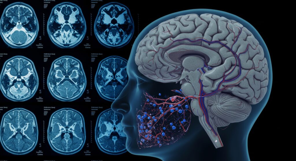
- In the 1970s, the first computed tomography (CT) scanner produced detailed cross-sectional images of the human body.
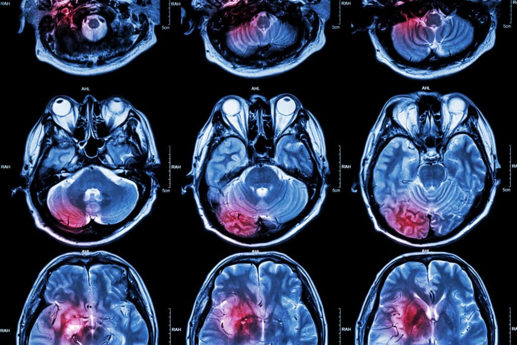
- In the 1980s, magnetic resonance imaging (MRI) applied magnetic fields and radio waves to generate detailed images of the body’s soft tissues.
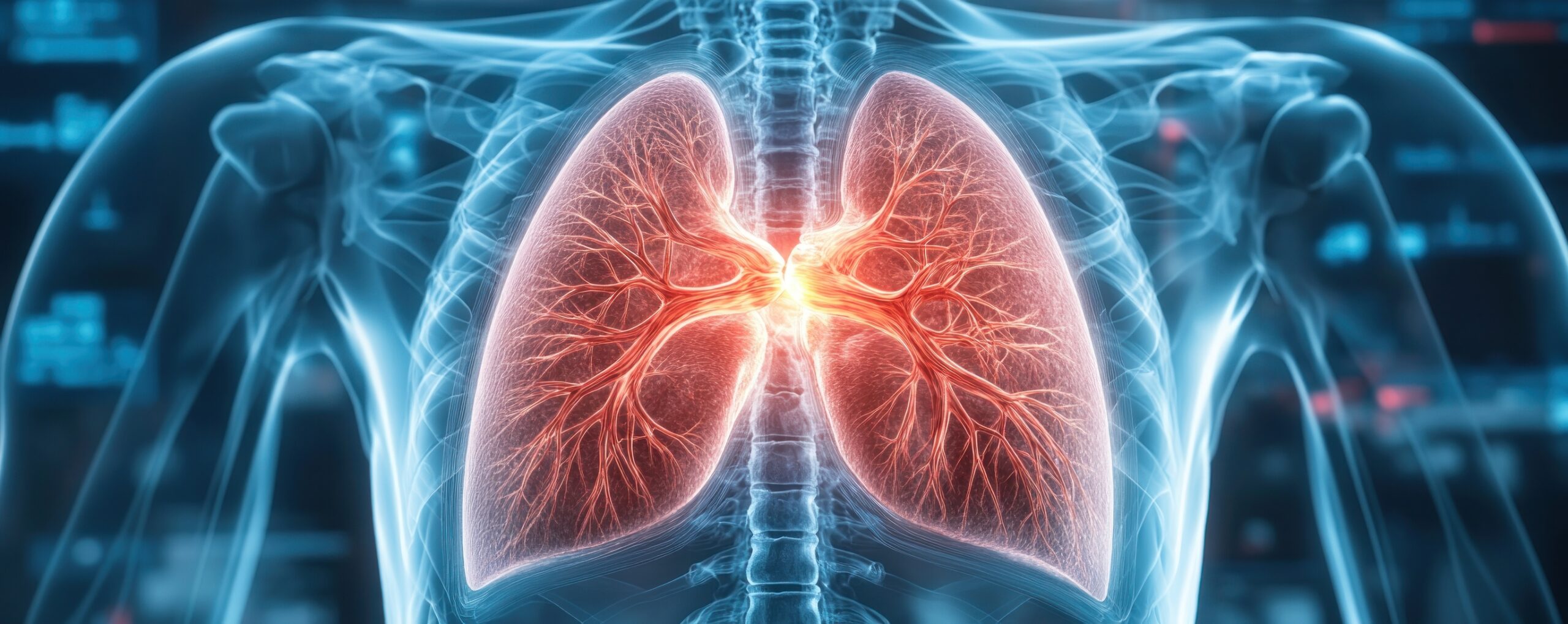
- In the 1990s, positron emission tomography (PET) became widely used to diagnose cancer and used a small amount of radioactive material to create images of metabolic processes.
Medical imaging continues to evolve with new techniques and technologies such as 3D imaging, digital radiography, and molecular imaging. These advances have greatly improved our ability to diagnose and treat various medical conditions.
Nuclear Medicine: A Vital Tool in Modern Healthcare
Nuclear medicine is a medical speciality that uses radioactive substances to diagnose and treat various medical conditions.
The history of nuclear medicine dates back to the early 20th century when scientists first discovered that certain substances could emit radiation. Then, in the 1930s, scientists began to explore using radioactive substances for medical purposes.
In the 1940s and 1950s, advances in nuclear technology led to the development of the first nuclear medicine techniques, including using radioactive isotopes to visualise internal organs and tissues. For example, in 1946, the first radioisotope tracer was used to diagnose a patient with thyroid disease. Then in 1951, the first radionuclide imaging scan was performed on a patient.
Over the next few decades, nuclear medicine technology continued to evolve with the development of new radiotracers and imaging techniques. Nuclear medicine diagnoses and treats various medical conditions, including cancer, heart disease, and neurological disorders.
Nuclear medicine procedures typically involve the injection or ingestion of a radiopharmaceutical, which contains a radioactive isotope. The radiopharmaceutical travels through the body and emits radiation, which is detected by a special camera that creates images of the internal structures.
Nuclear medicine has become an essential tool in modern medicine, providing clinicians with valuable information about the internal structures and functions of the human body. While the use of radioactive substances in medical practice carries some risks, the benefits of nuclear medicine have been shown to outweigh the risks for most patients.
The Invisible Light: A Journey Through the History of X-rays
The discovery of X-rays by the German physicist Wilhelm Conrad Röntgen conducted experiments using cathode rays in 1895. He observed that a screen coated with a fluorescent material placed near a cathode ray tube emitted a bright green glow when the tube was functioning.
Röntgen understood this glow was generated by a new type of ray emitted from the cathode ray tube. He called these rays ‘X-rays’, as he did not know what they were and wanted to denote their unknown nature.
Röntgen conducted further experiments to understand the properties of X-rays, including their ability to penetrate different materials and their effects on living tissue. He soon concluded that X-rays could be a powerful tool for medical diagnosis, as they allowed doctors to see inside the body without requiring invasive procedures.
The discovery of X-rays revolutionised medical imaging and profoundly impacted medical practice. Within months of Röntgen’s discovery, X-ray machines began to be utilised in hospitals and clinics worldwide, and the field of radiology was born.
CT Scans: A Breakthrough in Medical Technology
Computed tomography (CT) uses X-rays and computer algorithms to generate detailed human body images.
The history of CT began in the late 1800s when scientists began experimenting with using X-rays to see inside the body. However, it wasn’t until the 1960s that British engineer Godfrey Hounsfield and American physicist Allan Cormack developed the first CT scanner.
Hounsfield and Cormack’s invention used X-rays to create multiple images of the human body from different angles, which were combined using computer algorithms to create 3-D images of the internal structures of the human body. The first CT scanner was installed at Atkinson Morley’s Hospital in Wimbledon, London, in 1972.
The development of CT revolutionised medical imaging, allowing doctors to see inside the body with much greater detail and accuracy than ever before. Over the years, CT technology has continued to evolve, with advances in computer processing power and imaging technology leading to faster and more detailed scans. Today, CT scans are widely used in medical practice and have become an essential tool for diagnosis and treatment in many fields of medicine.
PET Scans: A Powerful Tool for Disease Detection and Treatment
The history of PET began in the 1950s when scientists first discovered positrons, which are the antimatter counterpart of electrons. Then, in the 1960s, scientists realised that positrons could be applied to study the body’s biological processes.
In 1975, Edward J. Hoffman and Michael E. Phelps developed the first PET scanner at the University of California, Los Angeles (UCLA). The scanner used a rotating ring of detectors to measure the distribution of positrons emitted by a radioactive tracer in the body. However, the first PET scan was performed on a human patient in 1976.
PET technology continued to evolve over the next few decades, with imaging technology improvements and new radiotracers’ development. Today, PET scanners can produce highly detailed images of the internal structures and biochemical processes, making them an important tool for diagnosing and treating various medical conditions, including cancer, heart disease, and neurological disorders.
MRI: Pioneering the Way to Advanced Medical Diagnostics
Magnetic resonance imaging (MRI) uses radio waves in a strong magnetic field to produce internal body images.
The history of MRI began in the 1930s when scientists began to study the phenomenon of nuclear magnetic resonance (NMR). In the 1970s, scientists working in the field of NMR started to realise this technology could be used to create images of the human body.
In 1977, the first human MRI scan was performed by British physicist Peter Mansfield and American chemist Paul Lauterbur. Mansfield and Lauterbur’s work used gradient magnetic fields to construct internal images of the body with much greater detail and accuracy than ever before.
Over the next few decades, MRI technology continued to evolve, with advances in computer processing power and imaging technology leading to faster and more detailed scans. MRI is often combined with other imaging modalities to form positron emission tomography (PET) hybrids to provide more detailed and precise information about the human body’s internal structures and functions. The continued development of MRI technology will lead to even more powerful and versatile imaging tools in the future.
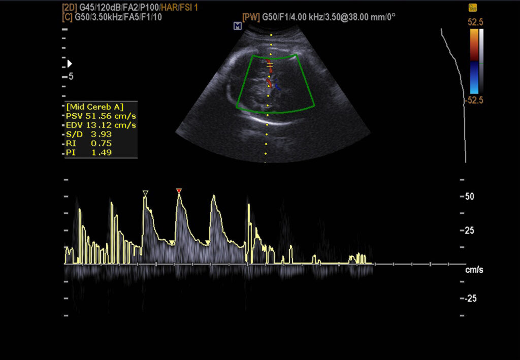
The Power of Sound: A Journey Through the History of Ultrasound
The imaging modality, ultrasound, uses high-frequency sound waves to produce images of the internal structures of the human body.
The history of ultrasound dates back to the early 19th century when scientists first discovered that sound waves could detect objects hidden underwater. In the 1940s, researchers began to explore using ultrasound for medical purposes.
In 1955, Scottish obstetrician Ian Donald used ultrasound to detect the presence of fluid-filled cysts in the human body. These observations led to the development of the first commercial ultrasound scanner in the 1960s, primarily used for obstetric imaging.
Over the next few decades, ultrasound technology continued to evolve, with improvements in imaging technology and new image processing and analysis techniques.
Ultrasound is used to help diagnose cardiovascular disease, musculoskeletal injuries, abdominal and pelvic disorders. In addition, it is commonly used during pregnancy to monitor foetal development and detect abnormalities.
The continued development of ultrasound technology will lead to even more powerful and versatile imaging tools, potentially revolutionising how many medical conditions are diagnosed and treated.
Conclusion
Modern medical imaging continues to be shaped by artificial intelligence and machine learning to analyse and interpret imaging data. These technologies can identify patterns and trends in large amounts of imaging data, helping clinicians make more accurate diagnoses and treatment decisions.
For example, molecular imaging visualises specific molecules within the body. In contrast, functional imaging can study how different organs and tissues function in real time.
In addition, advances in imaging technology, such as the development of higher-resolution scanners and faster data processing, are expected further to improve the accuracy and speed of medical imaging. These imaging modalities will allow clinicians to diagnose and treat medical conditions more quickly and effectively, leading to better patient outcomes.
Overall, the future of medical imaging is bright, with continued technological advancements expected to drive improvements in the accuracy, speed, and versatility of imaging techniques to diagnose various diseases.
Disclaimer
The content provided in Revolutionising Healthcare: A Historical Perspective on Medical Imaging by Open Medscience is for informational and educational purposes only. It is not intended to serve as medical advice, diagnosis, or treatment. The historical overview, technological descriptions, and clinical references are based on publicly available information and general scientific knowledge as of the date of publication (18 March 2023).
While efforts have been made to ensure the accuracy of the information presented, Open Medscience makes no representations or warranties regarding the completeness, accuracy, or reliability of any information contained in the article. Readers should consult qualified healthcare professionals for advice or information related to specific medical conditions or imaging procedures.
Open Medscience disclaims any liability for any loss, damage, or injury that may result from the use or reliance on the information provided in this publication.