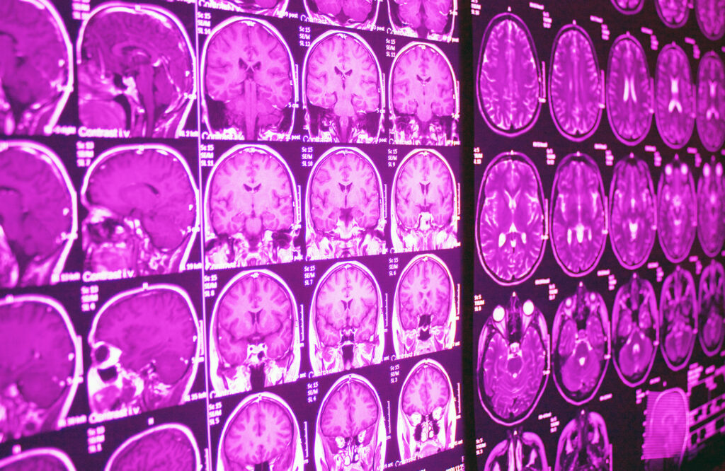
Magnetic resonance imaging, or MRI for short, is a medical imaging technology commonly used in the field of radiology. MRI machines utilize strong magnetic fields and computer-generated waves to produce comprehensive, high-resolution, anatomical images of a patient’s organs, tissues, and skeletal system. Today, from diagnosing strokes to detecting diseases such as cancer, MRI has become a literal life-saver around the world, and an indispensable tool for health.
So, how did MRI come about and what is the history behind this technology that has become such an integral part of modern medicine? If you’re a healthcare professional, a prospective student interested in the medical field and looking into online accelerated BSN programs, or even just someone interested in history and medicine, keep reading to get a slice of the rather fascinating story of MRI and to see how far we have come in medical technology.
1882: The Roots
The discovery of the rotating magnetic field by Serbian physicist, Nikola Tesla, was key groundwork for modern physics and technology. Tesla described how magnetic fields function and what sort of properties they have. Today, his contributions have been immortalized as a unit for measuring magnetic field strength — MRI scanners are usually 1.5 or 3 Tesla units.
1938: Incipience
The foundational building blocks of MRI technology are traced back to the discovery of the nuclear magnetic resonance (NMR) phenomenon in 1938 by Polish-American physicist and Professor at Columbia University, Isidor Rabi. NMR forms the main predecessor and basic principle of MRI technology. The phenomenon allows doctors to look ‘inside’ molecules and understand their internal structure and behaviors without actually having to damage or break them apart.
1946: Expanding the Foundations
Swiss-American physicist at Stanford University, Felix Bloch, and American physicist at the Massachusetts Institute of Technology, Edward Purcell, expanded the foundations laid by Rabi. They applied NMR to observe magic resonance properties on liquids and solids. This was fundamental to MRI’s later ability to scan the body’s water content. In addition, Bolch and Purcell also demonstrated that atomic nuclei were able to absorb radio frequency waves at the same frequency the nuclei were vibrating at.
1969-1970: Scanning for Cancer
By this time, a second generation of scientists would inherit the legacy left by Rabi, Bolch, and Purcell. Until the 1970s, MRI technologies were only used for physical and chemical analysis. However, Dr. Raymond Vahan Damadian, Professor at the State University of New York (SUNY) Downstate Medical Center and often called the ‘Father of MRI’ due to being the first to propose an MRI body scanning machine, made a groundbreaking experiment by using NMR to differentiate cancerous (malignant) and non-cancerous (benign) cells during this time.
It was successfully demonstrated with rats, to which Dr. Damadian found that hydrogen signals in cancerous tissue differ from that of healthy tissue as malignant tumors contain more water. Cancerous tissues also had longer response lengths than non-cancerous ones. Detecting cancer cells in humans could now be done without radiation.
1972-1974: Designing Machines and First Images
By 1972, Dr. Damadian lodged the first patent on MRI technology. Between 1973 and 1974, Dr. Damadian began designing and creating the first full-body MRI machine called the ‘Indomitable’. At the same time, in 1973, chemist and associate at the SUNY, Paul Lauterbur, produced the first magnetic resonance image of a water sample in a test tube. Immediately after, Lauterbur scanned the first live object — a clam. Lauterbur’s key contribution was that he was the first to observe that gradient magnetic fields can allow scientists to take multi-dimensional images of an object.
1977: Scanning Humans
The first MRI image of the human body was produced in 1977 by Dr. Damadian. While the image is rather crude compared to contemporary standards, at that time it was a monumental feat for the system’s development. Larry Minkoff, one of Dr. Damadian’s post graduate assistants and research team members, volunteered to be the main experimentee. A two-dimensional image of Minkoff’s heart, lungs, vertebrae, and musculature was produced by the machine.
1978-1985: To the Markets and Beyond
Dr. Damadian founded the FONAR Corporation in 1978. By 1980, FONAR produced the first commercially available MRI machine. In 1983, another company, General Electric Healthcare (GE) developed its first scanner called the SIGNA. Across the Atlantic, the German company Siemens also produced its first commercial scanner called the MAGNETOM, later coming into operation at the Mallinckrodt Institute in St. Louis, Missouri in August 1983. In 1985, the FONAR MRI scanner became the world’s first MRI scanner in which an interventional surgical procedure was performed. During the same year, FONAR also developed the first mobile MRI machine.
1995-1997: Patent Wars
The story of MRI is not without controversy and conflict, particularly for Dr. Damadian who is often credited as being the first to have piqued the interest of developing the modern MRI scanner across the wider science sphere. In 1995, FONAR sued Siemens for MRI patent infringement, though eventually extinguished when FONAR dropped the case in exchange for a lump-sum monetary repayment and ongoing royalties. In 1997, FONAR launched another case against GE for MRI patent infringement, with the court ruling in FONAR’s (and therefore Dr. Damadian’s) favor, leading GE to pay $128.7 million to FONAR in damages.
The 21st-Century: Continued Innovation
Since the 1990s and the turn of the 21st-century, MRI has advanced concomitantly with the progress made in technology as a whole. In 1983, an MRI scanner would be 140m2 with an average of ten liquid helium refills per year; today, it is 28m2 with only two liquid helium refills per year. The first ever scan made by Dr. Damadian in 1977 took nearly 5 hours to produce a full body scan; today, it only takes between 15 minutes to one hour.
While MRI has become an essential part of healthcare, its innovations have never slowed down. With recent developments in artificial intelligence (AI), MRI will only continue to become faster, more reliable, and more efficient in the future.
Invented or Developed?
From an objective standpoint, the invention of MRI technology is not attributable to any single individual. In fact, we can see that the development of MRI depended much on the blood, sweat, and tears of many scientists across the entire late 19th to 20th centuries. The reality is that the system was given through an accumulated series of discoveries made at various points in modern history.
Controversy however persists over things such as the 2003 Nobel Prize, which awarded Lauterbur instead of Dr. Damadian for contributions made towards the MRI technology development. Obviously, for Dr. Damadian, he felt that he was snubbed from the awards. Some believed that the Nobel Prize brushed Dr. Damadian aside due to his religious beliefs; whereas others viewed that while Dr. Damadian made major contributions to MRI development, it still did not meet the standards of receiving a Nobel Prize.
Nevertheless, the immortalization of figures like Tesla in the magnetic field metric used to measure MRI scanners reveals a different image – that the technology, in every practical sense, is a symbol of the leaps and bounds our civilization has collectively made over the past century. So regardless of whether you think one person should take more credit than the other, MRI is here saving lives and improving health outcomes more than ever for patients across the world – and it is all because of our own human ingenuity.
Disclaimer
The information presented in this article, A Short History of Magnetic Resonance Imaging (MRI), is intended for general educational and informational purposes only. While every effort has been made to ensure accuracy, Open Medscience does not guarantee the completeness, timeliness, or reliability of any content provided. This article does not constitute professional medical, scientific, or legal advice, nor should it be used as a substitute for professional consultation with qualified experts in radiology, medicine, or history of science.
References to specific individuals, companies, or events are included to provide historical context and do not imply endorsement or affiliation. Any views expressed in this article are those of the authors and do not necessarily reflect the official policy or position of Open Medscience or its partners.
Readers are encouraged to conduct their own research and consult relevant professionals for advice or information specific to their needs.
home » blog » medical imaging modalities »


