
Introduction
Modern dental implant treatment depends on accurate planning as much as surgical skill. A successful implant is not simply placed — it is designed, based on a detailed understanding of a patient’s bone, nerves, and surrounding anatomical structures. Advanced three-dimensional imaging has become one of the most important tools supporting this design process, allowing clinicians to plan with a level of precision that was simply not possible with older imaging methods. The result is safer surgery, fewer intraoperative surprises, and implant outcomes that are far more predictable over the long term.
Why 3D Imaging Matters
Traditional two-dimensional X-rays have supported dental diagnosis for decades, and they still provide useful baseline information. However, a flat image can only show so much. It compresses a three-dimensional jaw into a single plane, which means depth, width, and the precise spatial relationship between structures are often estimated rather than measured.
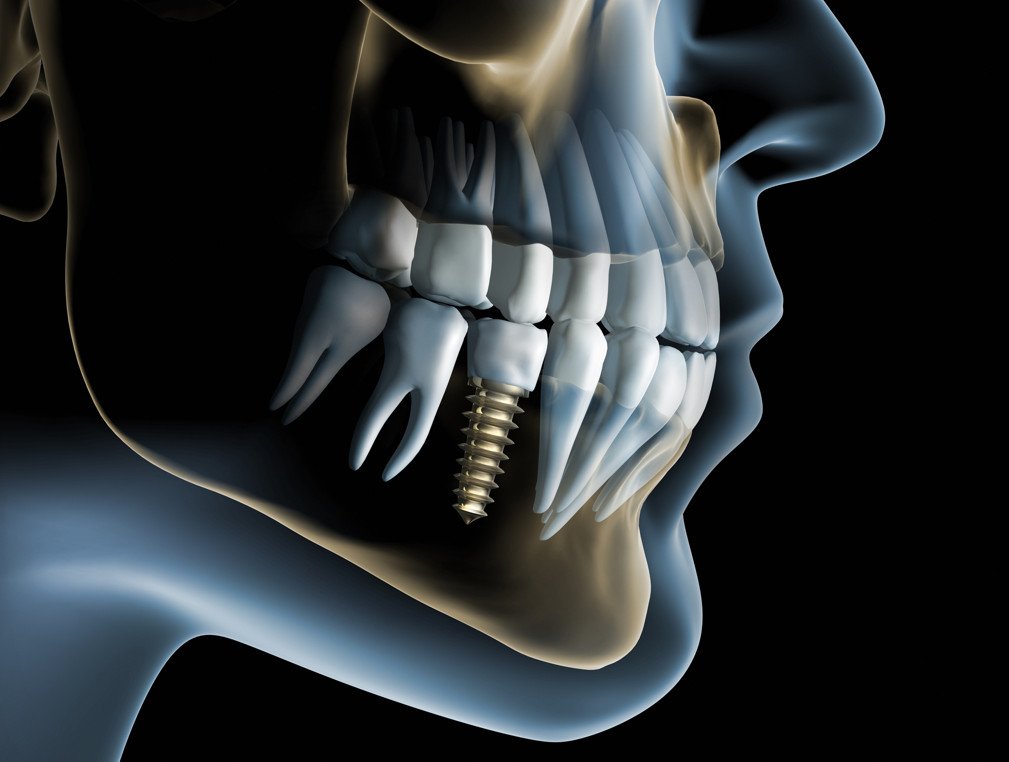
Cone Beam Computed Tomography (CBCT) changes this. By capturing a full three-dimensional volume of the jaw in a single scan, CBCT allows clinicians to rotate, section, and examine the anatomy from any angle. This makes it possible to measure bone volume and density with precision, locate the inferior alveolar nerve canal, assess the position of the maxillary sinus floor, and identify any pre-existing pathology before a single incision is made.
Better Planning, Better Results
The real value of CBCT imaging is realised when it is paired with digital implant-planning software. Once a scan is captured, a dentist can build a virtual model of the patient’s jaw and simulate implant placement before ever picking up a surgical drill. Implant size, angle, and depth can be tested and adjusted virtually, reducing guesswork and improving the odds of long-term implant stability.
This digital workflow supports several key advantages:
- More precise implant placement — implants can be positioned exactly where bone quality and quantity are optimal.
- Better protection of nerves and nearby teeth — critical structures are mapped in advance, reducing the risk of nerve injury or damage to adjacent teeth.
- Improved fit for crowns and restorations — accurate 3D data supports better-fitting final restorations, which can mean less chair time and fewer adjustments.
- Greater confidence in complex cases — patients with limited bone volume, prior extractions, or unusual anatomy benefit from a plan built on precise measurements rather than assumptions.
- More predictable healing — when an implant is placed in well-planned position and angulation, the surrounding bone and soft tissue are more likely to heal as expected.
Patients researching dental implants in Arlington VA often ask whether advanced imaging is included as part of treatment planning. It typically is, because CBCT-based planning supports a personalised approach rather than a one-size-fits-all placement strategy — something that matters greatly given how much anatomy can vary from one patient to the next.
Personalized Treatment for Every Patient
No two mouths are identical. Bone density, jaw shape, sinus position, and the effects of previous dental work — including old extractions, periodontal disease, or prior implant failures — all influence how and where an implant can safely be placed. A three-dimensional scan allows a clinician to evaluate these individual factors directly rather than relying on generalised assumptions.
This detailed anatomical picture helps determine whether bone grafting or sinus lift procedures are necessary before implant placement, identify potential surgical challenges early in the process, and select the implant size, length, and angle best suited to each patient’s unique anatomy. For patients with lower bone volume or more complex medical histories, this level of individualised assessment can be the difference between a straightforward procedure and one that requires significant revision. A customised, imaging-guided approach can reduce surgical risk, improve patient comfort during and after the procedure, and support stronger long-term functional outcomes.
Frequently Asked Questions
Is CBCT imaging safe? Yes. Dental CBCT uses a comparatively low radiation dose relative to medical CT scanning, and it is prescribed selectively — only when the additional diagnostic information is expected to meaningfully benefit treatment planning.
Does every implant patient need a 3D scan? Not necessarily in every case, but it is recommended for the majority of implant patients because it provides anatomical detail that standard two-dimensional X-rays simply cannot capture, particularly around nerve position and bone volume.
Can 3D imaging shorten treatment? It often does. By reducing unexpected findings during surgery and allowing much of the planning work to happen beforehand, CBCT-guided workflows tend to streamline the overall treatment timeline and reduce the likelihood of mid-procedure adjustments.
Does advanced imaging improve implant success? Implant success depends on multiple factors, including bone quality, oral hygiene, and overall health, but accurate diagnosis and precise pre-surgical planning are consistently associated with more predictable outcomes. According to guidance from the American Academy of Implant Dentistry, thorough diagnostic imaging is considered a core component of responsible implant treatment planning.
Conclusion
Advanced 3D imaging has changed dental implantology by making treatment more accurate, more individualised, and safer for patients. Rather than relying on estimation, clinicians can now build a treatment plan around precise, patient-specific data — from bone density and volume to the exact position of nerves and sinuses. Combined with careful clinical evaluation and experienced surgical care, this technology gives patients a clearer, more confident path toward a stable, long-lasting implant outcome.
Disclaimer: This article is provided for general educational and informational purposes only and should not be considered dental or medical advice. The suitability of dental implants, CBCT imaging, bone grafting, sinus lift procedures, or other treatments depends on individual clinical circumstances and should be assessed by a suitably qualified dental professional. Imaging techniques involve benefits, limitations, and potential risks, including exposure to ionising radiation, and should be used only when clinically justified. Treatment outcomes vary between patients, and no particular result or implant longevity can be guaranteed. References to dental practices, organisations, products, services, or external websites are provided for informational purposes and do not constitute an endorsement by Open MedScience. Readers should consult an appropriately qualified dental professional for personalised advice, diagnosis, and treatment.