Artificial Intelligence will play a vital role in the analysis of vasts amounts of medical imaging data.
Artificial intelligence: the future of medical imaging
Radiology can trace its roots back to the Nobel Laureate Wilhelm Conrad Röntgen who discovered X-rays in 1895. Consequently, this discovery led to the imaging of the human body which contributes to assisting with the diagnosis of various disease states.
Today, X-ray planer imaging is still being used for example, in mammography or chest X-rays in the clinical setting. However, the next generation of diagnostic imaging tools such as computed tomography (CT), magnetic resonance imaging (MRI), ultrasound (US) and Nuclear Medicine (NM) are now being used to produce 3-D anatomical and functional images of the human body.
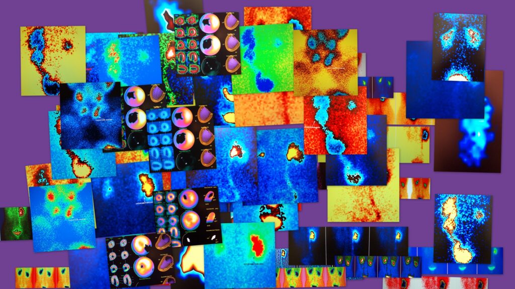
Accordingly, Nuclear Medicine departments make use of gamma emitters for single photon emission computed tomography (SPECT) and positron emitters for positron emission tomography (PET).
At present, X-ray scans account for 60% of all medical imaging in the Healthcare sector although 25% are rejected due to poor image quality. The objective is to decrease these X-ray rejection rates and facilitate resources to improve the quality of patient treatment through Artificial Intelligence.
The phrase Artificial Intelligence was first coined in 1956 by computer scientists at IBM and refers to a machine or computer that demonstrates intelligence in performing a task.
The market for Artificial Intelligence tools to process and analyse medical imaging studies is considered to push past $2 billion by 2023. The future of healthcare will depend on deep learning machines to provide solutions for the diagnosis of disease states, especially in larger ageing populations. The advancement of algorithms will drive clinical support towards patients by the assistance of radiologists, pathologists to implement theranostics. Artificial Intelligence will create data-driven platforms for decision-making in the diagnosis of disease states.
The relationship between the physician and patient will change through various Artificial Intelligence platforms which will take time to implement in the healthcare system. The existing workflow will contain hidden changes regarding the patient experience, provider productivity and diagnostic accuracy of medical imaging modalities.
Medical imaging neural networks
Artificial Intelligence will complement the vast number of digital images generated in hospitals which are the product of the next generation of imaging scanners especially the hybrids which include: MRI, CT, PET and SPECT. Advancements in the technology of CT and MRI scanners are making it possible to utilise even thinner slices of the human body to create more detailed 3-D and 4-D images.
These powerful images require vast amounts of digital data to help in the diagnosis of the patient. This has led to some hospitals exceeding 50 petabytes of data per year. To put this data into context 50 petabytes could store the entire written works of mankind! Medical imaging accounts for 90% of all healthcare data and astonishingly 97% of the acquired data will never be analysed.
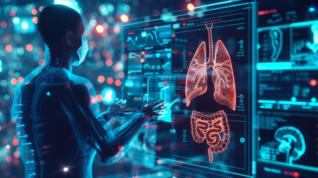
Consequently, Artificial Intelligence will play a vital role in the analysis of medical imaging data and this will only be viable if Artificial Intelligence and healthcare professionals interact to embrace deep thinking platforms such as automation in the diagnosis of the disease state of the patient.
Artificial Intelligence will help radiologists to analyse the medical images with regards to current trends and design treatment programmes which are personalised to the patient. This approach aims to provide the best care and treatment in the most cost-effective manner.
The Artificial Intelligence revolution in medical imaging will only work with the cooperation of healthcare providers and especially equipment manufacturers to incorporate the necessary software to advance the initiatives: these will include the development of data analytics tools to help consolidate vast amounts of generated digital images from complex algorithms in the clinical setting.
Several imaging providers are developing the next generation of imaging technology with the objective to transform hospitals to become more efficient in the management of patient treatment plans. This will be achieved by producing faster imaging and reducing radiation dosages through the PET and SPECT imaging modalities. This will allow medical imaging professionals to evaluate the anatomical images at a faster rate. The data cluster from these images can be translated into small data packages that can be accessed by Healthcare departments to give the professional a real-time insight into patient care and the required intervention.
Conclusion
Artificial Intelligence requires barriers to be broken down for it to become widely accepted and trusted in the mainstream medical imaging setting. For example, the regulatory process remains challenging to new instrumentation regarding Artificial Intelligence products. This is because validation studies are required to demonstrate the performance of deep learning algorithms in the clinical setting.
However, the main obstacle is to ensure the confidence of the radiologist in using Artificial Intelligence. It is paramount that algorithm developers partner with imaging technologists to provide solutions that are integrated with the workflow of the radiologists. In other situations, Healthcare providers are hesitant to purchase Artificial Intelligence tools from various software developers. This is because of vendor-specific integration and implementation which bring many challenges for hospitals and healthcare professionals. To circumvent these issues, the developers need to form Artificial Intelligence platforms to address the algorithms already used by Healthcare organisations to allow the costs to be kept in perspective.
Disclaimer
The content presented in this article, Artificial Intelligence in Cancer Imaging by Open Medscience (22 November 2018), is intended for informational and educational purposes only. It does not constitute professional medical advice, diagnosis, treatment, or recommendations of any kind.
The discussion on artificial intelligence (AI) and its applications in medical imaging reflects developments and perspectives available at the time of writing. While every effort has been made to ensure accuracy, Open Medscience does not guarantee the completeness or timeliness of the information provided, especially as the field of AI and medical imaging is evolving rapidly.
Any reliance placed on this material is strictly at the reader’s own risk. Readers are encouraged to consult qualified healthcare professionals or regulatory authorities before making decisions based on the content of this publication.
Open Medscience accepts no liability for any loss or damage arising from the use of, or reliance on, the information contained in this article. Furthermore, references to commercial entities, products, or services do not imply endorsement or recommendation by Open Medscience.
home » blog » artificial intelligence »


