
Introduction
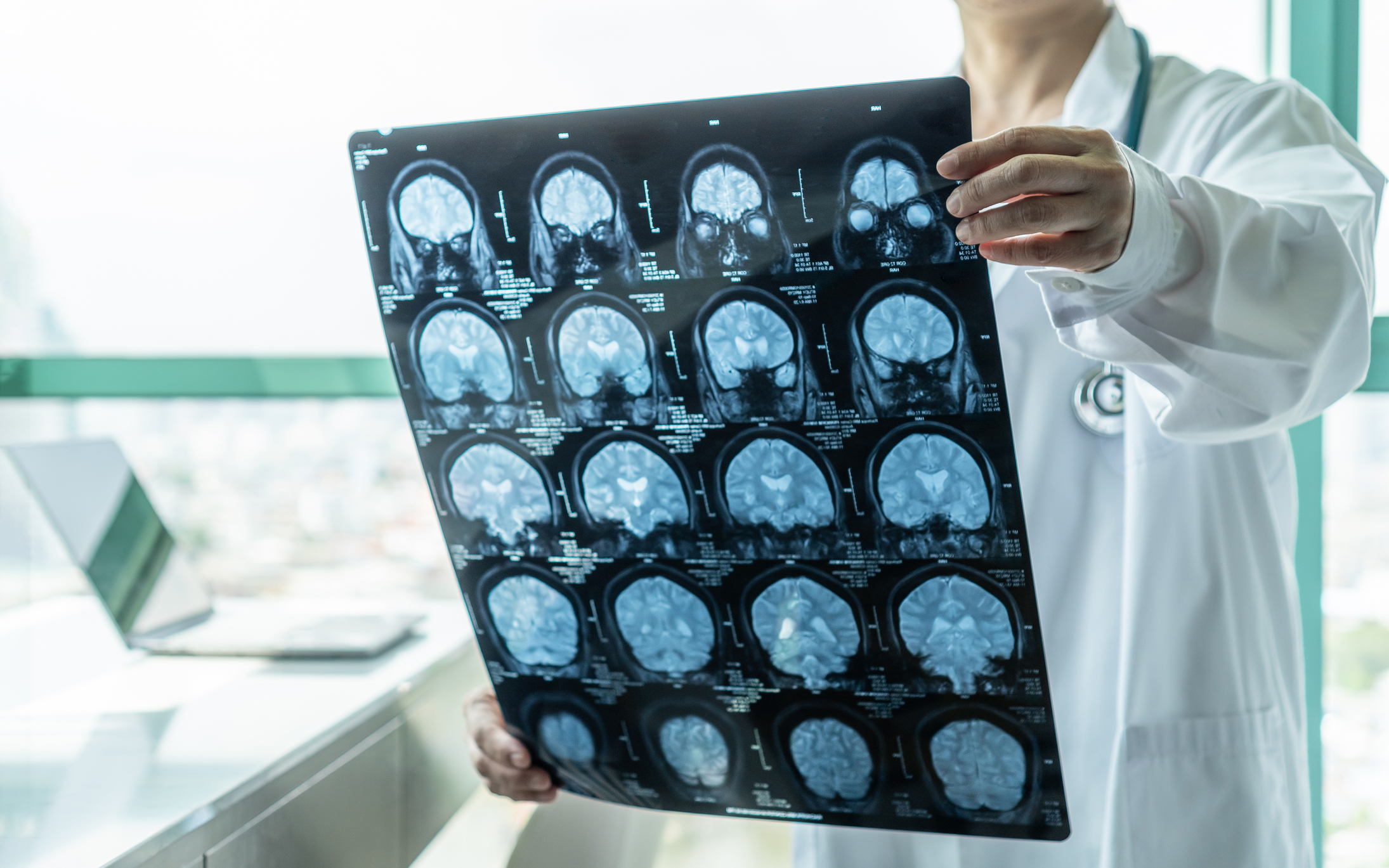
In clinical diagnostics, the focus has always been on what the doctor can see. A fracture may be visible on an X-ray, while CT can identify many structural abnormalities in greater anatomical detail. These visual indicators allow clinicians to quickly verify a pathology and establish the correct treatment plan.
A major problem arises when patients have severe symptoms, yet standard medical scans appear to show a perfectly healthy patient. This frequently occurs when patients experience an abnormality on a microscopic, cellular or metabolic level. This results in a gap between how a patient experiences their condition and how that condition can be verified on conventional medical imaging.
How Doctors Approach ‘Invisible Illnesses’
Many patients who suffer from long term viral issues, mild traumatic brain injuries or chronic nerve disorders do not show physical structural abnormalities. Conventional magnetic resonance imaging (MRI) and CT scans are designed to detect anatomical issues such as tumours, haemorrhages, or tissue atrophy. Since these methods look for structural rather than functional problems, they often fail to capture the underlying pathophysiology of complex conditions.
The absence of a visible abnormality creates genuine uncertainty in diagnostic care. A frequently cited study of primary care patients found that only 16% of symptoms presenting in ambulatory care had a clear organic cause, 10% were judged psychological, and the origin of roughly 75% could not be determined either way.
Historically, the absence of visible structural pathology has complicated the clinical workflow, sometimes leading to prolonged uncertainty regarding a diagnosis or to attributing symptoms prematurely to psychological causes. For clinical documentation and prolonged care, establishing objective biomarkers is essential to better map these complex conditions and guide therapeutic strategies.
Advanced Imaging Technology and The Diagnostic Gap
The latest developments in medical imaging may address this diagnostic gap. Instead of capturing a snapshot of an organ, new technologies are able to examine the physiological process in real-time. While highly promising, it is important to note that many of these applications are currently optimised for research purposes only, and using them to diagnose individual patients remains an ongoing area of clinical development.
Diffusion Tensor Imaging (DTI)
Conventional MRI offers a view of structure, but Diffusion Tensor Imaging (DTI) maps the diffusion of water molecules along white matter tracts. This technique allows for the assessment of the microstructural integrity of white matter in the brain.
Research indicates that DTI can detect subtle alterations in water diffusion patterns, which may reflect axonal injury or disruption in neural pathways following a TBI – at the group level. While DTI demonstrates strong sensitivity to microstructural changes at a group-level analysis, individual variance in white matter makes it difficult to use as a standalone diagnostic tool for a single patient.
A 2022 review concluded that diagnosing axonal injury in one individual patient requires several conditions to align and that further research is needed before DTI can serve as a standalone diagnostic test for a single patient. DTI is not currently an established clinical diagnostic tool for individual TBI patients and remains a research and adjunctive tool.
Positron Emission Tomography (PET)
Positron Emission Tomography (PET) measures metabolic and molecular activity. Since brain cells use glucose as their primary fuel source, fluorodeoxyglucose (FDG-PET) imaging allows clinicians to observe metabolic variations on a regional level.
PET imaging has revealed localised areas of decreased glucose use, which may correlate with neuroinflammation and cognitive symptoms that are experienced as ‘brain fog’. However, the evidence is inconsistent rather than convergent: one study found no significant difference in brain glucose metabolism between long COVID patients and healthy controls, concluding that fatigue itself is the factor at play. A separate study showed no abnormal FDG-PET findings at all, even among those patients who present neurocognitive symptoms.
Functional Magnetic Resonance Imaging (fMRI)
Functional MRI (fMRI) measures brain activity by detecting changes in blood flow. This is often referred to as measuring blood-oxygen-level-dependent, or BOLD signals.
In patients with chronic pain and central sensitisation disorders, fMRI studies have shown altered functional connectivity and hyperactive signaling patterns in the brain circuits that process pain. Recent reviews confirm that fMRI models predicting experimentally evoked pain generalise reasonably well, but models for diagnosing an individual patient’s pain remain far less validated, and none is currently accepted as a standalone diagnostic test.
BOLD signals are highly sensitive to daily physiological variations, the patient’s cognitive state, and even caffeine intake. This limits the reliability of fMRI scans for confirming a single patient’s diagnosis.
The Role of Quantitative Therapy and Digital Health Tracking
Most chronic conditions are highly fluid by nature, and diagnostic images only provide a snapshot in time. More and more clinicians are layering objective imaging data with longitudinal data acquired through digital health monitoring to develop a more comprehensive clinical portrait.
Objective imaging and biometric data are regularly obtained through wearable monitors, heart rate detectors, and custom software in clinical and treatment facilities to track physiological responses in real time. This data has real but limited value: studies comparing consumer wearables against ECG and polysomnography find substantial variation between devices, with heart-rate-variability accuracy ranging from strong to poor, and sleep-stage agreement with polysomnography as low as 50-65% for specific stages.
When combined with neuroimaging, this type of continuous objective data collection helps in providing a clear, multi-dimensional profile of a patient’s functional capacity over a longer period of time. This evidence-based approach aids treatment teams in personalising the treatment plans for patients and provides supplementary documentation that may be useful if a patient’s care team or a disability insurance lawyer needs to substantiate a claim alongside a clinical evaluation.
Technology Supports Patients and Caregivers
Ultimately, for doctors and caregivers, the introduction of both functional imaging and wearable tracking devices offers the ability to add objective, longitudinal detail to conditions that are otherwise difficult to determine from a single scan or visit. This gives medical professionals a deeper understanding of how these complex conditions are functioning and, in turn, allows for a more focused treatment plan.
Disclaimer: This article is provided for general educational and informational purposes only. It does not constitute medical, diagnostic, or legal advice. The imaging techniques discussed, including diffusion tensor imaging (DTI), positron emission tomography (PET), functional magnetic resonance imaging (fMRI), and wearable health technologies, have varying levels of clinical validation and should not be interpreted as standalone diagnostic tools unless supported by current clinical guidelines and specialist assessment. Diagnostic decisions should always be made by qualified healthcare professionals based on a patient’s complete medical history, physical examination, and appropriate investigations. References to disability insurance or legal claims are intended solely to illustrate potential uses of medical documentation and should not be regarded as legal advice. Patients concerned about chronic symptoms or their diagnosis should seek guidance from an appropriately qualified healthcare professional.
home » blog » health matters »