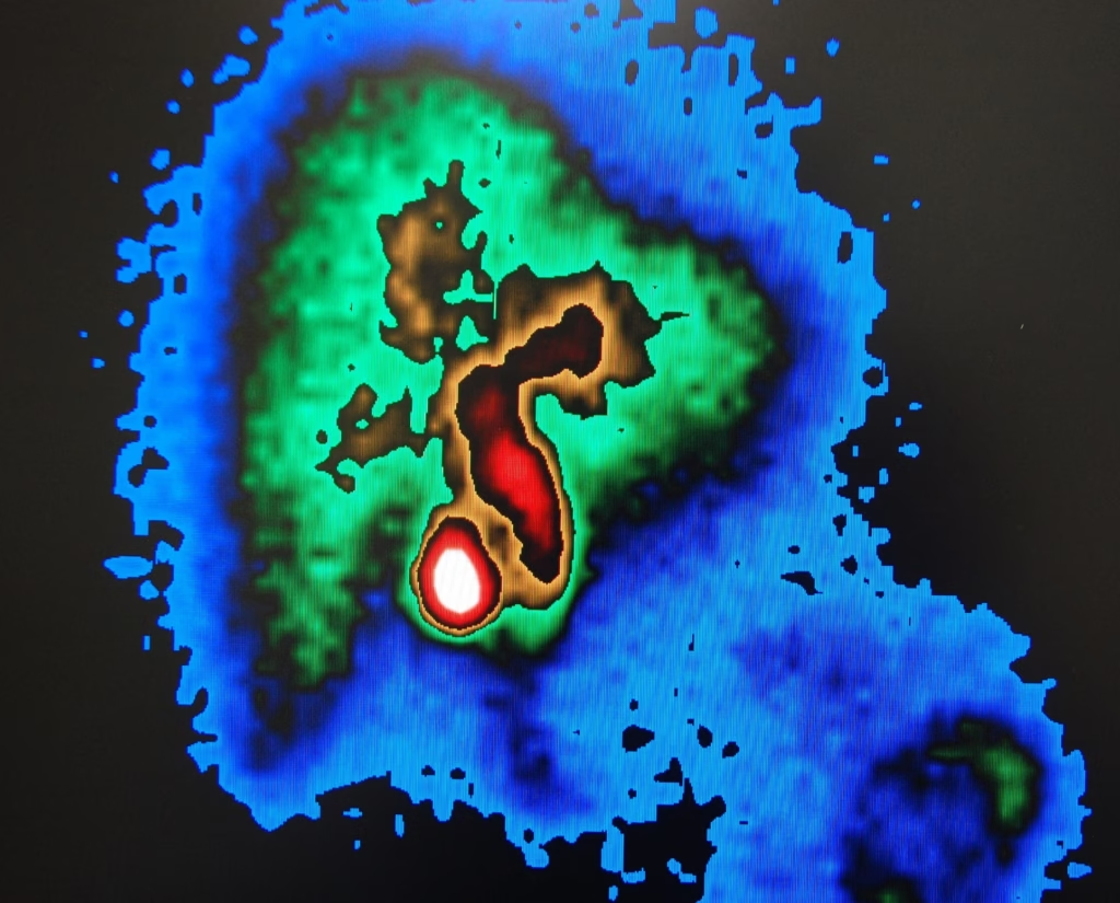
Advanced imaging, such as MRI and OCT, significantly enhances the diagnosis and treatment of optic nerve diseases.
Exploring the Visual Gateway
The optic nerve, a pivotal component of the visual system, serves as the primary conduit for visual information from the retina to the brain. It is a bundle of more than a million nerve fibres that transmit visual signals from the retina (a layer of light-sensitive tissue at the back of the eye) to the visual cortex of the brain. These signals are interpreted as images, and the health and integrity of the optic nerve are crucial for clear vision.
Common conditions affecting the optic nerve encompass glaucoma, optic neuritis, and optic nerve atrophy. Glaucoma, a major cause of vision loss, results from heightened intraocular pressure that can harm the optic nerve. Optic neuritis, characterised by inflammation of the optic nerve, is often linked to autoimmune diseases such as multiple sclerosis. Optic nerve atrophy involves irreversible harm and degradation of optic nerve fibres, culminating in diminished vision. In routine care, and if you are in the Hernando MS area, a trusted local optometrist can coordinate OCT and visual field tests to triage referrals.
Medical imaging plays a pivotal role in diagnosing and managing conditions affecting the optic nerve. Techniques such as Magnetic Resonance Imaging (MRI), Computed Tomography (CT) scans, and Optical Coherence Tomography (OCT) are essential in determining the structure of the optic nerve. These imaging methods help in detecting abnormalities, evaluating the level of damage, and monitoring the progression of diseases. With advancements in medical imaging, early detection and precise assessment of optic nerve conditions have become possible, allowing for timely intervention and better management of these conditions.
Anatomy and Function of the Optic Nerve
The optic nerve, also known as cranial nerve II, is a crucial element of the visual system. It’s a complex structure that begins at the retina, where photoreceptors convert light into electrical signals. These signals are then transmitted by various retinal cells before being transmitted to the ganglion cells. The axons of these ganglion cells converge to form the optic nerve. The optic nerve enters the eye at the optic disc, passes through the optic canal in the skull, and eventually reaches the brain. Each optic nerve contains approximately 1.2 million nerve fibres, indicating its role in transmitting numerous visual information.
In terms of its role in vision, the optic nerve is the primary pathway for visual data travelling from the eye to the brain. Once the optic nerve transmits these signals to the brain, specifically the visual cortex, they are interpreted as images. This process allows us to perceive our surroundings, recognise faces, and perform tasks that require visual input.
Diseases affecting the optic nerve can significantly impair its function, leading to a range of visual disturbances. For instance, increased intraocular pressure can damage the nerve fibres in glaucoma, leading to progressive vision loss and even blindness if untreated. Optic neuritis, often seen in multiple sclerosis, can cause temporary or permanent damage to the optic nerve, resulting in symptoms like blurred vision, pain with eye movement, and colour vision deficits. Optic nerve atrophy, which can be due to various causes, including genetic conditions, trauma, or ischemia (lack of blood flow), leads to the gradual loss of nerve fibres, resulting in decreased visual acuity and field loss. In all these conditions, the ability of the optic nerve to transmit visual information efficiently and accurately is compromised, thereby affecting vision.
Overview of Medical Imaging Techniques
Medical imaging techniques such as MRI, CT, Ultrasound, and OCT (Optical Coherence Tomography) offer various methods to detect and diagnose optic nerve conditions. Each of these techniques has unique principles and applications:
- MRI (Magnetic Resonance Imaging):
- MRI employs strong magnets and radio waves to create images of organs and tissues inside the body without using ionising radiation.
- How it Works: The patient lies inside a large magnet. Radio waves temporarily realign hydrogen atoms in the body without causing any chemical changes. When these atoms return to their original alignment, they emit varying levels of energy, which are detected and transformed into cross-sectional images.
- CT (Computed Tomography) Scan:
- Principle: CT scans use X-rays to create detailed images of the body. Unlike a regular X-ray, a CT scan captures images of internal organs, bones, soft tissue, and blood vessels.
- How it Works: The CT scanner rotates around the patient, sending X-rays through the body from different angles. These are detected on the opposite side, and a computer then uses this information to create a cross-sectional image, like “slices,” of the inside of the body.
- Ultrasound:
- Principle: Ultrasound imaging uses high-frequency sound waves to produce images of structures inside the body.
- A small transducer probe sends sound waves, which bounce off tissues and return to the probe. The machine then translates these reflected sound waves into live images, allowing real-time visualisation of organs.
- OCT (Optical Coherence Tomography):
- OCT is a non-invasive imaging technique that utilises light waves to capture cross-sectional images of the retina.
- How it Works: Similar to ultrasound imaging, OCT uses light instead of sound. The device emits light waves that reflect off the retinal layers. The varying time delays and magnitude of these reflections are measured and used to construct detailed images of the retina, helping in the assessment of conditions affecting the optic nerve, such as glaucoma and macular degeneration.
Each of these imaging techniques offers unique advantages in assessing the optic nerve and other structures of the eye, enabling healthcare providers to detect and manage a wide range of ocular conditions effectively.
Applications of Each Imaging Technique
Each medical imaging method – MRI, CT, Ultrasound, and OCT – has specific applications in evaluating the optic nerve, suited to various diseases and conditions. Here’s a detailed analysis of their uses:
- MRI (Magnetic Resonance Imaging) in Optic Nerve Evaluation:
- Usage: MRI is highly effective for visualising the optic nerve and surrounding tissues due to its excellent soft tissue contrast. It’s particularly useful for detecting demyelinating diseases, tumours, and inflammatory conditions.
- Preferred Conditions: MRI is preferred in cases of optic neuritis, multiple sclerosis, optic nerve glioma, and other intracranial pathologies affecting the optic nerve.
- Example: In a patient with optic neuritis, an MRI might reveal inflammation and demyelination of the optic nerve, aiding in diagnosis and treatment planning, particularly in the context of multiple sclerosis.
- CT (Computed Tomography) Scan in Optic Nerve Evaluation:
- Usage: CT scans are used when bone pathology, trauma, or foreign bodies are suspected. They provide excellent details of the bony orbit and are faster than MRI.
- Preferred Conditions: CT is often the go-to method for evaluating traumatic injuries, fractures of the orbit, or to detect calcifications within optic nerve tumours.
- Example: In cases of orbital fractures, a CT scan can reveal the extent of the fracture and any involvement of the optic nerve.
- Ultrasound in Optic Nerve Evaluation:
- Usage: While not a primary tool for optic nerve evaluation, ultrasound can be helpful in assessing the optic nerve sheath diameter, particularly in cases of increased intracranial pressure.
- Preferred Conditions: Optic nerve sheath diameter measurement in suspected intracranial hypertension and for optic nerve head drusen evaluation.
- For example, ultrasound can measure the optic nerve sheath diameter to support the diagnosis in a patient with papilledema (swelling of the optic nerve head due to increased intracranial pressure).
- OCT (Optical Coherence Tomography) in Optic Nerve Evaluation:
- OCT is highly beneficial for the assessment of the retinal nerve fibre layer (RNFL), which is directly related to the health of the optic nerve. It provides detailed cross-sectional pictures of the retina and optic nerve head.
- Preferred Conditions: OCT is indispensable in the management of glaucoma, as it can detect thinning of the RNFL and is also useful in diagnosing and monitoring optic atrophy, macular oedema, and other retinal conditions.
- Example: In glaucoma, OCT can track the progression of RNFL thinning over time, guiding treatment decisions.
Each imaging method uniquely contributes to the assessment and management of optic nerve conditions. Their appropriate use, often in combination, offers a comprehensive approach to diagnosis and monitoring, allowing for targeted treatments and improved patient outcomes.
Advantages and Limitations
Each imaging method used in optic nerve evaluation has its unique strengths but also comes with certain limitations and challenges:
- MRI (Magnetic Resonance Imaging) Strengths and Limitations:
- Strengths:
- Excellent soft tissue contrast, providing detailed images of the optic nerve and surrounding structures.
- Non-invasive and does not use ionising radiation.
- Ideal for identifying demyelinating diseases, inflammatory conditions, and tumours.
- Limitations:
- Expensive and not as widely available as other imaging modalities.
- It cannot be used in patients with certain metal implants.
- Requires the patient to remain for an extended period, which can be challenging for some.
- Strengths:
- CT (Computed Tomography) Scan Strengths and Limitations:
- Strengths:
- Provides detailed images of the bony structures surrounding the optic nerve.
- Faster than MRI, making it suitable for emergency situations.
- Effective in detecting fractures, calcifications, and foreign bodies.
- Limitations:
- Uses ionising radiation, which carries a risk, particularly in children or with repeated exposures.
- Less effective than MRI in differentiating soft tissue structures.
- Strengths:
- Ultrasound Strengths and Limitations:
- Strengths:
- Quick, portable, and relatively inexpensive.
- Useful for assessing the optic nerve sheath diameter and diagnosing conditions like optic nerve head drusen.
- Non-invasive and safe, with no radiation exposure.
- Limitations:
- Operator-dependent and can have variability in results.
- Lower resolution compared to MRI and CT, limiting its use in detailed optic nerve evaluation.
- Strengths:
- OCT (Optical Coherence Tomography) Strengths and Limitations:
- Strengths:
- Provides high-resolution images of the retinal layers and the optic nerve head.
- Non-invasive and quick to perform.
- Essential in diagnosing and monitoring glaucoma and other retinal diseases.
- Limitations:
- Primarily focuses on the retina and optic nerve head, so it’s unsuitable for evaluating the optic nerve’s entire length.
- It can be expensive, impacting accessibility in some settings.
- Strengths:
While each imaging technique has its own advantages in evaluating the optic nerve, it also has specific limitations and challenges, such as cost, accessibility, and suitability for different clinical scenarios. A combination of these methods is often used to comprehensively evaluate the optic nerve.
Recent Advances in Optic Nerve Imaging
The medical imaging field is constantly evolving, with recent technological advancements significantly enhancing the diagnosis and treatment of optic nerve diseases. Here are some key developments:
- High-Resolution Imaging and 3D Reconstruction:
- Advancement: The development of higher resolution imaging in modalities like MRI and OCT allows for more detailed visualisation of the optic nerve structure. 3D reconstruction techniques further enhance this, providing a comprehensive view of the nerve and surrounding tissues.
- Impact: These advancements enable earlier and more accurate detection of optic nerve pathologies, such as small tumours or subtle changes in glaucoma, leading to timely and precise treatment interventions.
- Artificial Intelligence and Machine Learning:
- Advancement: AI and machine learning algorithms are increasingly being integrated into medical imaging. These technologies can analyse vast amounts of imaging data, identify patterns, and assist in diagnosis.
- Impact: AI can improve optic nerve disease diagnosis accuracy and efficiency, including predicting disease progression in conditions like glaucoma. It also aids in personalised treatment planning.
- Functional MRI (fMRI):
- Advancement: fMRI goes beyond structural imaging to assess the functional activity of the optic nerve and visual pathways in the brain by measuring blood flow changes.
- Impact: This technique is valuable in understanding the functional impact of optic nerve diseases and evaluating the effectiveness of therapeutic interventions.
- Advanced Optical Coherence Tomography (OCT):
- Advancement: Enhanced OCT technologies, such as swept-source OCT and OCT angiography, provide even higher-resolution images and can image blood flow without the need for dye injection.
- These advancements allow for a more detailed assessment of the optic nerve head and retinal nerve fibre layer, which is crucial in preventing glaucoma and other retinal diseases.
- Portable and Accessible Imaging Devices:
- Advancement: The development of portable imaging devices, including handheld OCT and smartphone-based fundus cameras, has made optic nerve imaging more accessible.
- Impact: This portability allows for optic nerve evaluation in diverse settings, reaching populations that previously had limited access to such diagnostic tools.
These technological advancements in medical imaging improve the diagnostic accuracy for optic nerve diseases and facilitate earlier intervention, better monitoring of disease progression, and more effective treatment planning. As these technologies continue to evolve, they provide further enhancements in patient care and outcomes for those with optic nerve disorders.
Future Directions
Future trends in optic nerve imaging are likely to be driven by a combination of technological advancements and a deeper understanding of optic nerve diseases. Here are some anticipated developments and emerging research areas:
- Integration of Nanotechnology:
- Future imaging might incorporate nanotechnology to enhance resolution and specificity. For example, nanoparticles could be used as contrast agents in MRI or CT scans, providing unprecedented detail and enabling early detection of subtle changes in the optic nerve.
- Advanced AI and Deep Learning:
- AI and machine learning algorithms will become more sophisticated, offering enhanced image analysis capabilities. This could include better predictive models for disease progression, particularly in conditions like glaucoma, and personalised treatment plans based on individual risk assessments.
- Molecular Imaging and Biomarkers:
- Research is focusing on identifying specific biomarkers associated with optic nerve diseases. Molecular imaging techniques that can target these biomarkers may offer a more precise diagnosis and understanding of disease mechanisms.
- Non-invasive Functional Imaging:
- Techniques like functional MRI (fMRI) and advanced OCT may evolve to provide more detailed functional information about the optic nerve and its connections to the brain. This can aid in understanding how diseases like glaucoma affect visual function beyond structural changes.
- Telemedicine and Remote Imaging:
- With the rise of telemedicine, remote imaging technologies will become more prevalent. Portable, user-friendly imaging devices could facilitate regular monitoring of optic nerve health, especially in remote or underserved areas.
- Personalised Medicine:
- Imaging techniques might be combined with genetic testing and other diagnostic tools to offer personalised medicine approaches. This could involve tailoring treatment based on an individual’s specific genetic makeup, lifestyle, and disease characteristics.
- Hybrid Imaging Systems:
- Future developments may see the integration of multiple imaging modalities into a single, hybrid system. This could provide comprehensive information – combining structural, functional, and molecular data – in one examination.
These future trends and emerging technologies hold the promise of transforming the landscape of optic nerve imaging, leading to earlier disease detection, more accurate diagnoses, and personalised treatment strategies that could significantly improve patient outcomes.
Conclusion
In summary, medical imaging plays a crucial role in evaluating and managing optic nerve conditions. Key points include:
- Diverse Imaging Techniques: Techniques like MRI, CT scans, ultrasound, and OCT each offer unique strengths in optic nerve evaluation. MRI provides detailed soft tissue contrast, CT scans excel in revealing bony structures and trauma, ultrasound is useful for assessing the optic nerve sheath, and OCT offers high-resolution images of the retina and optic nerve head.
- Application in Disease Diagnosis and Management: These imaging modalities are essential in diagnosing and managing a variety of optic nerve conditions, such as glaucoma, optic neuritis, and optic nerve atrophy. They aid in detecting abnormalities, assessing disease progression, and guiding treatment.
- Advancements in Technology: Recent advancements in medical imaging technology, including higher resolution imaging, AI and machine learning, functional imaging, and portable devices, have significantly improved the accuracy and efficiency of optic nerve evaluations.
- Future Trends: Future developments in optic nerve imaging are expected to involve nanotechnology, advanced AI analysis, molecular imaging, non-invasive functional imaging, telemedicine, personalised medicine, and hybrid imaging systems.
- Importance in Patient Care: The ongoing evolution of medical imaging technologies is enhancing our ability to diagnose and treat optic nerve diseases more effectively. These advancements underscore the importance of medical imaging in providing high-quality patient care, allowing for early detection, accurate diagnosis, tailored treatment plans, and improved patient outcomes in optic nerve conditions.
The role of medical imaging in managing optic nerve conditions is indispensable. As technologies advance, they offer the potential for even more precise and personalised care, significantly impacting the prognosis and quality of life for patients with optic nerve disorders.
Disclaimer
The content of this article, “Medical Imaging Techniques for the Optic Nerve”, is intended for informational and educational purposes only. It is not a substitute for professional medical advice, diagnosis, or treatment. While every effort has been made to ensure accuracy, Open Medscience makes no representations or warranties regarding the completeness, reliability, or applicability of the information provided.
Medical imaging techniques, technologies, and clinical practices are constantly evolving. As such, the descriptions and interpretations of imaging methods and their applications to optic nerve conditions should not be construed as exhaustive or universally applicable. Any diagnostic or therapeutic decision should be made in consultation with a qualified healthcare professional, taking into account the specific medical circumstances of each individual patient.
Open Medscience does not endorse or recommend any particular imaging device, software, method, or treatment mentioned in this article. References to specific technologies, procedures, or conditions are for illustrative purposes only and should not be interpreted as medical advice.
By reading this article, you acknowledge that you understand and agree that Open Medscience is not liable for any decisions or actions taken based on the content herein.
home » blog » human anatomy »


