Modern anatomy has evolved rapidly through today’s technology, and human anatomical structures have been more understood since the publication of Frankenstein.
Brief History Of Anatomy
To study the internal structures of the human body, anatomists require a lawfully donated body to a medical school. However, medical history has shown this is not always the case and resulted in limiting the knowledge about the internal anatomy of the human body.
During the late medieval period, the anatomist Leonardo da Vinci (1452-1519) and Andreas Vesalius (1514-1564) published detailed anatomical drawings to express an interest in human anatomy. These drawings set the foundation for medical schools to teach anatomy by human dissection supported by illegally obtaining corpses.
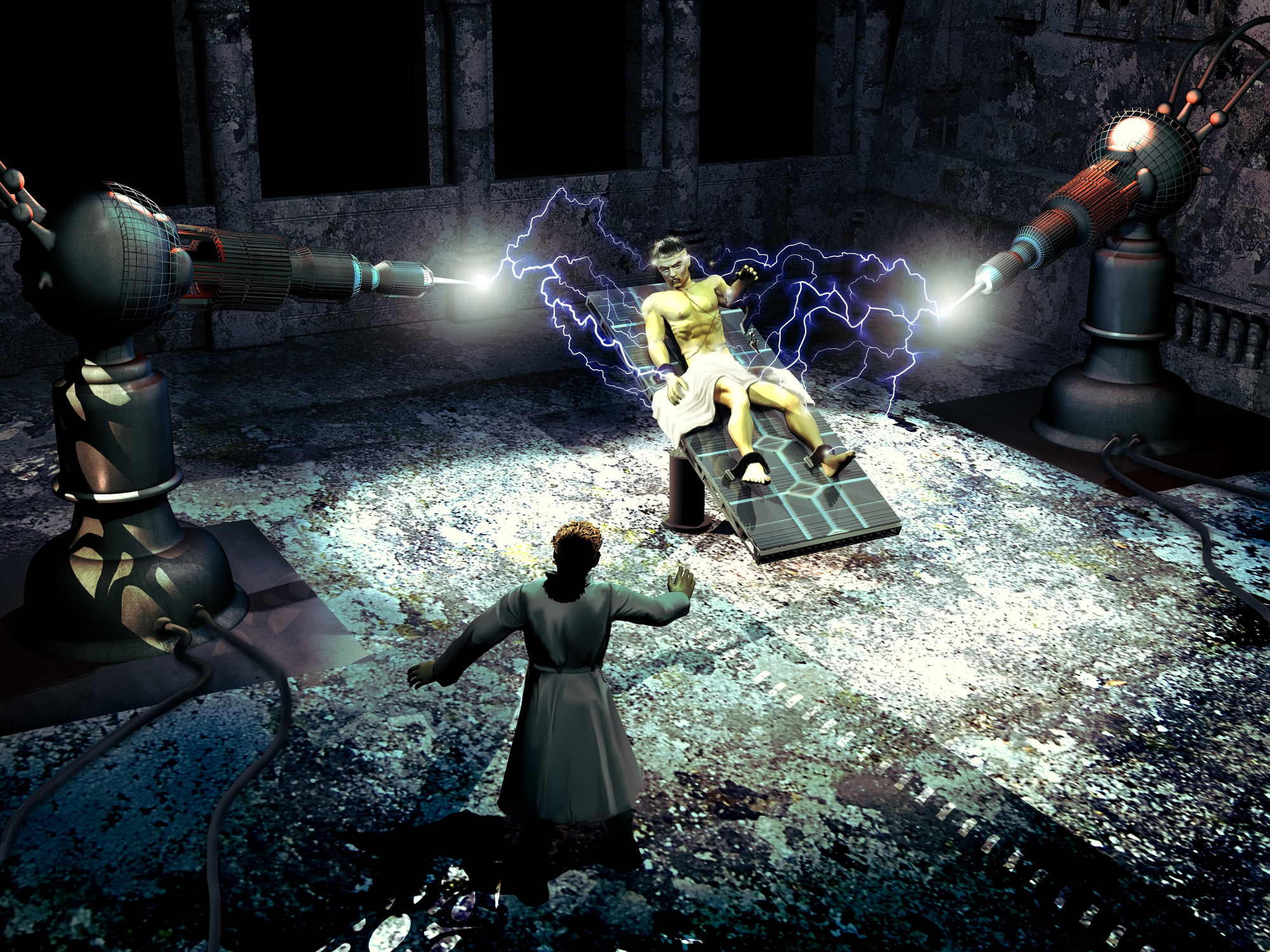
Interestingly, the human anatomy has always been part of fictional stories; for example, Victor Frankenstein was a young scientist who created ‘The Monster’ using unconventional scientific experiments. This story was created by the English author Mary Shelley (1797-1851) in the eighteenth century. Frankenstein created the monster over two years by meticulously constructing the body using anatomical parts brought to life.
During the study of anatomy in England, the Age of Enlightenment become infamous for body snatching from graveyards to provide an adequate supply of corpses. Consequently, it was a factor because the teaching of medicine and surgery was becoming more established through medical schools. There was a shortage of corpses legally available for anatomy classes. The lack of corpses forced the anatomy teachers to pay for bodies from London gangs who dug them up from graveyards.
The Anatomy Act 1832
To help and circumvent the shortage of corpses, the 1752 Murder Act was passed by Parliament to allow the bodies of executed murderers and be used to study anatomy.
In the notorious case of William Burke and William Hare murdered 16 people over about ten months in Edinburgh in 1828. They sold these corpses to the renowned lecturer of anatomy Robert Knox (1791-1862), at the University of Edinburgh for dissection lectures.
However, these murders met the requirements for bodies for medical research and contributed to the passing of the Anatomy Act of 1832. This Act of Parliament authorised physicians, anatomy lecturers and medical students to dissect donated bodies. It was legislated in response to the public disgust at the illegal trade in corpses.
The Anatomy Act 1832 was first presented in 1828 because acquiring bodies for medical research was scrutinised by the House of Commons, but an initial attempt at legislation failed.
However, the Anatomy Act of 1832 gave surgeons and medical students legal access to bodies from workhouses, hospitals and prisons that were unclaimed 48 hours after death.
Modern Anatomy
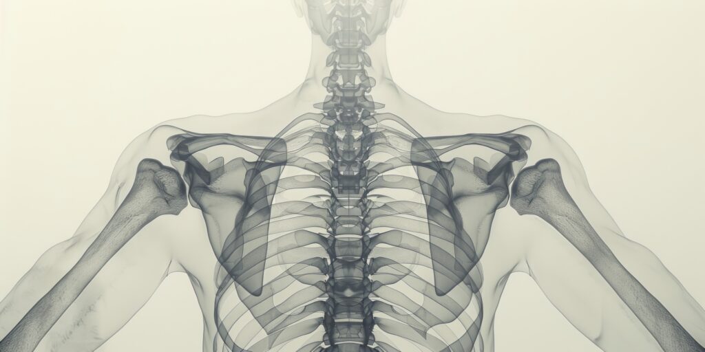
Modern anatomy has evolved rapidly through today’s technology, and human anatomical structures have been more understood since the publication of Frankenstein. For example, the muscles and bone structures are more understood, and plastic surgery has improved through skin grafts being more successful operations. Furthermore, the secrets of the nervous system on how electricity can cause muscle spasms since the time of Galvani.
Since Shelley’s writing of Frankenstein, the understanding of human anatomy may be accurate at the surface, but little was known about the human body’s inner workings. However, Frankenstein created a sinister monster that is today recognised throughout the world over 200 years ago.
[anatomy]
Anatomy Timeline
The advancement of the microscope by Anton van Leeuwenhoek (1632-1723) and Marcello Malpighi helped to progress anatomical research and led to some scientific discoveries:
- Van Leeuwenhoek was able to magnify the fine details of various tissues and was the founder of microscopic anatomy known as histology.
- Robert Hooke (1635-1703) was the first to recognise and name cells in the tissues.
- Robert Brown (1773-1858) recognised the presence of nuclei.
- In the 1830s, Theodor Schleiden and Matthias Schwann proposed that cells are universal in all tissues and play a vital role. This theory is the basis for modern histology, embryology and pathology concepts.
- In 1761, Giovanni Battista Morgagni, an Italian researcher, made several discoveries and was the first pathologist.
Today, anatomical education has been more constructive and beneficial since the Age of Enlightenment due to the advancements in digital technology, web-based resources and computer-aided learning. For example, a wide range of 3-D virtual reality models such as Visible Body; Primal Pictures; 3D4Medical; Cyber Anatomy HolographicTM; BodyViz; are the most prevalent and influential.
This technology platform enables teachers and students of anatomy to engage in the illustrations and information they need to conduct anatomical research.
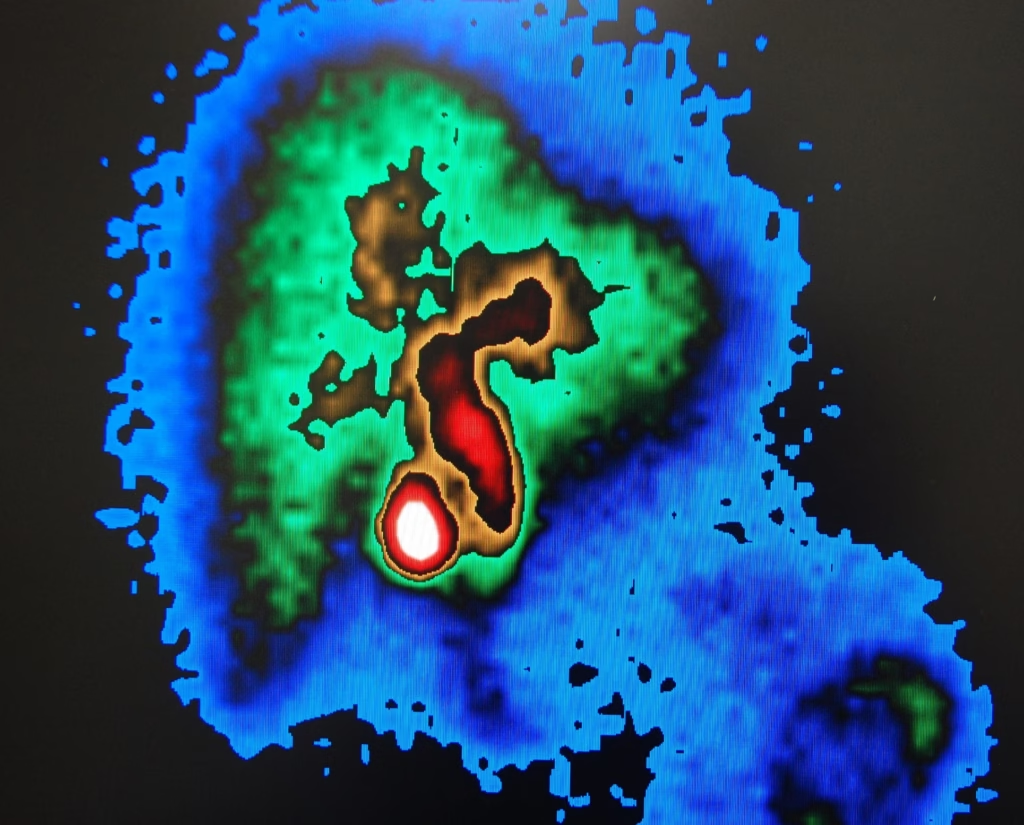
In the twentieth century, further advances in radiological techniques have permitted researchers to make remarkable connections between anatomy and physiology. In addition, these imaging modalities allowed for the integration of anatomy with genetics, biochemistry and biophysics.
Furthermore, microscopy and the discovery of x-rays have set the foundation of advanced technologies such as PET, MRI and CT scanners to allow a non-invasive look inside the human body.
Furthermore, anatomy books are much more different from when Gray’s Anatomy was first published in 1858. An earlier book on anatomy by Claudius Galenus was a 15-volume collection on De Anatomicis Administrationibus (Galen on Anatomical Procedures). This book gave an account of the achievements and failures of surgeons between 129 and 198 AD. The book provided experimental details on the phrenic nerves and the diaphragm from numerous dissections.
The history of anatomy, especially in the medieval period, was reported by the Italian anatomist Mondino de Luzzi (1270-1326) from the University of Bologna. His teachings on dissection even influenced Leonardo da Vinci. Furthermore, Andreas Vesalius (1514-1564) published the De humani corporis fabrica (On the Structure of the Human Body), and other ancient texts of Aristotle and Galen were still used in medical schools of Europe. However, Vesalius discovered inaccuracies in the ancient texts, and the De humani corporis fabrica became an authoritative textbook on anatomy.
The De humani corporis fabrica contained over 200 engravings by various artists, including Jan Steven van Calcar (1499-1546). One of these illustrations shows Vesalius lecturing to a large crowd while dissecting a corpse.
Conclusion
Modern medical students use the modern Gray’s Anatomy with its colour artwork, which has become the gold standard. In addition, the modern Gray’s contains MRI, X-ray, and PET scans which would have been unimaginable in Henry Gray’s time.
Interestingly, the American educator Abraham Flexner (1866-1959) wrote a famous report on medical education reform and the importance of the basic medical sciences. The report concluded that anatomy is an essential science for basic medical training. Today, the human body or parts are preserved techniques to ensure adequate material for future medical students.
Disclaimer
The content of this article, “Frankenstein to Modern Anatomy of the Human Body”, is intended for general informational and educational purposes only. It explores the historical and cultural evolution of human anatomical study, including references to literature, scientific development, and historical practices.
The article references historical events and figures, some of which involve illegal or unethical practices from earlier periods. These references are included purely for historical context and do not reflect or endorse such actions. Additionally, the fictional character of Frankenstein is used symbolically to highlight changes in scientific understanding over time, and should not be interpreted as a factual or medical source.
While efforts have been made to ensure accuracy, Open Medscience does not guarantee the completeness or reliability of the information presented. Readers are encouraged to consult academic or clinical sources for formal anatomical or medical education. Any use of this content for academic, clinical or professional purposes should be supported by current, peer-reviewed literature and official guidelines.
Open Medscience is not liable for any decisions made based on this article. For questions relating to medical practice or education, please consult a qualified medical professional or educational institution.
home » blog » human anatomy »