Visual Evoked Potentials (VEPs) are electrical signals generated by the brain in response to visual stimuli. The signals are recorded through electrodes placed on the scalp and can provide information about visual processing and function. VEPs are used in clinical settings to diagnose neurological and visual disorders and in research to study brain function and cognitive processes.
The Neural Basis of Visual Evoked Potentials
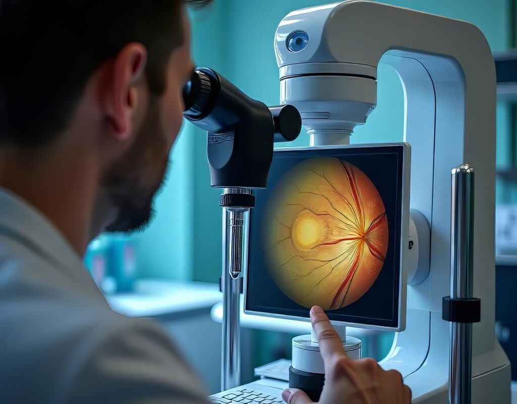
Visual evoked potential (VEP) is an electrophysiological test that measures the electrical activity of the visual system in response to visual stimuli, typically flashes or light patterns. The test evaluates the integrity and function of the visual system, including the retina, optic nerve, and visual pathways in the brain.
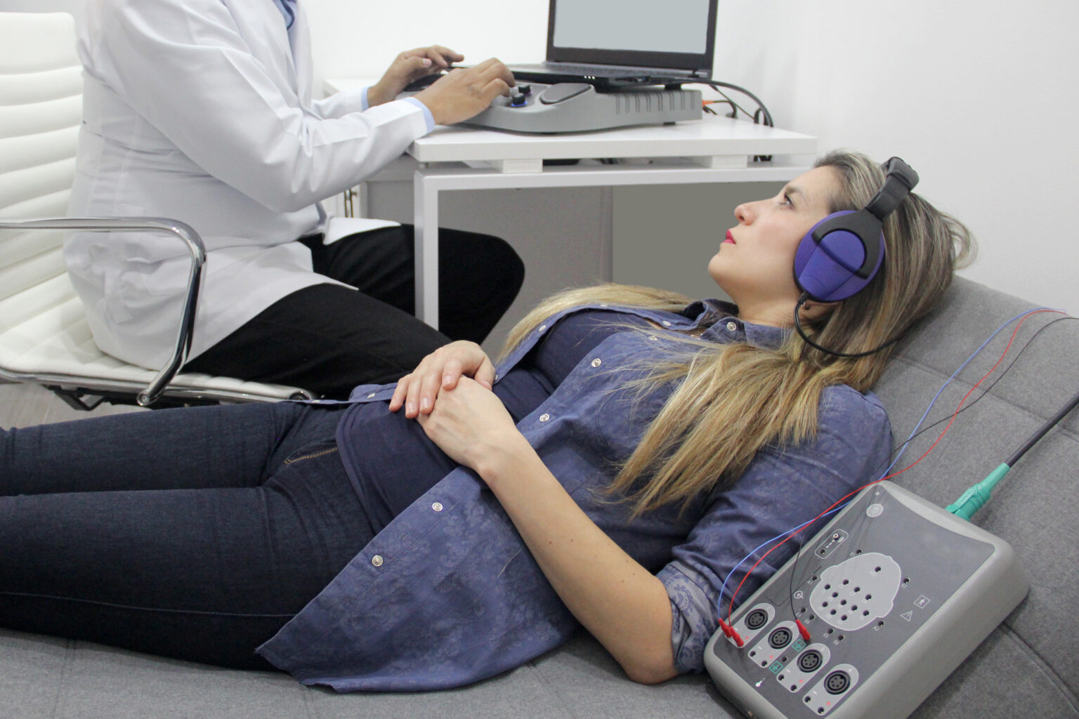
During the VEP test, the patient wears a special electrode cap or electrodes placed on the scalp to record electrical signals from the brain’s visual cortex. Then, the patient is subjected to visual stimuli, such as flashes of light or patterns on a computer screen, and the electrical responses from the visual cortex are recorded.
The recorded signals are then analysed to determine the latency and amplitude of the response, which can provide information about the health and function of the visual system.
Visual Evoked Potentials in Multiple Sclerosis
VEP testing can help diagnose various conditions that affect the visual system, including optic neuritis, multiple sclerosis, glaucoma, and amblyopia. Optic nerve inflammation can result in vision problems resulting in blurred or double vision, including pain with eye movement.
During VEP testing for MS, the patient is shown a series of visual stimuli, such as flashes of light or checkerboard patterns. At the same time, electrodes record the electrical signals from the brain’s visual cortex. The recorded signals are then analysed to determine the latency and amplitude of the response.
In MS patients with optic neuritis, VEP testing can show delayed latency and reduced visual response amplitude, indicating that the optic nerve and visual pathways are not functioning correctly. However, VEP testing alone is insufficient to diagnose MS and should be combined with other clinical and diagnostic tests.
Clinical Applications of Visual Evoked Potentials
Visual evoked potential (VEP) testing can diagnose various conditions affecting the visual system, for example:
- Amblyopia, known as lazy eye, is when the brain ignores or suppresses input from one eye. VEP testing can evaluate the function of each eye and determine if there is a problem with the visual pathways in the brain.
- Glaucoma damages the optic nerve, which can lead to vision loss. VEP testing can help diagnose and monitor the progression of glaucoma by measuring the electrical responses from the visual cortex in response to visual stimuli.
- Brain tumours affecting visual pathways can cause vision problems. VEP testing can help diagnose and monitor the progression of these tumours by measuring the electrical responses from the visual cortex.
- VEP testing can also diagnose and monitor certain retinal disorders, such as optic disc drusen and macular degeneration, by evaluating the electrical responses from the visual cortex to visual stimuli.
Assessment of Visual Function Using Visual Evoked Potentials
Visual evoked potential (VEP) data is typically analysed by measuring the latency and amplitude of the electrical responses from the visual cortex in response to visual stimuli. The analysis of VEP data involves several steps, including:
- VEP signals are recorded using an electrode cap or individual electrodes placed on the scalp. The recorded signals are then amplified and filtered to remove noise.
- VEP signals are usually averaged over multiple trials or visual stimulus presentations.
- The latency of the VEP response is measured as the time from the onset of the visual stimulus to the peak of the evoked response. Latency analysis can provide information about the speed of neural transmission along the visual pathways.
- The amplitude of the VEP response is measured as the magnitude of the peak-to-peak voltage of the evoked response. Amplitude analysis can provide information about the strength of neural activity in the visual cortex.
- VEP data is often compared to normative data from healthy individuals to determine any abnormalities in the VEP response. Deviations from normal values can indicate underlying pathology or dysfunction of the visual system.
The analysis of VEP data is a complex process that requires specialised equipment and expertise. As a result, trained healthcare professionals or specialised laboratories often perform it.
The Future of Visual Evoked Potentials in Ophthalmology
The future of VEP testing looks promising, as it continues to be an essential tool for evaluating the visual system’s function and diagnosing a wide range of conditions.
While VEPs and MRI are different types of tests, they can be used together to gain a complete picture of brain function. For example, researchers may use VEPs to study the brain’s visual processing pathways while using MRI to identify the certain brain regions involved in those pathways.
In some cases, VEPs can also be used during an MRI scan to help map the visual cortex and identify brain areas that may be affected by disease or injury. This type of technique is known as functional MRI (fMRI) and can provide valuable information for research and clinical applications.
- VEP testing may be used for the early detection of Alzheimer’s and Parkinson’s disease.
- VEP testing can better understand individual differences in the visual system, which can help healthcare professionals tailor treatment plans to each patient’s unique needs.
- Advances in technology, such as high-density electrode arrays and wearable devices, may improve the accuracy and efficiency of VEP testing and make it more accessible to patients.
- VEP testing may be integrated with other medical imaging modalities, such as positron emission tomography PET and magnetic resonance imaging, to evaluate the visual system and brain function.
Therefore, combining VEPs and MRI can provide a powerful tool for studying brain function and identifying abnormalities or changes in the brain’s visual processing pathways.
Disclaimer
The information presented in this article, Visual Evoked Potentials and Their Clinical Applications, is provided for general informational and educational purposes only. It is not intended as medical advice and should not be relied upon as a substitute for consultation with a qualified healthcare professional.
While every effort has been made to ensure the accuracy and reliability of the content, Open Medscience does not guarantee the completeness, timeliness, or accuracy of the information provided. Clinical decisions should always be based on a combination of test results, clinical judgment, and the patient’s individual circumstances.
Any references to diagnostic methods, including Visual Evoked Potentials (VEPs), are intended to support awareness and understanding and not to replace clinical training or professional medical advice. If you have questions about a medical condition or procedure, please consult a healthcare provider or specialist.
Open Medscience does not accept any responsibility for any loss, injury, or damage incurred by the use or reliance on the information contained in this publication.
home » blog » optical imaging »