
Molecular imaging has altered the way disease is visualised, staged and monitored. Rather than relying solely on anatomical changes, nuclear medicine can reveal biological processes at the cellular and molecular level. Within this landscape, radiolabelled peptides have become an important class of investigational and clinical tools, linking the specificity of peptide-receptor binding with the sensitivity of PET and SPECT imaging.
Their value lies in a relatively simple concept: a peptide can be designed or selected to recognise a biological target, while a radionuclide allows that interaction to be detected externally. When carefully matched to the right radionuclide, chelator and biological target, radiolabelled peptides can provide information that conventional imaging may not capture with the same functional precision.
This is particularly relevant to precision medicine, where patient selection, treatment planning and response assessment increasingly depend on measurable disease biology rather than broad diagnostic categories alone.
Why Peptides Are Well Suited To Molecular Imaging
Peptides occupy a useful middle ground between small molecules and larger biologics. They are typically small enough to penetrate tissues and clear from the bloodstream relatively quickly, yet structurally capable of binding with high affinity to specific receptors or transporters. This combination can produce favourable imaging contrast when the target is sufficiently expressed in diseased tissue and background uptake remains manageable.
In radiopharmaceutical development, these characteristics matter. An imaging agent must reach its target, bind with adequate specificity, clear from non-target tissues and remain stable long enough for acquisition. Peptides can often be modified to improve metabolic stability, alter pharmacokinetics, or enable radiolabelling through a chelator without completely disrupting target recognition.
The best-known clinical examples involve somatostatin receptor imaging in neuroendocrine tumours. Radiolabelled somatostatin analogues demonstrated that receptor expression could be visualised in vivo, supporting diagnosis, staging and therapeutic decision-making. This model has helped establish a wider framework for peptide-based theranostics: identify the target, image the target, and, where appropriate, use a related radioligand to deliver therapeutic radiation.
From Anatomical Imaging To Biological Characterisation
CT and MRI remain indispensable in oncology, cardiology, neurology and many other areas of medicine. However, anatomical imaging often shows structural consequences of disease rather than the underlying molecular behaviour. Molecular imaging adds another layer by asking different questions: Which receptors are expressed? Is a lesion metabolically active? Does the target remain present after treatment? Is there heterogeneity between tumour sites?
Radiolabelled peptides can contribute to this biological characterisation when receptor expression is clinically meaningful. In oncology, peptide-based PET or SPECT imaging may help identify lesions expressing a relevant target, assess disease extent, or support selection for radionuclide therapy. In research settings, the same approach can be used to study target distribution, pharmacodynamics and disease mechanisms.
This is not only a matter of detecting more lesions. It is about understanding whether a patient’s disease biology aligns with a particular diagnostic or therapeutic strategy. Precision medicine depends on this alignment. A highly selective therapy is unlikely to benefit a patient whose disease does not express the intended target.
The Radiochemistry Behind The Signal
A radiolabelled peptide is not simply a peptide with a radioactive atom attached. Its performance depends on the full molecular design.
The radionuclide determines the imaging or therapeutic application. Positron emitters, including gallium-68 and fluorine-18, are widely associated with PET imaging. Gamma emitters, including technetium-99m and indium-111, are used in SPECT. Therapeutic radionuclides, including lutetium-177 and actinium-225, are studied or used for targeted radionuclide therapy, depending on the agent, indication and regulatory context.
The chelator, when required, must hold the radiometal securely under physiological conditions. Instability can lead to off-target radiation exposure and poor image quality. The linker can influence receptor binding, internalisation, hydrophilicity and clearance route. Even small structural changes can alter biodistribution.
This is why radiopharmaceutical translation requires close collaboration between radiochemists, biologists, imaging specialists, clinicians, physicists and regulatory experts. A promising receptor-binding peptide is only the starting point. It must become a reproducible, stable, safe and clinically interpretable imaging agent.
Theranostics And Patient Selection
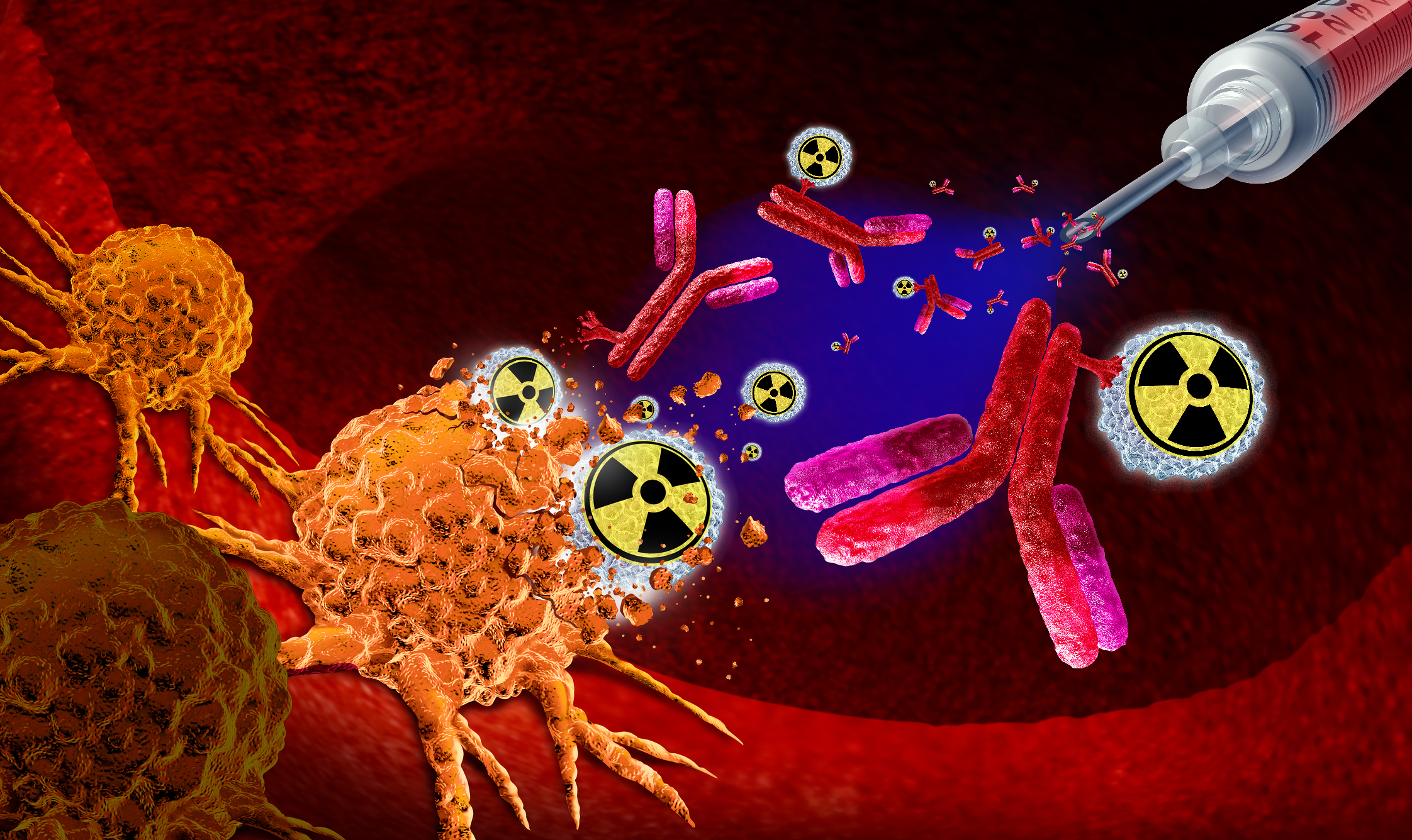
One of the most important contributions of radiolabelled peptides is their role in theranostics, where diagnostic imaging and targeted therapy are connected through shared biological targeting. In this model, an imaging scan can help determine whether the target is sufficiently present before a therapeutic radiopharmaceutical is considered.
Peptide receptor radionuclide therapy in neuroendocrine tumours is the clearest example. Somatostatin receptor imaging can help demonstrate target expression, while therapeutic radiolabelled analogues deliver radiation to receptor-positive disease. Although not every patient is eligible and outcomes vary, this approach illustrates how molecular imaging can guide treatment selection more intelligently than anatomy alone.
Researchers are also exploring other targets, including gastrin-releasing peptide receptors, fibroblast activation protein and receptors associated with specific tumour types or microenvironmental features. Some candidates are further along than others, and early imaging promise does not always translate into clinical benefit. Target density, internalisation, normal tissue uptake, dosimetry, tumour heterogeneity and toxicity all influence whether a peptide-based approach is viable.
Interpreting Peptide Research With Scientific Caution
The growing public interest in peptides has created a need for better scientific literacy. The word “peptide” can refer to many different entities: endogenous signalling molecules, diagnostic radiotracers, therapeutic analogues, research compounds, cosmetic ingredients and unregulated products sold online. These categories should not be conflated.
In molecular imaging, peptide research is grounded in target biology, radiochemistry, imaging physics, dosimetry and clinical validation. That is very different from consumer-facing claims about performance, ageing, recovery, or general wellness. Readers moving between clinical literature and broader peptide discussions need to assess context carefully: What is the compound? Is it approved, investigational, or preclinical? Is the evidence from cell studies, animal models, early human trials, or established clinical use? Is the endpoint imaging uptake, symptom change, survival, safety, or something else?
For readers who want a broader educational orientation to peptide terminology and research categories outside the formal radiopharmaceutical literature, Peptide Insider’s peptide education hub can serve as a research-focused reference point, provided it is not treated as a substitute for medical advice, regulatory guidance, or peer-reviewed clinical decision-making.
Challenges In Translation
Despite their promise, radiolabelled peptides face practical and scientific challenges. Receptor expression may vary between patients, between lesions in the same patient and over time. Kidney uptake can be a concern for some peptide radiopharmaceuticals because renal clearance may increase radiation exposure to sensitive tissue. Metabolic degradation can reduce target delivery. Manufacturing and quality control must be reliable, especially for short-lived radionuclides.
There are also interpretive challenges. High uptake is not always synonymous with aggressive disease, and low uptake does not necessarily mean disease absence. Imaging findings must be assessed alongside clinical history, pathology, laboratory data and other imaging modalities. Precision medicine is not achieved by a tracer alone; it emerges from integrating multiple sources of patient-specific information.
Regulatory pathways also require robust evidence. A radiolabelled peptide may show attractive biodistribution in early studies, but clinical adoption depends on demonstrating safety, reproducibility, diagnostic value and, in some cases, impact on patient management or outcomes.
The Future of Peptide-Based Molecular Imaging
The next phase of peptide-based imaging is likely to be shaped by better target selection, improved radiochemistry and more sophisticated data integration. Artificial intelligence may assist with image reconstruction, lesion quantification and pattern recognition, although these tools require careful validation. Total-body PET could improve kinetic modelling and low-dose imaging protocols. New chelators, linkers and radionuclide combinations may expand what is possible for both diagnosis and therapy.
Equally important will be patient stratification. As medicine becomes more biologically precise, imaging agents that identify target expression non-invasively may help reduce uncertainty before treatment. Radiolabelled peptides are well placed within this shift because they can connect molecular recognition with whole-body visualisation.
Their future, however, should be viewed with balance. Not every target will prove clinically useful. Not every promising tracer will become a routine imaging agent. The field advances through careful chemistry, rigorous validation and cautious interpretation.
Conclusion
Radiolabelled peptides illustrate the strength of molecular imaging: the ability to visualise disease biology rather than anatomy alone. By pairing peptide-based targeting with radionuclide detection, researchers and clinicians can investigate receptor expression, refine patient selection and support the development of theranostic strategies.
For precision medicine, this is a significant direction of travel. The aim is not simply to image more, but to image more meaningfully. As radiochemistry, target biology and clinical evidence continue to mature, peptide-based radiopharmaceuticals are likely to remain central to the evolution of molecular imaging and personalised care.
Disclaimer: This article is provided for educational and informational purposes only and does not constitute medical advice, diagnosis, treatment guidance, or a recommendation to use any peptide, radiopharmaceutical, radionuclide, or investigational compound. Radiolabelled peptides and peptide-based radiopharmaceuticals may be approved for specific clinical indications, under investigation, or limited to research settings, depending on the compound and jurisdiction. Clinical decisions should be made by appropriately qualified healthcare professionals in accordance with current regulatory guidance and individual patient circumstances. References to external educational resources are provided for general information only and do not imply endorsement of any product, treatment, or medical claim by Open MedScience.