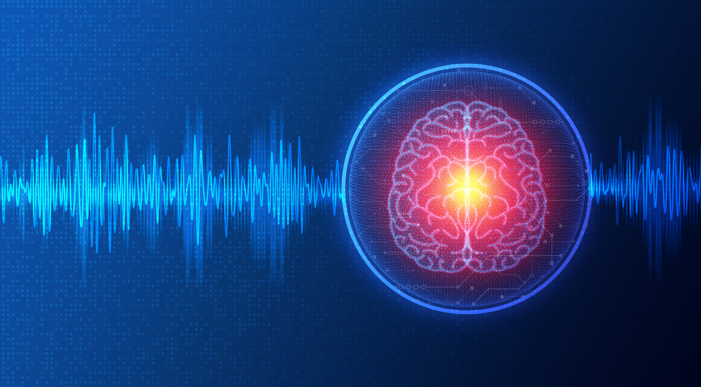
Deep Brain Imaging
Deep brain imaging is a rapidly advancing field that enables researchers and clinicians to visualise and study the structure and function of the brain at unprecedented depths and levels of detail. This cutting-edge technology is pivotal in understanding complex neural mechanisms, diagnosing neurological disorders, and guiding surgical interventions.
At its core, deep brain imaging refers to techniques that allow for the observation of brain regions buried deep beneath the cortical surface. These areas, including the thalamus, hippocampus, and basal ganglia, are crucial for processes such as memory, emotion, motor control, and sensory integration. Traditional imaging techniques, such as computed tomography (CT) or standard magnetic resonance imaging (MRI), often lack the resolution or specificity required for detailed examination of these regions. Innovations in imaging technology, however, are overcoming these limitations.
One of the most transformative advancements in this area is functional magnetic resonance imaging (fMRI). fMRI measures brain activity by detecting changes in blood flow, offering insights into which regions are activated during specific tasks. With enhancements in magnetic field strength and pulse sequences, fMRI has become a cornerstone of deep brain imaging, capable of probing functional connectivity at a macro level. Similarly, diffusion tensor imaging (DTI), a specialised MRI technique, maps white matter tracts, shedding light on the intricate wiring of deep brain structures.
Positron emission tomography (PET) is another key modality in deep brain imaging. By using radiotracers, PET provides metabolic and molecular information, making it invaluable in studying conditions such as Parkinson’s disease and Alzheimer’s disease. Recent advances, such as hybrid PET/MRI systems, allow for simultaneous metabolic and anatomical data acquisition, enabling a more comprehensive understanding of deep brain pathophysiology.
Optical techniques, like multiphoton microscopy and optogenetics, have revolutionised the study of deep brain function in animal models. These methods use light to stimulate and record neural activity with extraordinary spatial and temporal resolution. While their application in humans is limited due to the skull’s opacity, innovations in implantable devices and adaptive optics hold promise for future translational applications.
Ultrasound-based methods, such as functional ultrasound (fUS) imaging, are also emerging as powerful tools for deep brain imaging. fUS detects changes in cerebral blood flow with high sensitivity and can be used in real-time applications, making it ideal for intraoperative monitoring.
Deep brain imaging is not without challenges. Achieving high resolution while maintaining safety and non-invasiveness is a primary concern. Additionally, the interpretation of complex data requires advanced computational methods, such as machine learning and artificial intelligence.
In summary, deep brain imaging is a transformative field that combines advances in physics, engineering, and neuroscience. As technologies continue to evolve, the potential to uncover the mysteries of the human brain and improve neurological healthcare grows exponentially.
home » Deep Brain Imaging

 By
By