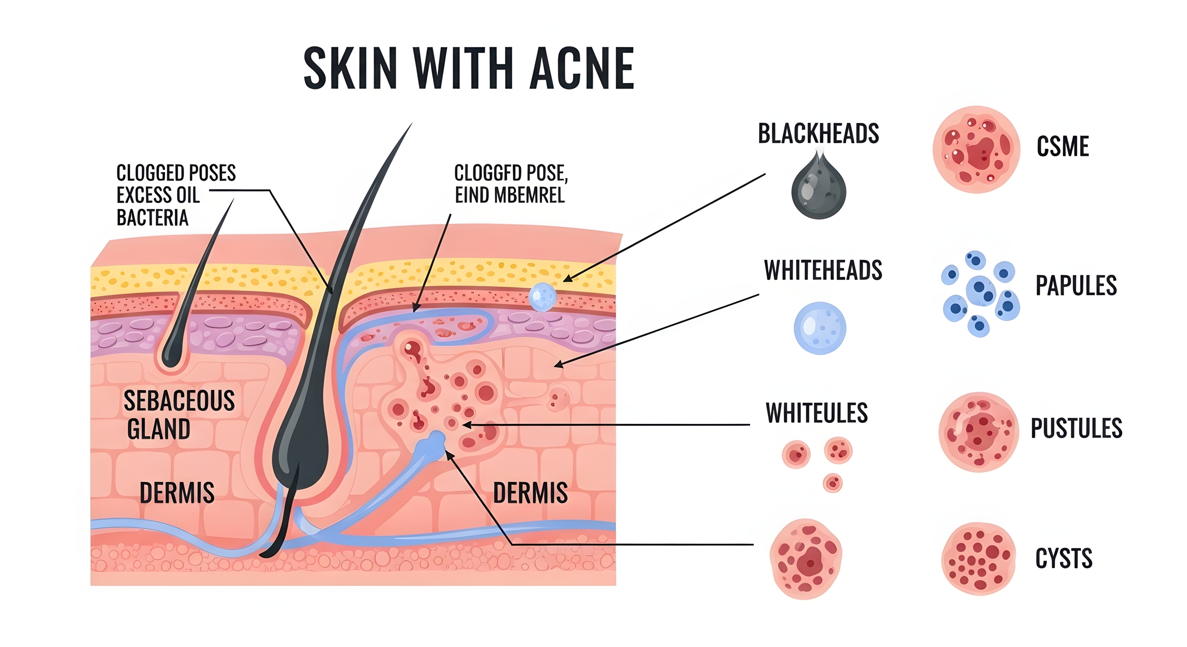
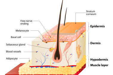
The Biology of Blemishes: Why Most Treatments Only Address Part of the Problem
 By
By
Learn about the complexities of blemishes and discover advanced blemish treatment strategies that truly work.
By
Learn about the complexities of blemishes and discover advanced blemish treatment strategies that truly work.
 By
By
Explore the importance of summer hydration and learn how to keep your body hydrated with fun and tasty drink options.
 By
By
Explore how creatinine pH specific gravity sample analysis ensures sample integrity and reduces contamination risks in labs.
 By
By
Learn about non-surgical aesthetic treatments and how they deliver significant results without the risks associated with surgery.
 By
By
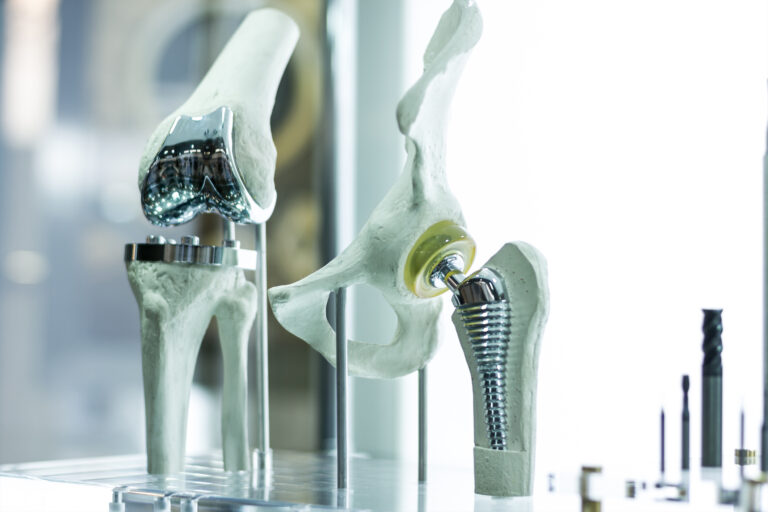
Discover the benefits of titanium in medicine, from prosthetics to surgical tools, and its impact on patient care.
 By
By
Discover how a nighttime supplement routine can help you achieve restful sleep and improve overall nightly recovery with ease. Image for illustration only. Person depicted is a model.
 By
By
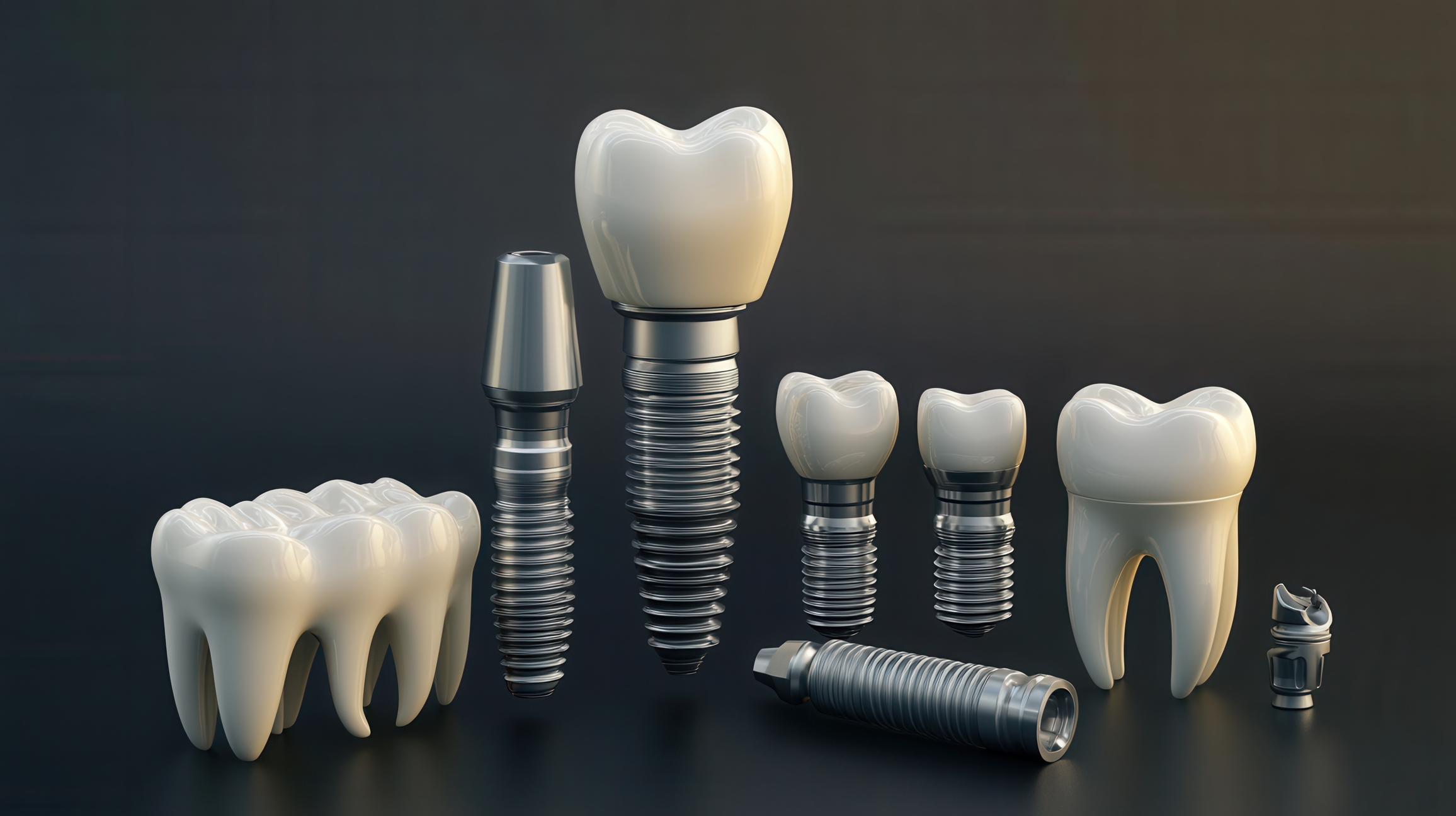
Comparing Tooth Replacement Methods helps patients understand bridges, dentures, and implants before making decisions.
 By
By
Learn how advanced limb restoration partners improve outcomes in rehabilitation through innovative technologies and compliance.
 By
By
Find out everything about anti-wrinkle injections treatment, including the process, expectations, and choosing the right practitioner.
 By
By
Learn about the importance of synthetic urine standards in laboratories to replicate human urine accurately and safely. Image for illustration only. Person depicted is a model.
 By
By
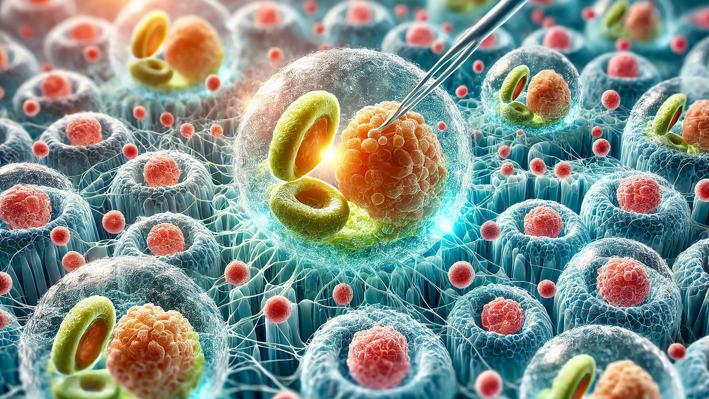
Stem-cell exosomes deliver powerful biological cues that improve skin structure, boost scalp vitality, and accelerate recovery.
 By
By
Uncover the critical role of anastrozole in research and its significance in advancing hormone-dependent cancer therapies.
 By
By
Find out how updated pressure injury prevention strategies can make a difference in healthcare facilities and patient safety. Image for illustration only. People depicted are models.
 By
By
Learn about the advantages of copper IUD control, a hormone-free method that ensures reliable pregnancy prevention. Image for illustration only. People depicted are models.
 By
By
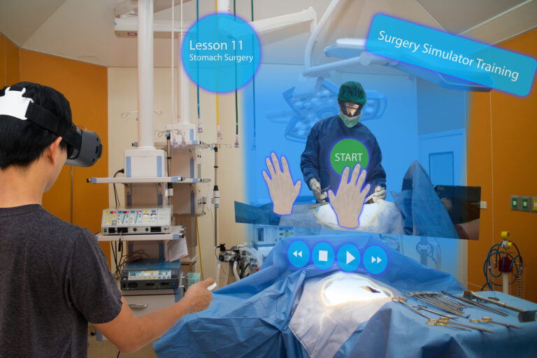
Learn how Virtual Reality Medical Training is revolutionising the way medical students learn and practise without patient risk. Image for illustration only. People depicted are models.
 By
By
Uncover the innovative insights of the World Transformation Movement and its impact on understanding patients’ emotional and cognitive needs.
 By
By
Low-speed crashes may look minor, but they often cause hidden spinal, ligament, and brain injuries over time.
 By
By
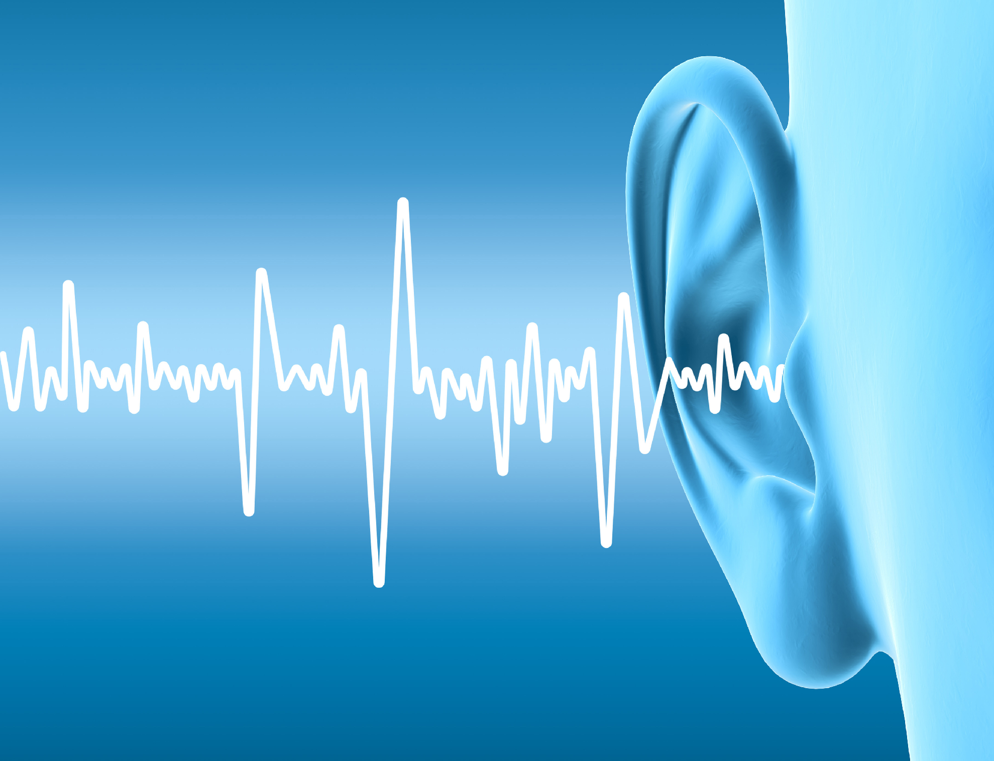
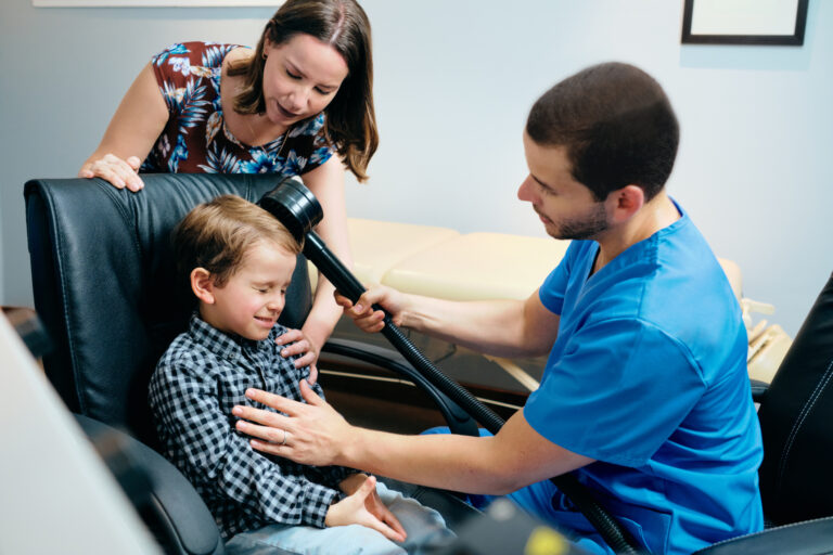
Discover the benefits of hearing aids in Malaysia and how they support individuals with hearing loss to engage fully in life.
 By
By
Learn how to enhance recovery after a minor car accident. Discover tips for coping with physical and emotional challenges. Image for illustration only. People depicted are models.
 By
By
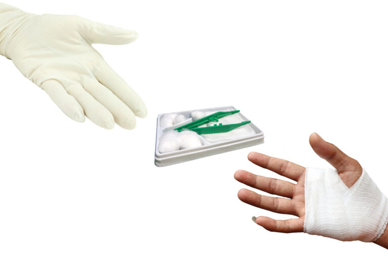
Clean and dress a wound properly to minimise infection, promote faster healing, and ensure better long-term recovery.
 By
By
Understand Follicular Unit Transplantation and its effectiveness in addressing hair loss while maintaining natural appearance. Image for illustration only. People depicted are models.
 By
By
Uncover the surprising medical benefits of Botox, including its effectiveness in treating migraines and hyperhidrosis.
 By
By
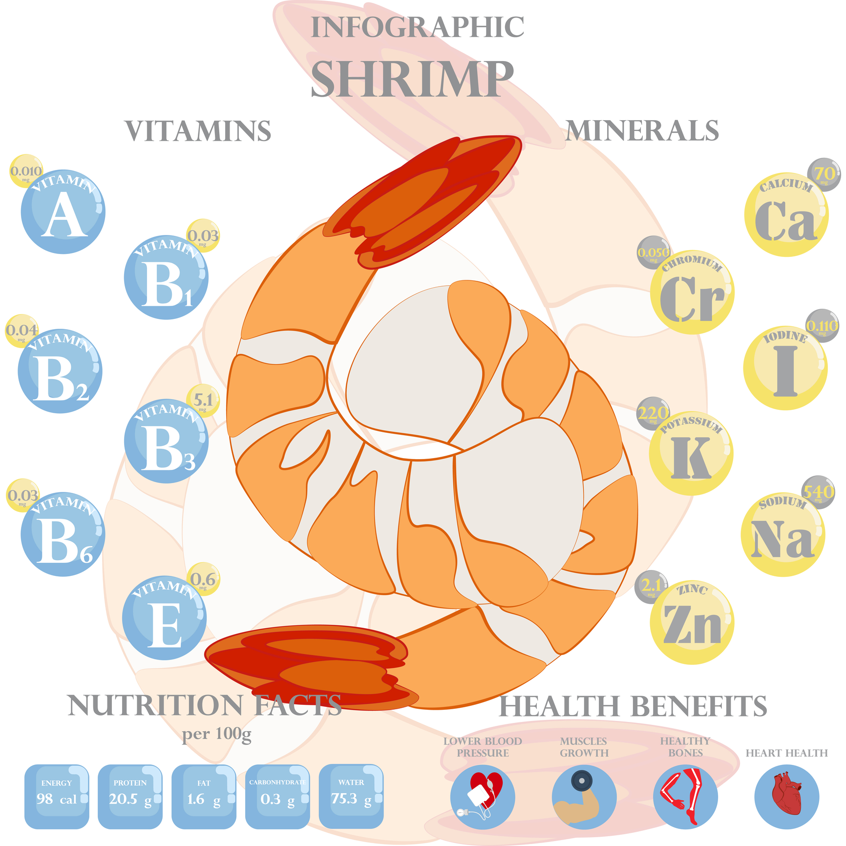
Shrimp’s zinc, selenium, and omega-3s support hormone balance, blood flow, and libido, enhancing sexual wellness.
 By
By
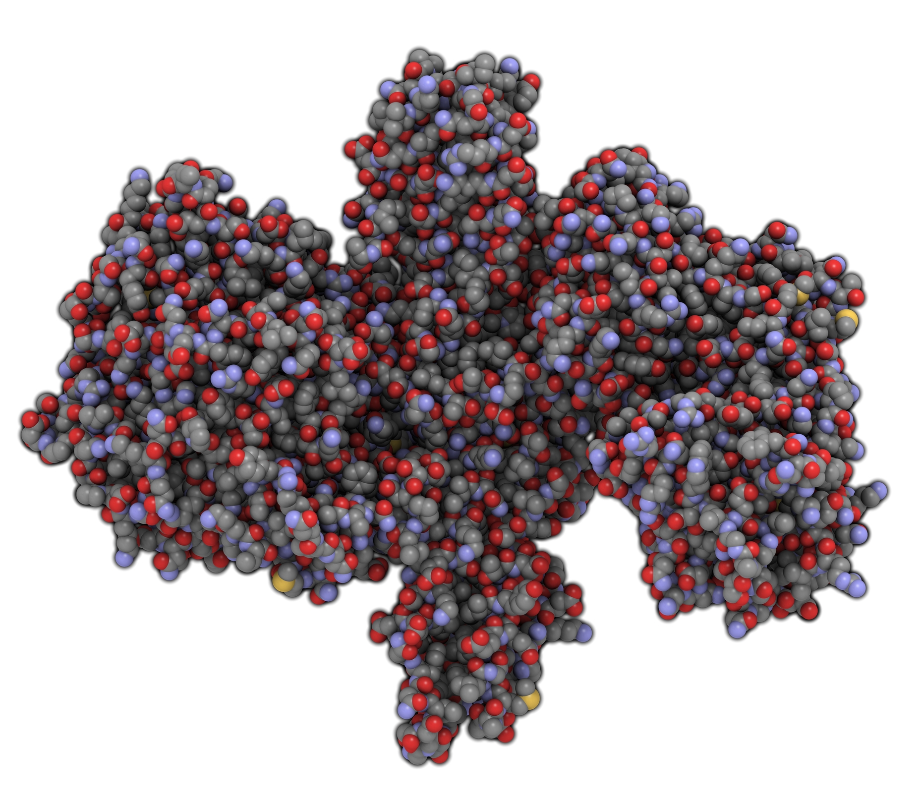
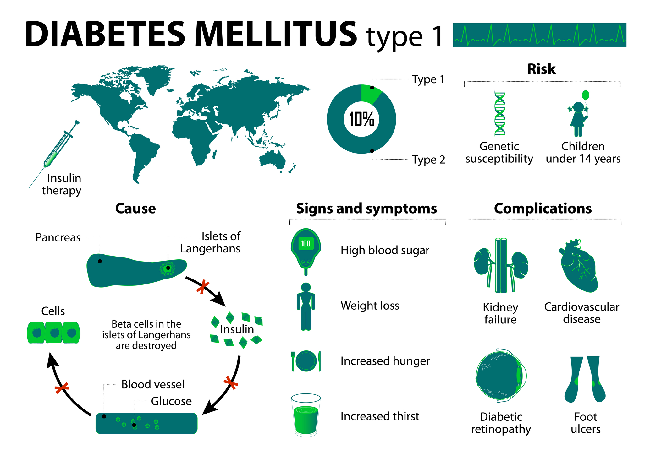
GLP-1 therapy uses receptor agonists to regulate blood sugar, reduce appetite, slow digestion, and support weight loss in patients.
 By
By
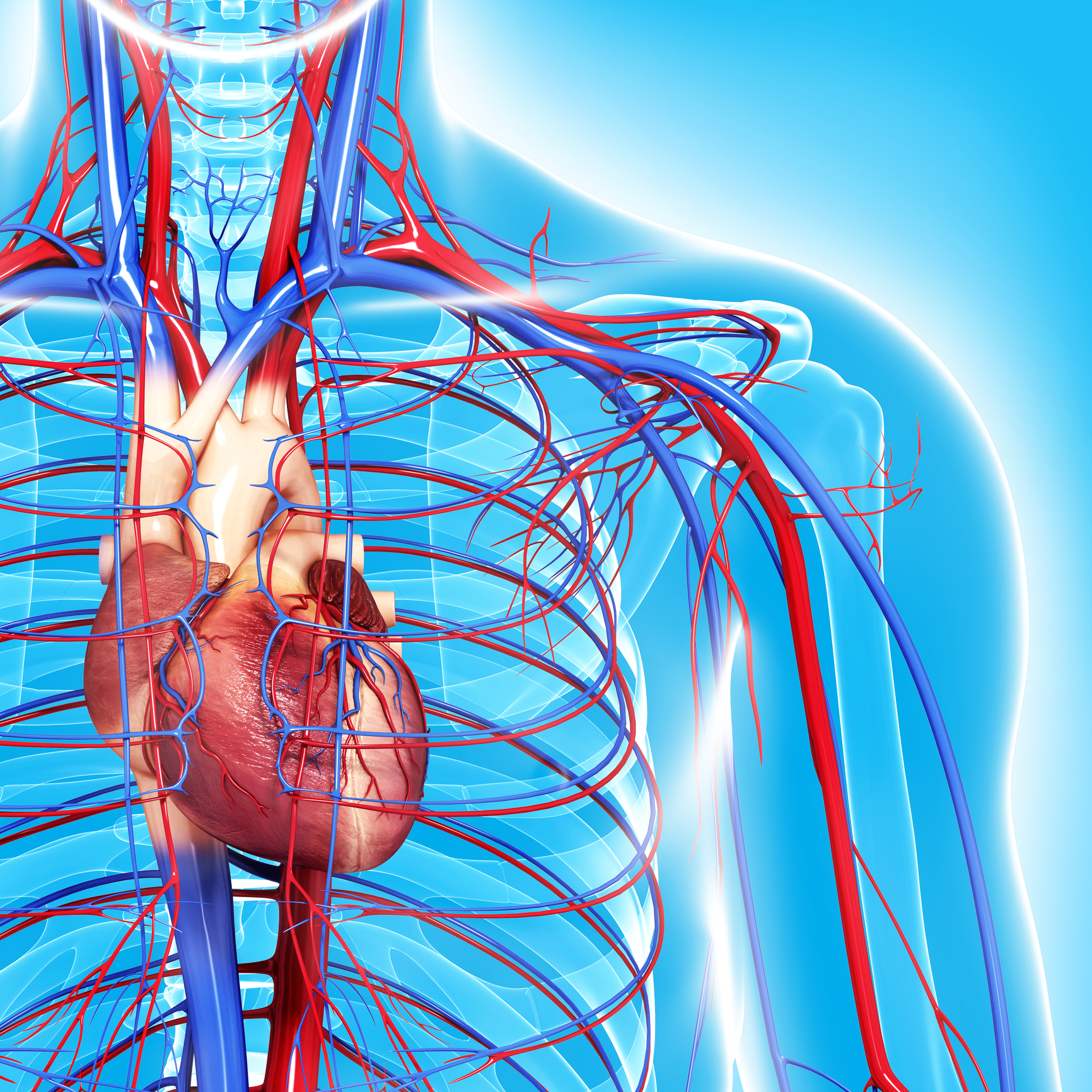
Movement and circulation are essential for maintaining healthy blood flow, reducing the risks of sedentary lifestyles.
 By
By
Knee health is essential for mobility, preventing pain, strengthening joints, improving flexibility, and maintaining an active, injury-free lifestyle daily.
 By
By
rTMS Therapy for Fibromyalgia uses magnetic pulses to reduce pain, improve mood, and enhance cognitive function. Image for illustration only. People depicted are models.
 By
By
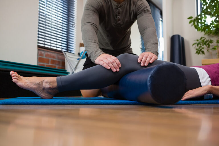
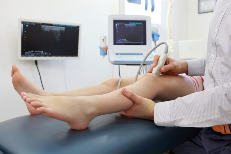
Sciatica nerve pain often radiates down the leg, but physiotherapy helps restore mobility and reduce discomfort effectively. Image for illustration only. Person depicted is a model.
 By
By
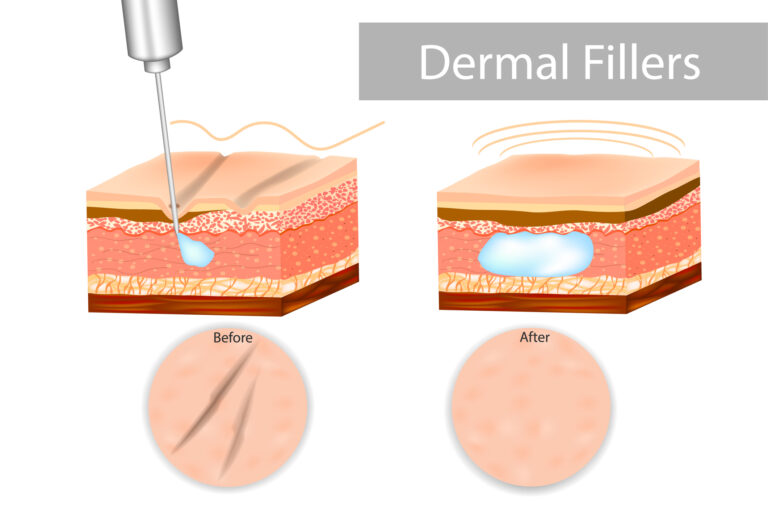
Hand and Neck Rejuvenation improves skin texture, reduces wrinkles, restores volume, and enhances a youthful, natural look.
 By
By
Viagra Cialis combo offers potential benefits for erectile dysfunction when used responsibly under medical supervision and guidance.