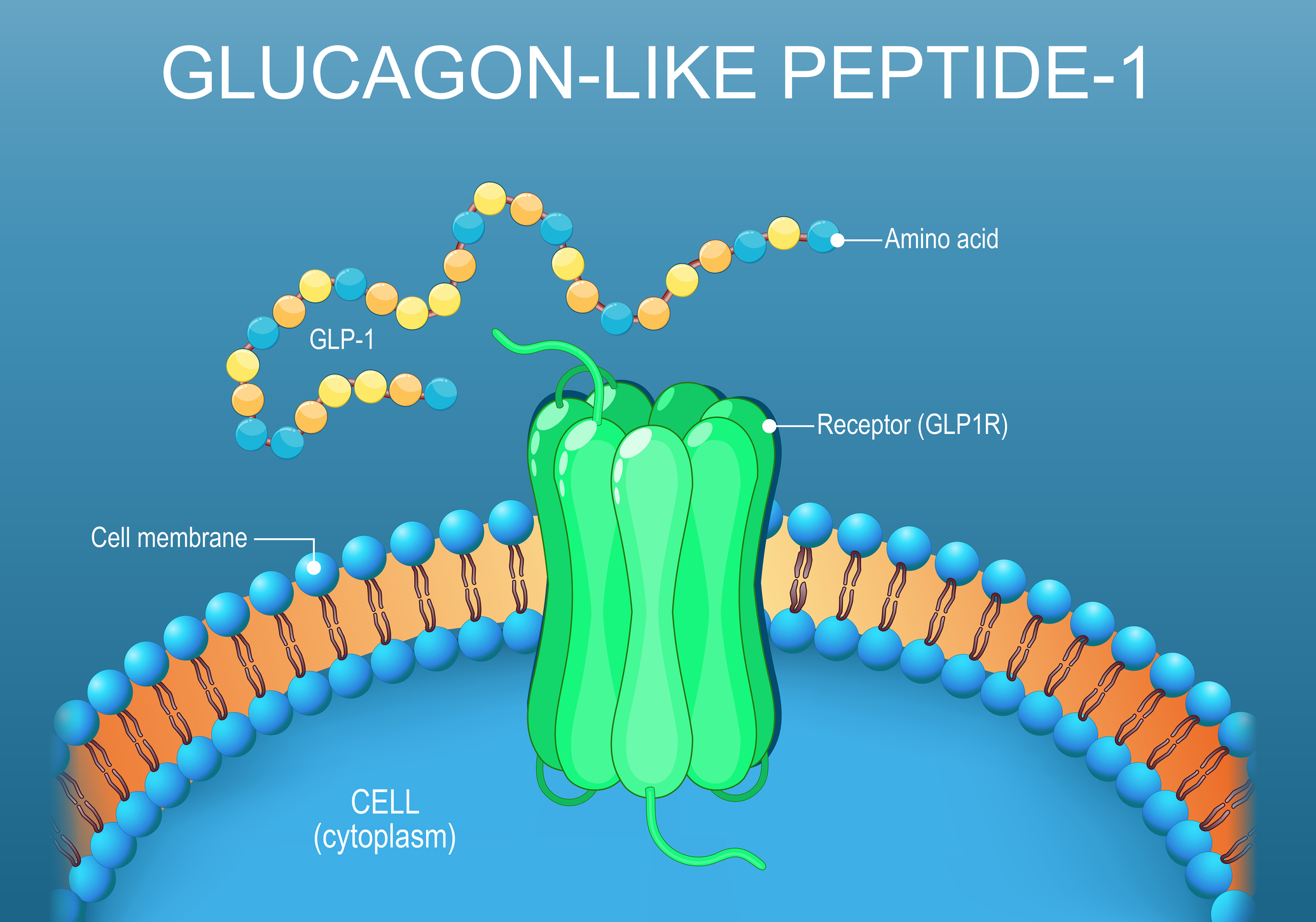
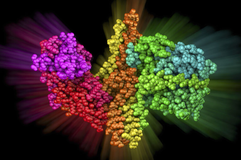
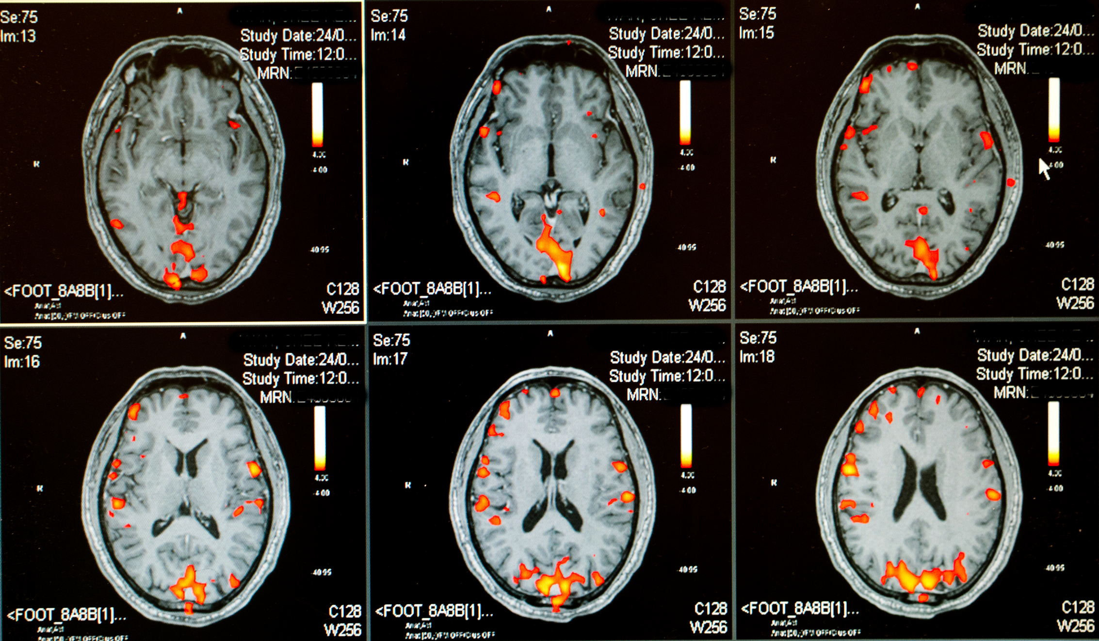
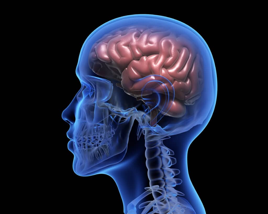
GLP-1 Receptor Agonists and the Clinical Evidence for Neuroprotection Beyond Diabetes
 By
By
Uncover the findings from the recent trials on GLP-1 receptor agonists neuroprotection and their implications for neurodegenerative diseases.
By
Uncover the findings from the recent trials on GLP-1 receptor agonists neuroprotection and their implications for neurodegenerative diseases.
 By
By
Learn about the challenges of radiology EMR and neurology EHR in critical decision-making situations. Data architecture matters.
 By
By
Learn how antidepressant discontinuation syndrome affects patients. Find strategies for managing symptoms when stopping treatment.
 By
By
Explore the potential of generative AI segmentation and its transformative impacts on data analysis and marketing strategies.
 By
By
Discover neuroimaging innovation transforming clinical practice, from advanced imaging methods to integrated treatment techniques.
 By
By
Learn how neuromorphic chips in brain research are revolutionising our approach to understanding intelligence and brain disorders.
 By
By
Uncover the role of botulinum toxin in healthcare. Learn how it treats chronic conditions and not just wrinkles.
 By
By
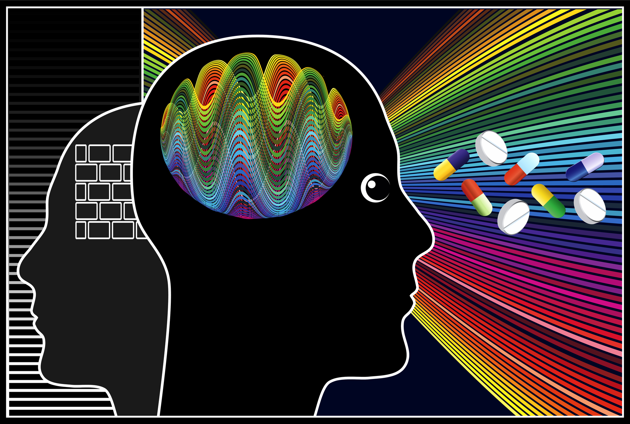
Learn about nootropics for brain health and how they can play a role in boosting your mental sharpness and cognitive abilities.
 By
By
Uncover the benefits of advanced MRI sequences in multiple sclerosis, offering deeper insights into disease mechanisms and treatments. Image for illustration only. Person depicted is a model.
 By
By
Discover the potential of oxygen therapy for sleep apnea. An option for patients who cannot tolerate conventional treatments. Image for illustration only. Person depicted is a model.
By
Discover how intracranial haemorrhage detection can be enhanced with AI: a breakthrough in emergency neuroradiology.
 By
By
Explore the profound effects of car accident trauma on mental well-being. Discover how psychological scars can last for years after a crash. Image for illustration only. Person depicted is a model.
 By
By
Minor injuries can have a significant long-term impact. Find out why you should never overlook these physical setbacks.
 By
By
Uncover the innovative insights of the World Transformation Movement and its impact on understanding patients’ emotional and cognitive needs.
 By
By
Stem cells offer creative approaches for illness therapy in precision medicine. Personalized treatment is made possible by the capacity of stem cells to develop into several cell types.
 By
By
Deep brain stimulation offers experimental hope for severe addiction cases unresponsive to conventional treatments and therapies.
 By
By
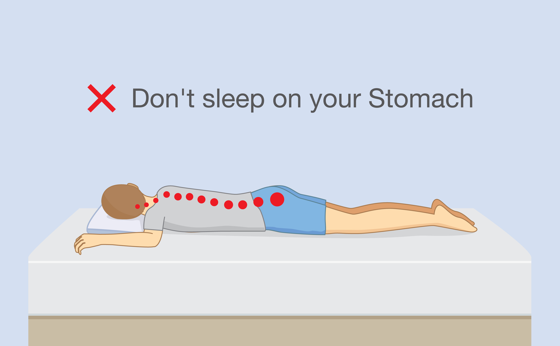
Sleeping on your stomach can lead to neck strain, back pain, breathing difficulties, nerve pressure, and long-term sleep quality issues.
 By
By
Neurology healthcare tools improve accuracy, enable early detection, enhance monitoring, and personalise treatment for better outcomes.
 By
By
Genomic research revolutionises healthcare by providing insights into diseases, enabling precise treatments, and improving patient outcomes worldwide.
 By
By
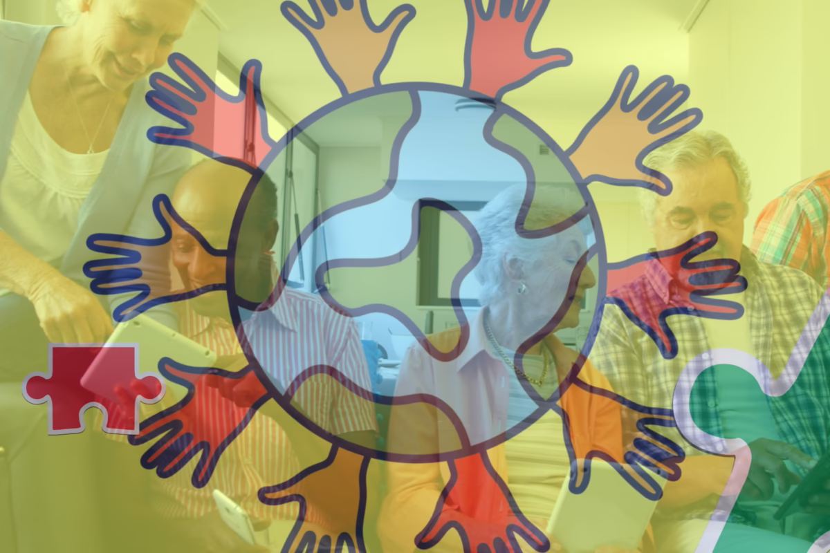
Global mental health initiatives are fostering greater access to treatment, reducing stigma, and promoting holistic care worldwide.
 By
By
Gene therapy offers unprecedented potential to treat and prevent genetic disorders by correcting defective genes or altering gene expression. Image for illustration only. Person depicted is a model.
 By
By
Diagnostic imaging in motor neurone disease (MND) is crucial for early detection, disease monitoring, and differentiating from other conditions.
 By
By
Revolutionizing rehabilitation empowers patients through advanced technology, personalized programs, and holistic, transformative recovery experiences.
 By
By
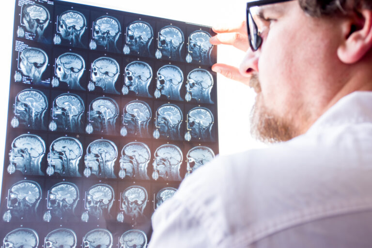
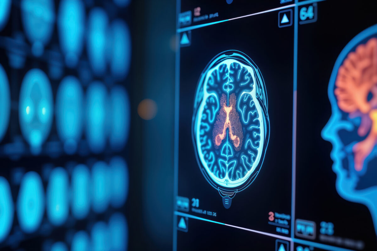
Neurological diagnostics have been transformed by advanced imaging techniques, enhancing accuracy in identifying brain disorders.
 By
By
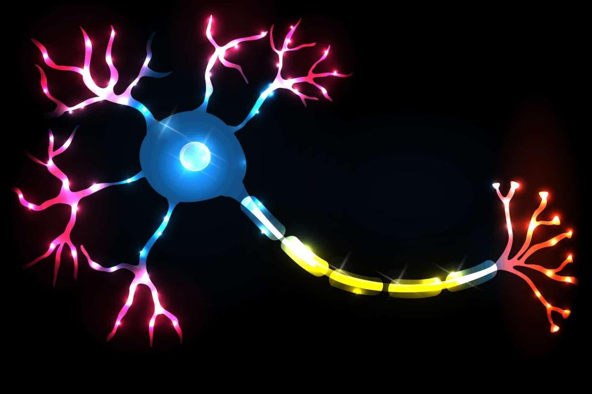
The central nervous system is a complex network that manages sensory processing, motor functions, and cognitive abilities.
 By
By
PET/MR scanners used in the area of neurological and psychiatric medicine.