Medical X-ray Imaging Using Computed Tomography
 By
By
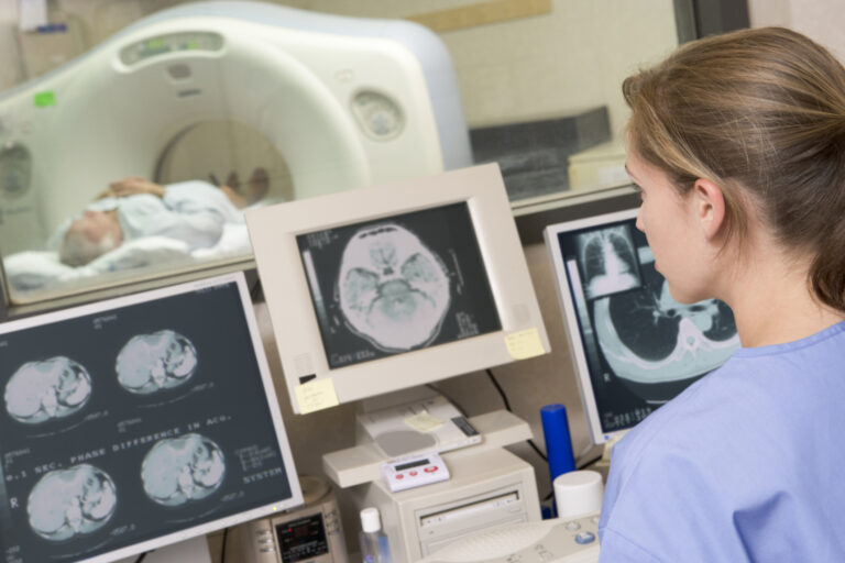
Conventional X-ray systems are based on an immovable X-ray tube whereas the CT scanner uses a rotational X-ray source. Image for illustration only. People depicted are models.
Hounsfield units (HU) are used in computed tomography (CT) to represent CT numbers in a standardised format of the resultant image. HU is derived from a linear transformation of the measured attenuation coefficients based on arbitrarily assigned air and pure water densities. Hence, the radiodensity of distilled water at standard temperature and pressure (STP) is 0 HU, and the radiodensity of air is -1000 HU.
Therefore, the HU ranges from -1000 HU for air to +~2000 HU for very dense bone. Also, the software (3-D slicer or Image J) of all CT scanners and picture archiving and communication systems (PACSs) can measure the density of a region of interest (ROI) by overlaying the electronic images. These HU are reported for an array of clinical applications.
For example, the evaluation of the fat content of the liver gives a value for the liver-to-spleen ratio in HU <1.0 and liver attenuation <40 HU; fat (HU= −120 to −90), cortical bone (HU= +1800 to +1900); white matter (HU= +20 to +30), grey matter (HU= +37 to +45), lung (HU= -700 to −600) and silver foreign objects (HU=+17,000).
The Hounsfield units are proportional to the degree of X-ray attenuation in the tissue for CT scanning. However, in cone beam computed tomography (CBCT), the degree of x-ray attenuation is represented by the grayscale (voxel value) and has several advantages over CT. Consequently, CBCT especially includes shorter acquisition times, lower radiation dosages for the patient, and submillimetre resolution. Moreover, the most commonly encountered artefact in CT scanning is beam hardening due to the perimeter of an object appearing brighter than the centre.
Beam hardening can scatter more radiation and, therefore, show incorrect Hounsfield units similar to a CT scan: this becomes a limitation for CBCT. Furthermore, cone beam computed tomography can be used to evaluate some features of bone density, distance to anatomical structures, and stability of implants. In conclusion, CBCT involves high levels of radiation scatter and artefacts, but there can be several disadvantages to estimating bone density.
In addition, several studies have shown a linear relationship between HU in CT scanning and grayscale in CBCT. This suggests that ascertaining CBCT voxel values can be used to estimate bone density.
home » Hounsfield units
Conventional X-ray systems are based on an immovable X-ray tube whereas the CT scanner uses a rotational X-ray source. Image for illustration only. People depicted are models.