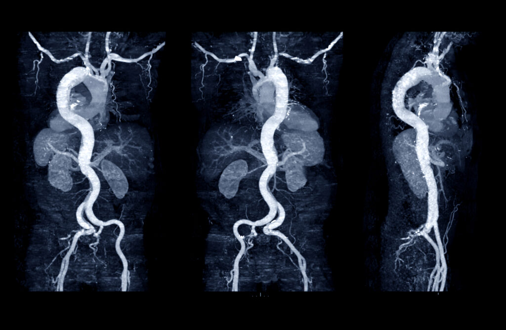
Perivascular Fat Density on CTA Linked to Abdominal Aortic Aneurysm Progression
 By
By
Discover the significance of the Aneurysm Progression Marker in predicting AAA growth with non-invasive perivascular fat density measurements.
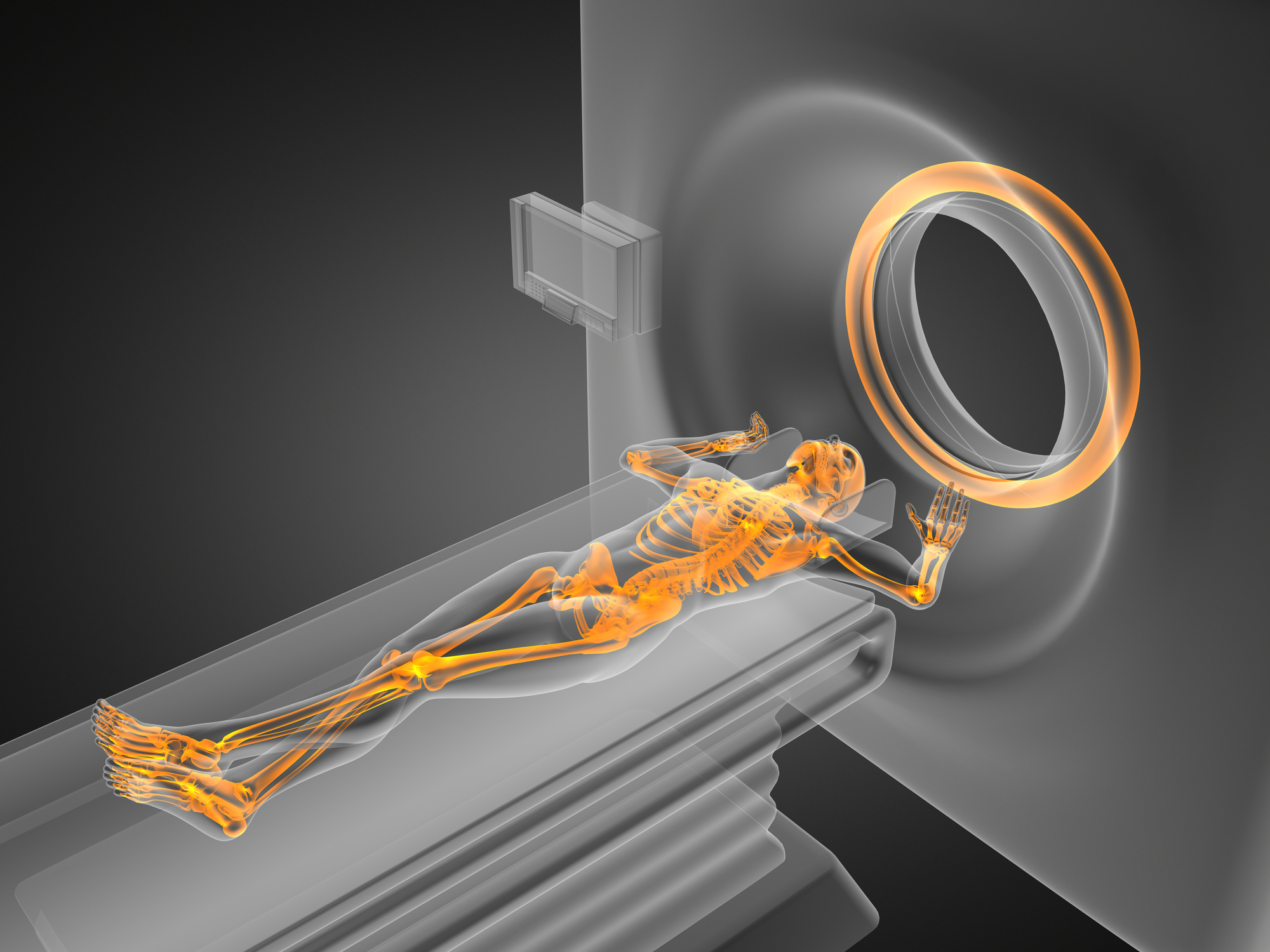
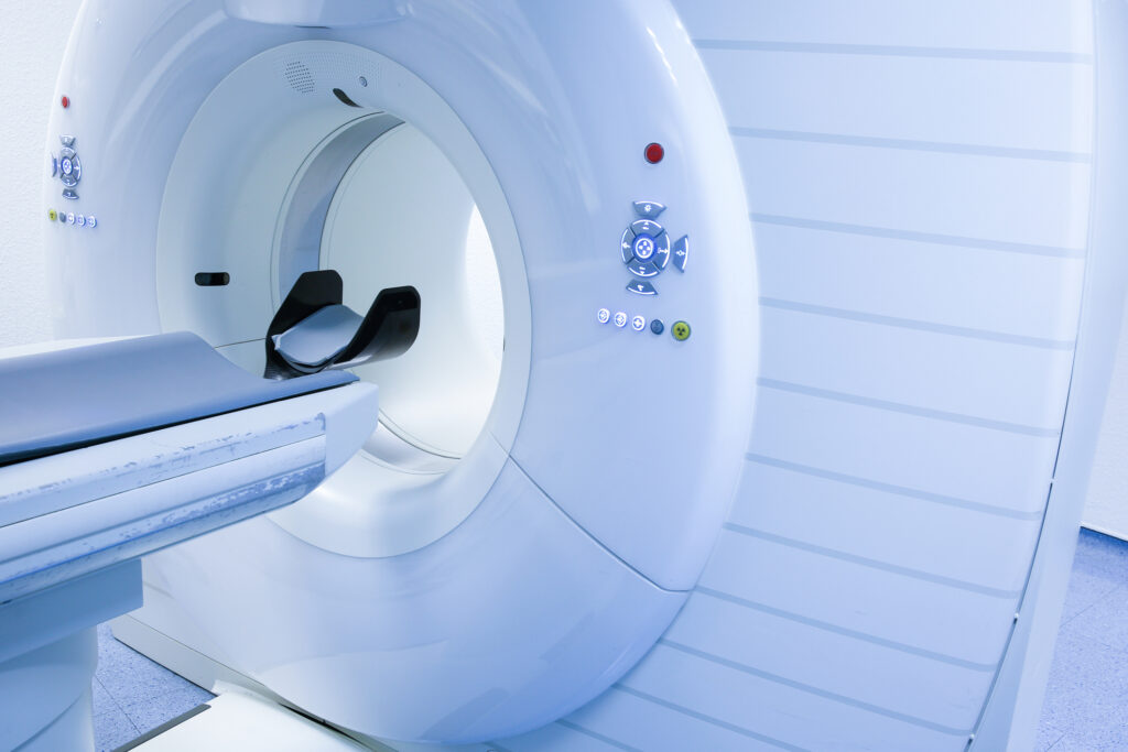
Computed Tomography (CT) was invented by Allan M. Cormack and Godfrey N. Hounsfield in 1972 and is a popular and essential tomographic medical imaging modality. Over the past decade, significant advancements in CT have been due to improvements in speed, radiation dose, slice count, and image quality.
The first CT scans lasted approximately 30 minutes, whereas today, the apex of CT scanners takes just 1-2 seconds to collect images. A pivotal breakthrough concerning radiation dose reductions of 70%- 80% in these modern CT scanners uses techniques based on hybrid Iterative image reconstruction methods. In addition, more dose-efficient CT detector materials with larger dimensions have assisted with lower CT patient radiation dosages without affecting image quality at the same time.
Computed Tomography is used in the clinical setting to perform CT Perfusion imaging to diagnose acute ischaemic stroke. Computed Tomography perfusion of a tumour is used to evaluate the impact of modern cancer therapy regimens.
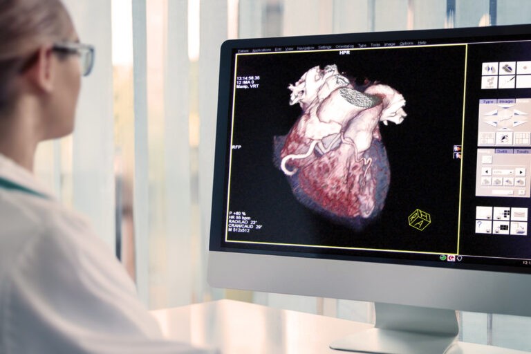
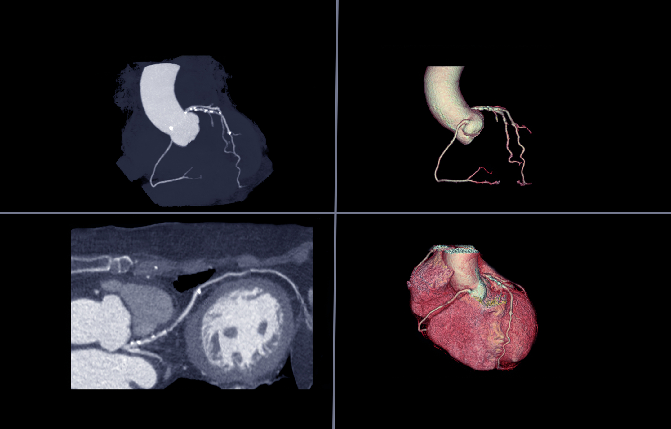
Single-heartbeat CT coronary angiography has shown very high specificity and sensitivity for coronary artery disease. PET/CT for cancer treatment planning allows physicians to have a much more accurate understanding of the extent of a patient’s disease.
Hybrid imaging will enable clinicians to evaluate metabolic and anatomical functions concurrently. These fusion images provide a rapid assessment of response to therapy or myocardial perfusion. Computed Tomography simulation systems include 3-D CT imaging and IMRT.
However, CT simulation has become essential in stereotactic radiosurgery by targeting radiation around sensitive structures.
Furthermore, radiologists are able to have CT data printed in 3-D to assist with surgical planning and implant prototyping.
home »
By
Discover the significance of the Aneurysm Progression Marker in predicting AAA growth with non-invasive perivascular fat density measurements.
 By
By
Proton beam therapy targets cancer accurately, minimising side effects but faces challenges in cost and accessibility.
 By
By
Radiotheranostics merges diagnostics and therapy, offering targeted cancer treatment amidst challenges like high costs and need for specialised facilities.
 By
By
X-rays, discovered in 1895 by Wilhelm Roentgen, revolutionised medical diagnostics and profoundly influenced science and technology.
 By
By
3D medical imaging transforms diagnostics and treatment, enhancing precision, patient education, and enabling AI-driven analysis and immersive experiences.
 By
By
Large vessel vasculitis and polymyalgia rheumatica are both inflammatory rheumatic diseases that can result in serious illness if not diagnosed.
By
Brachial plexus imaging is vital for diagnosing nerve conditions; ultrasound’s potential challenges MRI’s traditional dominance in this area.
By
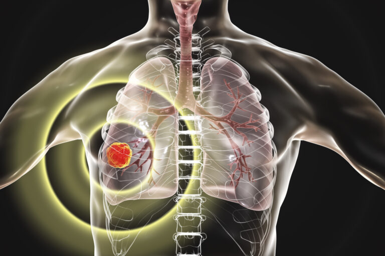
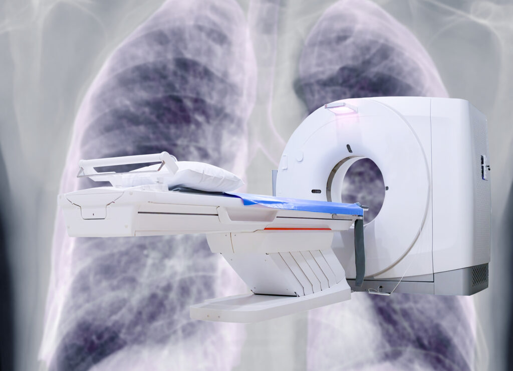
Lung cancer’s high mortality demands early detection; advances like low-dose CT and photon-counting CT improve noninvasive, accurate screening outcomes.
 By
By
Medical imaging of the human skeleton enables accurate diagnosis, treatment, and monitoring of diverse bone and joint conditions.
 By
By
Advanced medical imaging technologies transformed liver disease detection, diagnosis and management, enhancing diagnostic accuracy and personalized treatments.
 By
By
Photon Counting Computed Tomography enhances image quality, tissue differentiation, radiation reduction, and material decomposition via precise photon detection.
 By
By
Medical imaging advancements, aided by deep learning reconstruction, enhance diagnosis accuracy and efficiency, overcoming traditional limitations.
 By
By
Dark Field Computed Tomography enhances medical imaging by utilising X-ray scattering for improved contrast and resolution in soft tissues.
 By
By
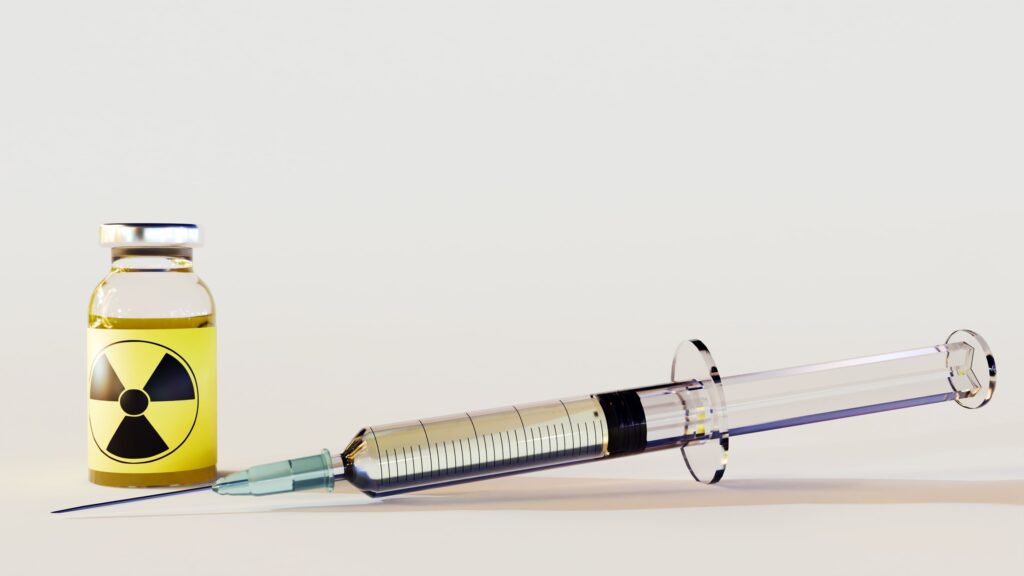
Copper-64 DOTATE, a diagnostic radiopharmaceutical, effectively targets neuroendocrine tumours, enabling precise imaging and personalised treatment plans for cancer patients.
 By
By
The new technologies emerging in the clinical setting include fractional flow reserve (FFR)-CT, CT perfusion imaging and coronary plaque assessment. Image for illustration only. Person depicted is a model.
 By
By
CTCA imaging has revolutionised how physicians detect coronary artery disease due to its exceptional sensitivity.
 By
By
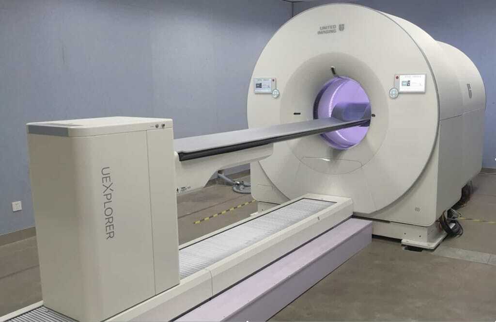
EXPLORER, the world’s first medical imaging scanner to produce a 3-D picture of the whole human body.
 By
By
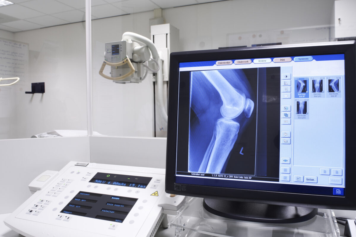
Conventional X-ray systems are based on an immovable X-ray tube whereas the CT scanner uses a rotational X-ray source. Image for illustration only. People depicted are models.
 By
By
Medical imaging modalities, including MRI, CT, and ultrasound, facilitate accurate diagnoses and treatments.
 By
By
Learn what medical imaging is and how techniques like X-ray, CT, and MRI are used for accurate diagnosis and assessment.