Angiogram Imaging
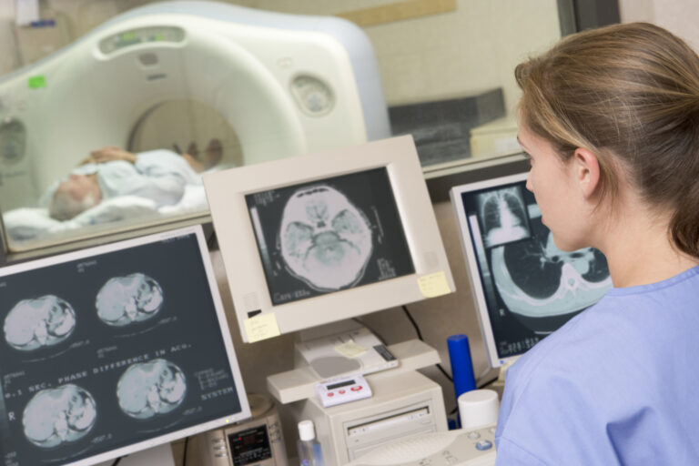
An angiogram uses X-rays to examine blood vessels within the human body. These X-ray angiography imaging systems have been developed to address the need to lower the patient’s X-ray dose. To address these issues, a new generation of X-ray tube combinations is incorporated into the next generation of angiogram machines. These changes are included in the Azurion 7 FlexArm systems, designed to enhance positioning flexibility for image-guided procedures.
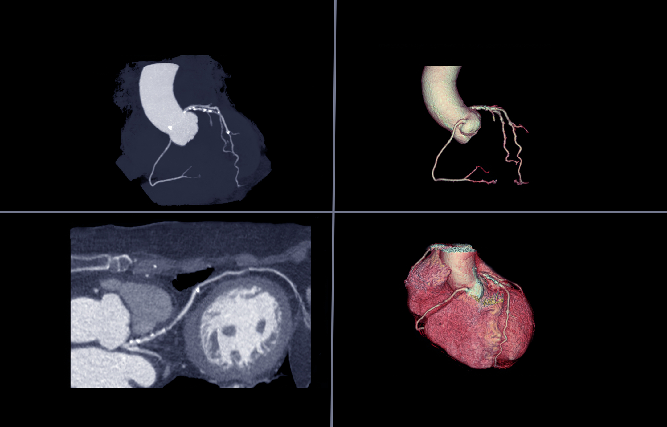
During the angiogram procedure, clinicians need to quickly visualise any problems and changes to the patient’s anatomy. The Azurion 7 system includes technology to perform 2-D and 3-D imaging, which is carried out using an image beam that automatically maintains alignment with the patient. This approach allows consistent visualisation, enabling the clinician to focus on the treatment.
Angiogram systems can be used for complex procedures in structural heart space, which require visualisation of the surrounding soft-tissue anatomy in a 3-D setting. Ultrasound imaging, such as a transesophageal echo, can be used during the angiogram. This ultrasound technology assists the operator in seeing real 3-D images of anatomy whilst also looking at the main screen in the cath lab.
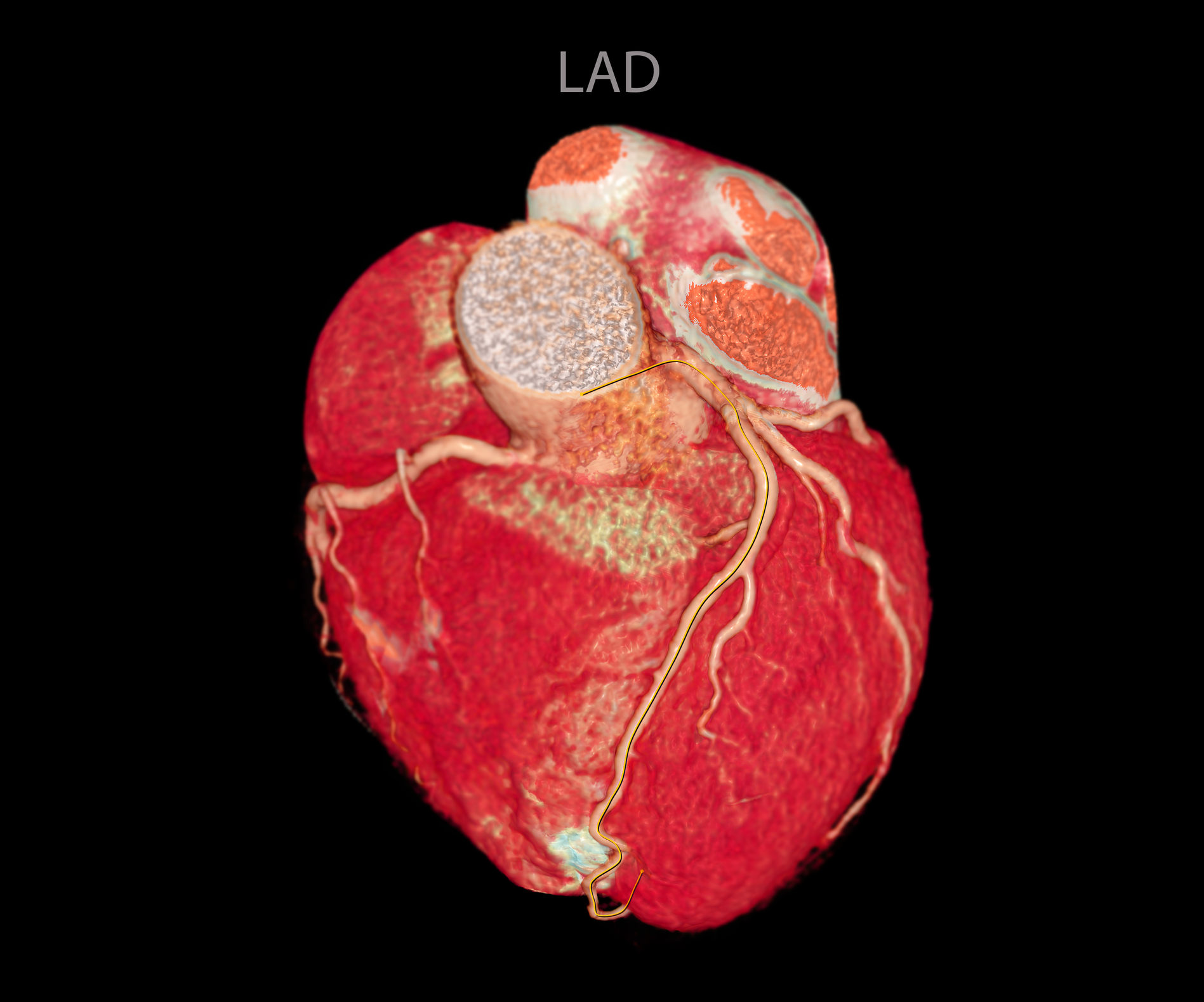
Furthermore, interventional cardiology has been developed to replace pressure wires: this allows fractional flow reserve (FFR) to be determined and evaluated if stenting is required. The next generation of angiogram machines, such as Liver Assist Virtual Injection technology, will use artificial intelligence algorithms to allow automation and analysis. This technology incorporates intelligence-enhanced software to identify the feeder blood vessels in liver tumours.
Artificial intelligence can enhance and map the blood vessels and demonstrate the direction of flow in each vessel segment by generating a 3-D rotational angiography image to aid navigation. These new angiogram machines aim to reduce the X-ray dosages during imaging and scatter radiation by using the modern patient table. Studies have shown that these patient tables gave scatter doses of between 640 and 2,040 μSv/h compared to standard shielding, which gives a reading of between 7,910 and 34,870 μSv/h.
home » angiogram
 By
By