Image Guided Brachytherapy
Image Guided Brachytherapy (IGBT) is a technique used to treat several types of cancer. It has been shown to improve survival rates in cancer patients and is used in a personalised approach to lower the risk of complications. The global cancer burden (GLOBOCAN) database estimated 18.1 million new cases and 9.6 million deaths in 2018. Hence, 20% of men and 17% of women worldwide develop cancer during their lifetime, whereas 13% of men and 9% of women die.
Therefore, with the rise in cancer cases worldwide, using image-guided brachytherapy poses a safe and effective treatment against prostate, breast and cervical cancers. Brachytherapy uses internal radiation based on radioactive sources and has been a treatment for many cancers for over a century. In the last decade, image-guided brachytherapy (IGBT) has been at the forefront of cancer treatment due to advances in medical imaging, dose delivery and personalised treatment planning.
Image-guided brachytherapy (IGBT) aims to maximise the radiation dose to kill cancer cells while sparing the surrounding healthy tissues and organs. The technique involves using detailed 3-D medical images to depict organ volume changes to personalise and optimise brachytherapy for the patient’s treatment plan. The internal images generated show the exact tumour size and location in relation to the organs.
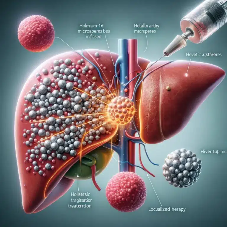
This will enable the oncology team to place the radioactive source directly next to or inside a tumour for treatment. The radioactive source can either be temporary—using a removable applicator—or permanent, known as seeds. These seeds remain indefinitely inside the body, losing their radioactivity over time and becoming harmless.
Furthermore, image-guided brachytherapy can tolerate higher doses of radiation and, therefore, can target a tumour directly. This targeted approach spares healthy tissues by receiving a lower radiation dose because the source is placed internally or close to the tumour.
You are here:
home » image guided brachytherapy

