Windows into the Brain: The Expanding Role of Medical Imaging in Neurological Disorders
 By
By
Discover how medical imaging in neurological diseases revolutionises diagnosis and management of conditions like Alzheimer’s and Parkinson’s.
Alzheimer’s disease (AD) is a progressive neurodegenerative disorder characterised by a gradual decline in cognitive function, memory loss, and personality changes. The pathological hallmarks of AD include the accumulation of amyloid-beta version (Aβ) plaques and neurofibrillary tangles in the brain. Therefore, early diagnosis of AD is crucial for implementing appropriate interventions and treatment strategies to delay the disease progression. Positron Emission Tomography (PET) amyloid imaging is a powerful, non-invasive diagnostic tool for detecting Aβ plaques in the brain, which can help confirm the presence of AD and monitor disease progression.
PET amyloid imaging uses small, radiolabeled molecules known as radiotracers that specifically bind to Aβ plaques in the brain. The most commonly used radiotracers include 18F-florbetapir, 18F-florbetaben, and 18F-flutemetamol. These radiotracers are injected into the patient’s bloodstream and travel to the brain, binding to Aβ plaques. A PET scanner then detects the emitted radiation from the radiotracers and produces a detailed, three-dimensional image of the brain. This image allows clinicians to visualise and quantify the presence and distribution of Aβ plaques in the brain.
PET amyloid imaging offers several advantages over other diagnostic tools for AD:
Despite its advantages, amyloid imaging is not without limitations. One significant drawback is the high cost of the procedure, which can limit its accessibility for many patients. Additionally, PET amyloid imaging can detect Aβ plaques but does not provide information on other pathological features of AD, such as neurofibrillary tangles or neuroinflammation. Furthermore, a positive PET amyloid scan does not guarantee the presence of AD, as Aβ plaques can also be found in cognitively normal older adults.
home »
By
Discover how medical imaging in neurological diseases revolutionises diagnosis and management of conditions like Alzheimer’s and Parkinson’s.
 By
By
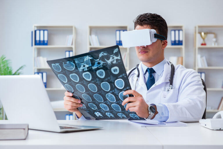
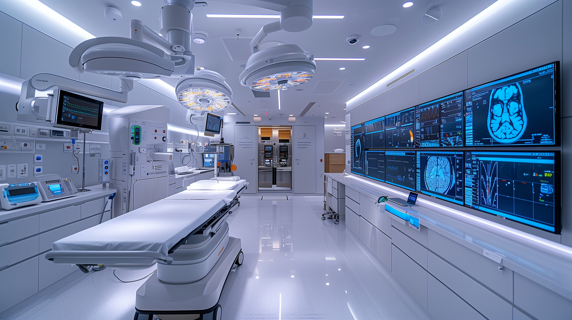
Medical imaging 2025 integrates advanced AI, precision tools, seamless data systems, personalised diagnostics, and enhanced patient experiences worldwide. Image for illustration only. Person depicted is a model.
 By
By
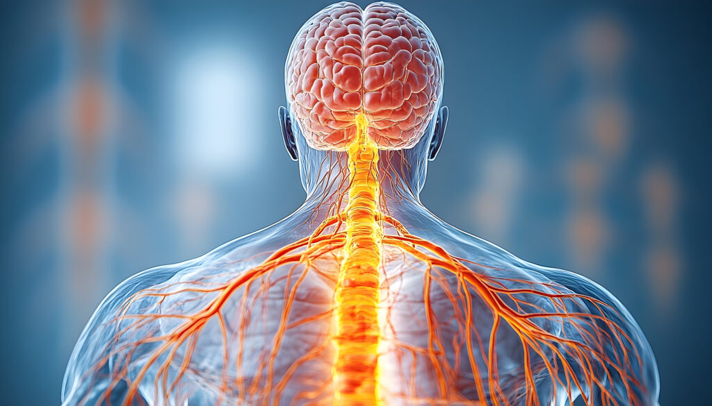
Neuroimaging in nuclear medicine allows for detailed visualisation of brain activity, aiding in diagnosing complex neurological conditions.
 By
By
Imaging of β-amyloid plaques are necessary for clinical and neuropsychological characteristics in Alzheimer’s disease.