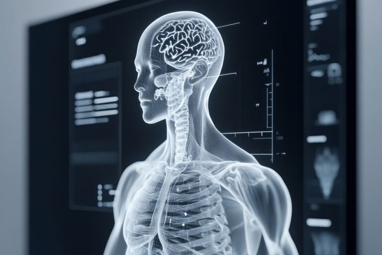
Bridging the Gap Between Medical Imaging and Neurological Care
 By
By
Discover the complexities of neurological care and the necessity for seamless information transfer in clinical practice.
By
Discover the complexities of neurological care and the necessity for seamless information transfer in clinical practice.
 By
By
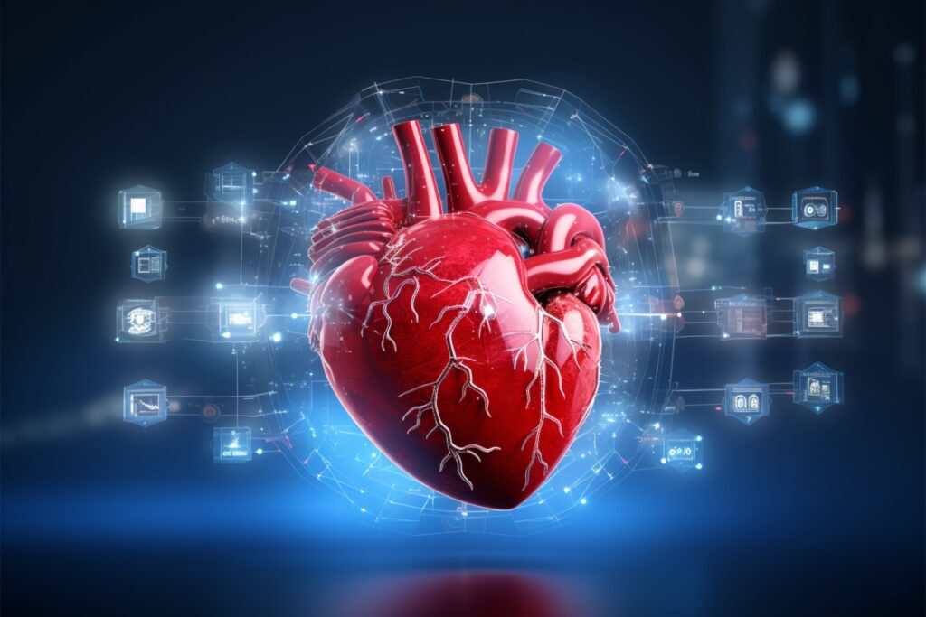
Cardiology forums and conferences in 2026 will highlight new cardiovascular research, technologies, clinical strategies, and healthcare leadership discussions worldwide.
 By
By
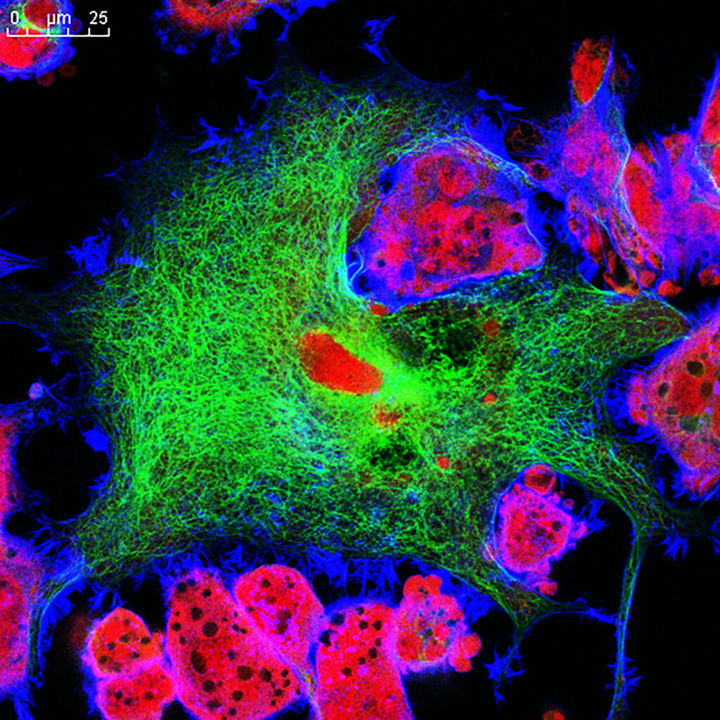
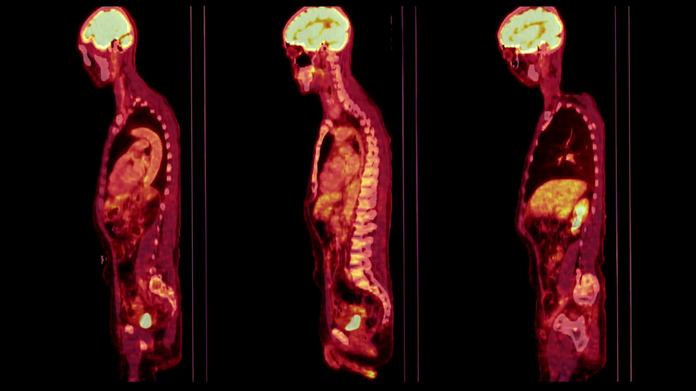
Discover how health and wellbeing imaging insights are reshaping our understanding of metabolic health and personal wellness.
 By
By
Discover how radiolabelling in 2025 is transforming clinical care with advanced technologies and precise therapeutic solutions.
 By
By
Discover how radiopharmaceuticals in medicine are transforming diagnostics and treatments with atomic precision for better health outcomes.
 By
By
Discover the role of generative artificial intelligence in medical imaging, improving workflows and data quality in healthcare.
 By
By
Discover the fundamental SPECT imaging principles and their vital role in diagnosing heart and brain conditions effectively.
 By
By
Iodine-123 in nuclear medicine enables accurate imaging, particularly for thyroid disorders, neuroendocrine tumours, and neurological conditions.
 By
By
Understand the strengths and limitations of Congenital neuroblastoma PET in assessing rare childhood tumors in this critical study.
 By
By
Uncover the key uses of Carbon-14 radiotracers. Learn how they contribute to environmental science and biochemical studies.
 By
By
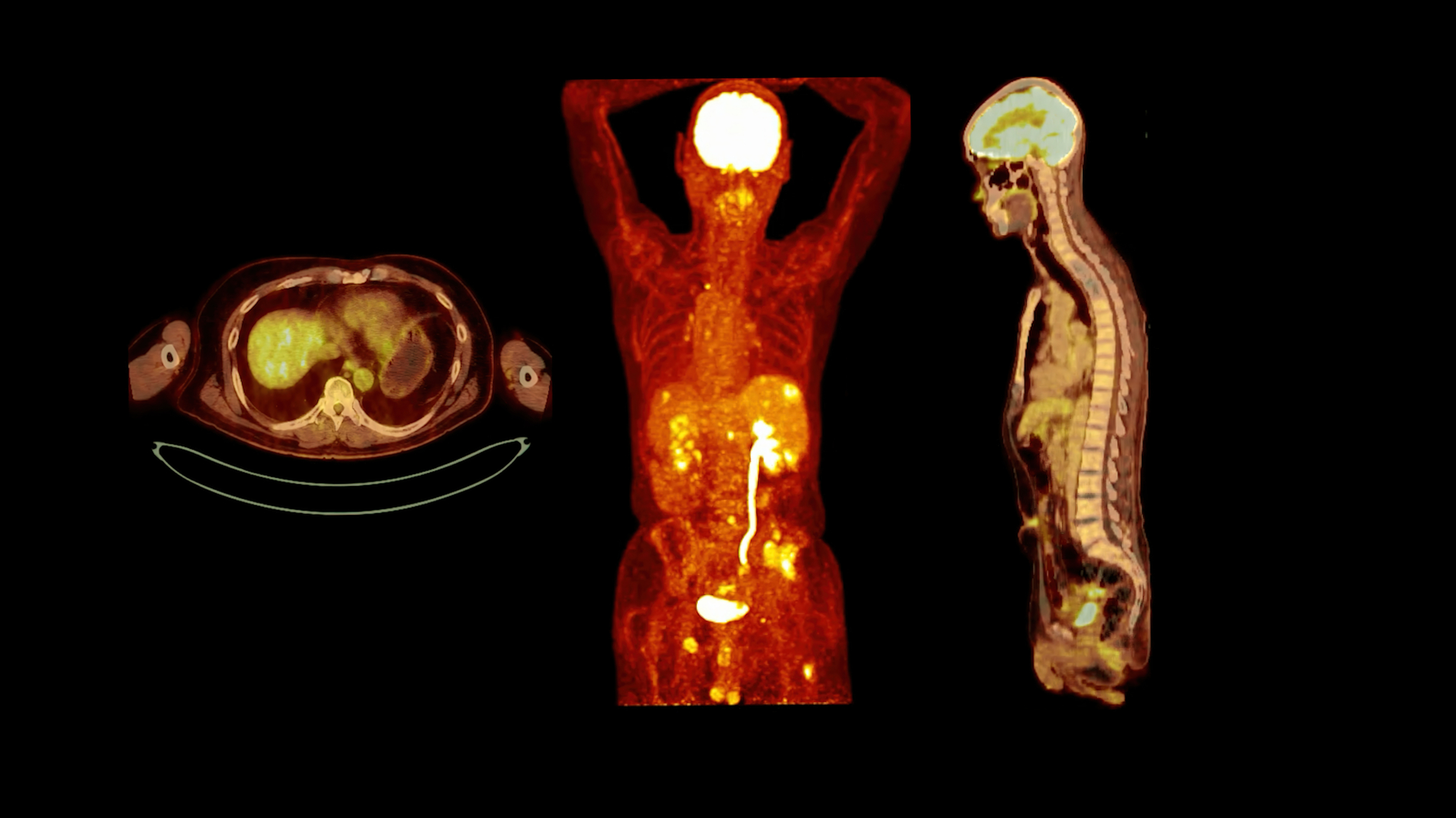
Uncover the benefits of radioactive imaging. Understand PET, SPECT, and their role in advanced medical imaging and research.
 By
By
Learn how bioengineered contrast agents enhance imaging clarity and accuracy in nuclear medicine. Discover their unique benefits.
 By
By
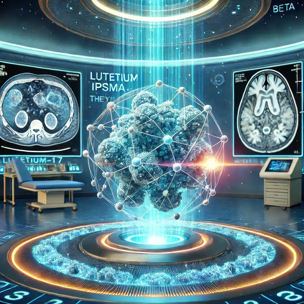
Lutetium-177 iPSMA revolutionises prostate cancer treatment, delivering targeted beta radiation to metastatic cells with precision and minimal side effects.
 By
By
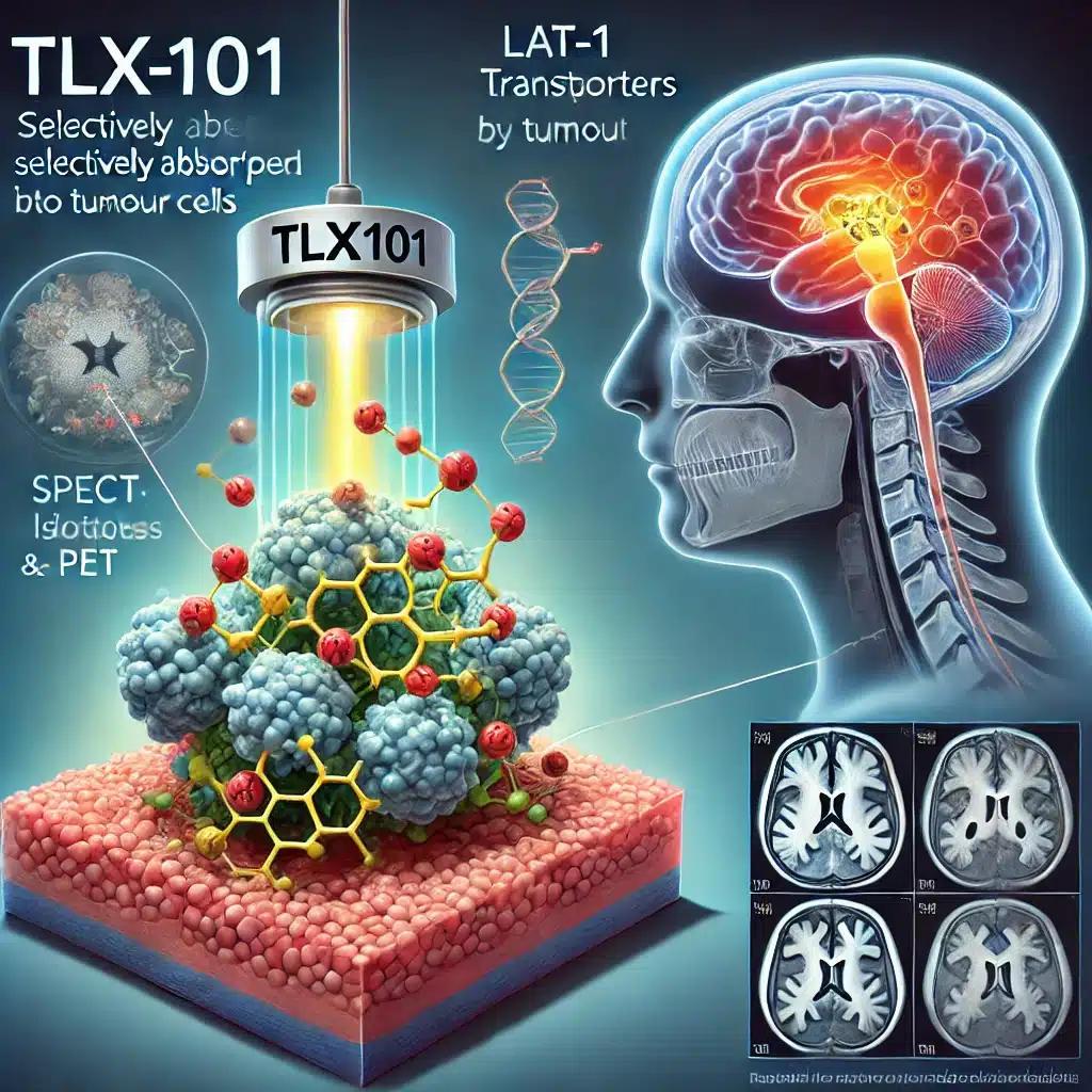
TLX101 is a revolutionary radiopharmaceutical targeting gliomas, offering dual imaging and therapy through tumour-selective uptake, enhancing treatment outcomes.
 By
By
Radiopharmaceuticals in diagnostics provide critical insights into disease processes, improving accuracy, patient care, and treatment outcomes significantly.
 By
By
Neuroimaging in nuclear medicine allows for detailed visualisation of brain activity, aiding in diagnosing complex neurological conditions.
 By
By
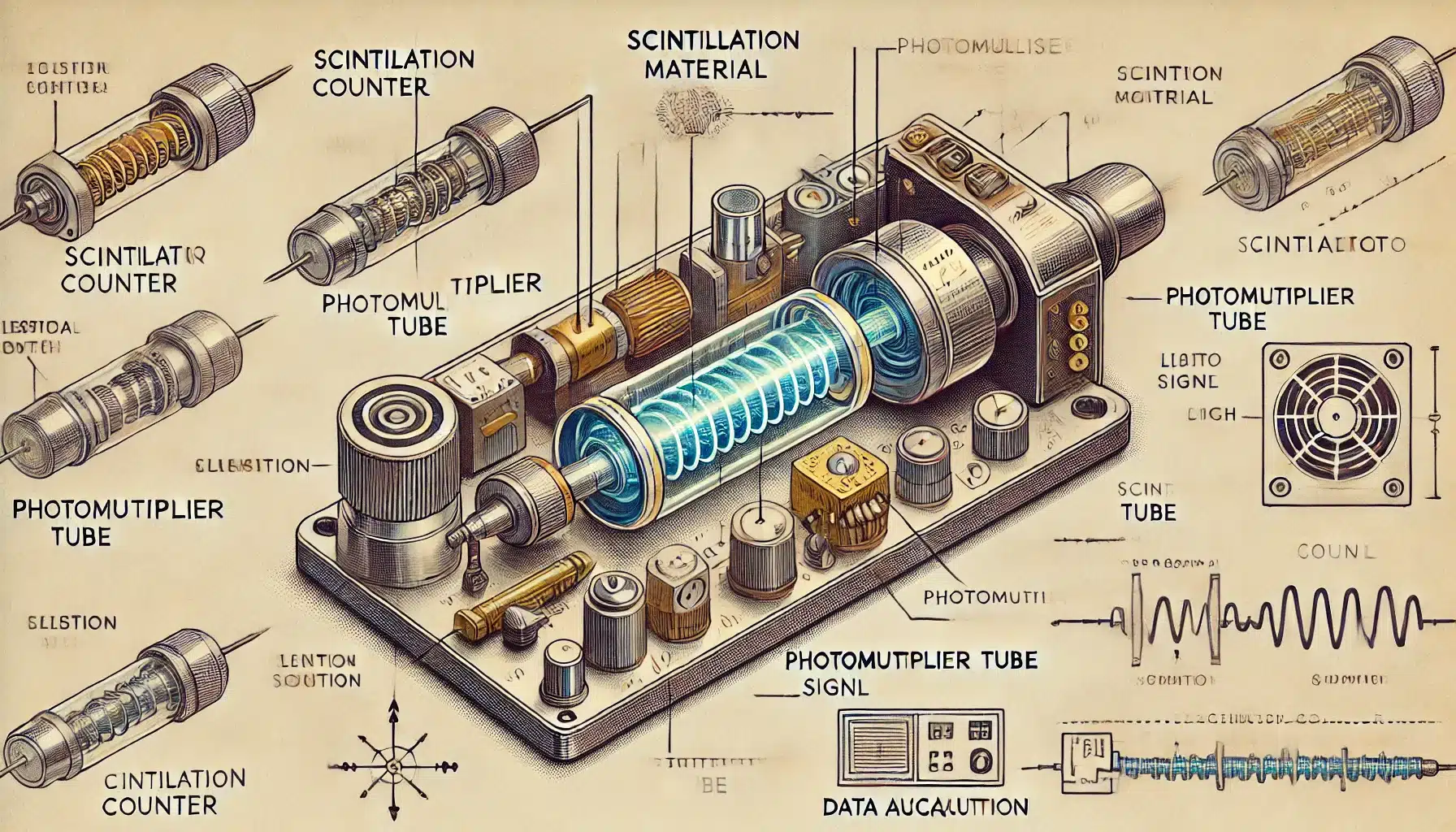
A scintillation counter detects ionising radiation, converting emitted light from scintillators into electrical signals for precise measurement.
 By
By
SPECT imaging combines nuclear medicine with computed tomography to deliver functional insights, revolutionising diagnosis and treatment across cardiology, neurology, and oncology.
 By
By
SPECT imaging, by combining functional and anatomical insights, significantly enhances diagnostic accuracy in various medical fields.
 By
By
Technetium-99m, discovered in 1937, transformed medical imaging with its versatile and safe diagnostic applications.
 By
By
Iodine-123 Ioflupane known as DaTscan aids in diagnosing Parkinson’s disease by visualising dopamine transporters using SPECT imaging.
By
Iodine-123 iobenguane is vital for detecting, staging, and monitoring neuroendocrine tumours, guiding treatment, and offering prognostic insights.
By
Gallium-67 citrate is a radiopharmaceutical used for imaging infections, inflammation, cancer, and fever of unknown origin.
 By
By
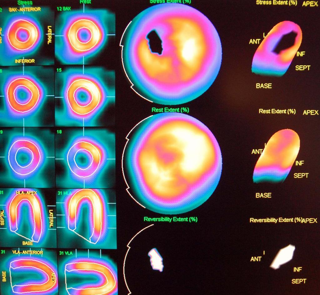
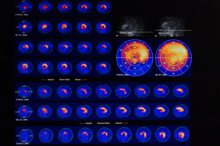
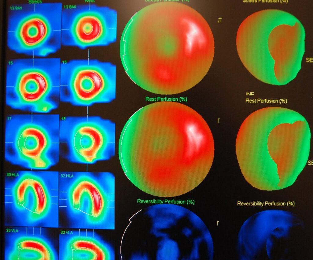
SPECT imaging is used to obtain a myocardial perfusion scan (SPECT scan) to investigate the function of the heart muscle.
 By
By
Overview on the central role of chelation in labelling radiocompounds.