Technetium-99m radiopharmaceuticals have transformed the field of medical imaging and nuclear medicine. Its history is a fascinating journey of scientific discovery and innovation.
Technetium: Tracing the Journey from Artificial Element to Diagnostic Powerhouse
Technetium, the first artificially produced element, was discovered by Italian scientists Carlo Perrier and Emilio Segrè in 1937. The element was named “technetium,” derived from the Greek word “technetos,” meaning artificial, reflecting its synthetic origins. However, it wasn’t until the 1950s that Tc-99m potential in medical imaging was recognised.
The discovery of Technetium-99m (Tc-99m) as a useful radioisotope for medical purposes can be largely credited to Walter Tucker and Margaret Greene at the Brookhaven National Laboratory in the United States. They identified the unique properties of Tc-99m in 1958. It emits gamma rays, which are ideal for medical imaging, and it has a short half-life of about 6 hours, which is long enough to conduct diagnostic tests but short enough to minimise radiation exposure to the patient.
The first clinical use of Tc-99m was in brain imaging, but it soon became evident that its real potential lay in its ability to be attached to various compounds (labelling), thereby targeting different organs or systems in the body. This versatility transformed diagnostic nuclear medicine. By the 1970s, Tc-99m had become the most widely used radioisotope in diagnostic imaging.
The development of Tc-99m-based radiopharmaceuticals expanded its applications. Compounds like Tc-99m MDP for bone scans and Tc-99m sestamibi for cardiac imaging have become standard tools in medical diagnostics. These radiopharmaceuticals allow for non-invasive visualisation of internal organs, blood flow, and metabolic activity, providing invaluable information for diagnosing and treating various diseases.
Further advancements in imaging technology, such as the development of Single Photon Emission Computed Tomography (SPECT), have leveraged the properties of Tc-99m to produce three-dimensional images, offering even more detailed insights.
Today, Tc-99m is used in millions of diagnostic procedures annually, from detecting cancer and heart disease to evaluating kidney function and bone disorders. Its discovery and subsequent development represent a significant milestone in medical technology, illustrating the profound impact of nuclear science on healthcare. The story of Tc-99m is not just a chapter in the history of medicine but also a testament to human ingenuity and the relentless pursuit of knowledge to improve lives.
The Vital Role of Technetium-99m in Modern Diagnostics
Diagnostic imaging with Technetium-99m (Tc-99m) represents a cornerstone in modern medical imaging, owing to its optimal physical and chemical properties. Tc-99m is a radioisotope that emits gamma rays, which are ideal for detection by a gamma camera, a device specifically designed to visualise the distribution of gamma-emitting radiopharmaceuticals within the body. This capability enables clinicians to create highly detailed images of internal structures and functions, providing invaluable insights into various medical conditions.
One of the most significant advantages of Tc-99m is its short half-life of approximately 6 hours. This brief half-life means that the radioisotope decays quickly, significantly reducing the duration of radiation exposure for patients. This feature is essential in medical imaging, as it minimises the potential risks associated with radiation, making the procedure safer, especially for repeat examinations or for use in vulnerable populations such as children.
Moreover, the gamma radiation emitted by Tc-99m is of a suitable energy level that facilitates easy detection and results in high-resolution images. This quality is crucial for accurately diagnosing and monitoring a range of medical conditions. The clear images produced by Tc-99m help clinicians in making precise assessments of bodily functions and structures, such as evaluating blood flow, identifying cancerous growths, or detecting bone abnormalities.
Technetium-99m: The Chameleon of Nuclear Medicine
Technetium-99m (Tc-99m) showcases remarkable versatility in the field of nuclear medicine, primarily due to its ability to form diverse radiopharmaceuticals by combining with various compounds. Each of these radiopharmaceuticals is tailored to target specific organs or functions, making Tc-99m a pivotal tool in diagnostic imaging.
One of the most common applications of Tc-99m is in bone scans, using Tc-99m Methylene Diphosphonate (MDP). This compound has a high affinity for bone tissues, particularly areas with increased osteoblastic activity. Consequently, it is highly effective in detecting bone metastasis, a common site for breast and prostate cancers. Tc-99m MDP is also crucial in identifying fractures, particularly those that are not easily visible on conventional X-rays, and in diagnosing bone infections like osteomyelitis, where it can highlight areas of inflammation.
Another significant application of Tc-99m is in cardiac imaging, particularly using Tc-99m labelled red blood cells. This radiopharmaceutical is instrumental in cardiac blood pool imaging, a technique that evaluates the functional aspects of the heart. It allows for the assessment of the heart’s ejection fraction, which is crucial in diagnosing and managing various cardiac conditions, including heart failure and cardiomyopathy. Furthermore, it plays a role in detecting shunts and evaluating the heart’s chambers’ size and movement, providing comprehensive insights into cardiac function.
These examples underscore the adaptability of Tc-99m in nuclear medicine, offering tailored diagnostic solutions for a range of medical conditions. Its ability to bind with different compounds to target specific areas of the body makes it an invaluable tool in the early detection and management of various diseases.
The Pivotal Role of Technetium-99m in Myocardial Perfusion Imaging
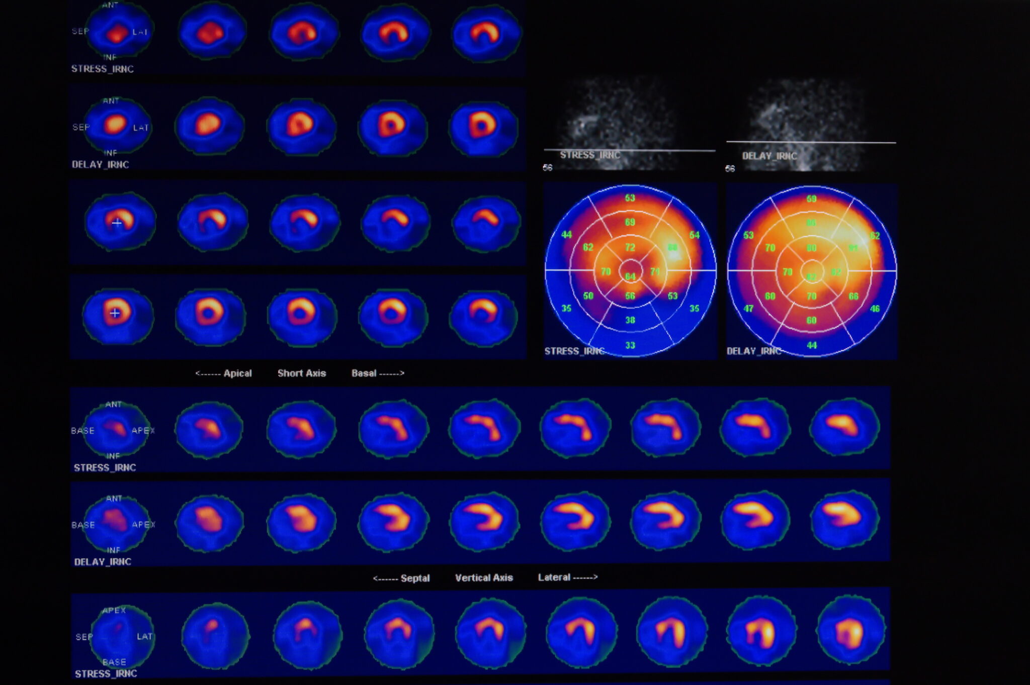
Technetium-99m (Tc-99m) plays a crucial role in myocardial perfusion imaging (MPI), a cornerstone of non-invasive cardiac imaging. MPI is instrumental in visualising blood flow through the heart muscle, offering vital insights into cardiac health. This imaging modality utilises Tc-99m-labeled radiopharmaceuticals, such as Tc-99m Sestamibi or Tc-99m Tetrofosmin, which are absorbed by healthy heart muscle cells.
A significant application of MPI is in diagnosing coronary artery disease (CAD), a leading cause of heart-related ailments. CAD occurs when the coronary arteries, which supply blood to the heart muscle, become narrowed or blocked. MPI with Tc-99m helps in detecting areas of the heart muscle that receive inadequate blood supply due to these blockages. Cardiologists can determine the presence and extent of CAD by comparing images taken during rest and after stress (either exercise or pharmacologic stress).
Moreover, Tc-99m MPI is invaluable in assessing the severity and impact of heart attacks (myocardial infarctions). After a heart attack, parts of the heart muscle may be damaged or scarred, affecting the heart’s ability to pump blood efficiently. MPI can identify these areas of damage and provide information about the size and location of a heart attack. This information is critical in guiding treatment decisions, such as the need for angioplasty, stenting, or coronary artery bypass surgery.
Tc-99m MPI also aids in evaluating the effectiveness of treatments for heart conditions, monitoring disease progression, and making prognostic assessments. The ability of Tc-99m MPI to provide detailed and accurate images of the heart’s perfusion under various conditions makes it an indispensable tool in diagnosing and managing heart diseases.
Technetium-99m in Oncology: A Vital Ally in the Detection and Staging of Bone Metastases
Technetium-99m (Tc-99m) radiopharmaceuticals are extensively utilised in the field of oncology for cancer diagnosis and staging, particularly in identifying bone metastases. Bone metastasis occurs when cancer cells spread from their original site to the bones, a common occurrence in various types of cancer, including breast, prostate, and lung cancers.
Tc-99m labelled compounds, such as Tc-99m Methylene Diphosphonate (MDP), are pivotal in bone scintigraphy, a diagnostic procedure specifically designed for detecting bone metastases. These radiopharmaceuticals are absorbed by bone tissue, with a preferential uptake in areas where the bone is remodelling or repairing itself — a process often stimulated by metastatic cancer cells. When injected into the body, Tc-99m MDP travels to these areas and highlights them on the resulting scans, providing clear images of skeletal abnormalities.
This imaging technique is highly sensitive in detecting bone metastases, often identifying them earlier than traditional X-rays. Early detection is crucial as it significantly impacts the treatment plan and prognosis. Bone scans with Tc-99m can also monitor the response to cancer treatments, such as chemotherapy or radiation therapy, by showing changes in the activity of bone lesions over time.
Furthermore, Tc-99m radiopharmaceuticals aid in staging cancer and determining the extent of its spread. This information is vital for oncologists to develop appropriate treatment strategies and predict the disease’s likely course. The ability of Tc-99m radiopharmaceuticals to provide detailed and specific images makes them an invaluable tool in the accurate diagnosis and staging of cancer, ultimately aiding in more effective patient management and treatment planning.
Thyroid Imaging: The Gateway to Understanding Thyroid Disorders and Guiding Effective Treatment
Thyroid imaging is a vital diagnostic tool in endocrinology, utilised primarily for evaluating thyroid function and morphology. This imaging technique is particularly useful in diagnosing various thyroid disorders, including hyperthyroidism, hypothyroidism, thyroid nodules, and thyroid cancer.
A small amount of radioactive iodine (usually I-123) or technetium-99m (Tc-99m) is administered to the patient during a thyroid scan. These isotopes are chosen due to their ability to mimic the behaviour of naturally occurring iodine, a key element the thyroid gland uses to produce hormones. Once administered, these isotopes are absorbed by the thyroid gland and emit gamma rays, which are detected by a gamma camera to create an image of the thyroid.
The resulting images provide valuable information about the thyroid gland’s size, shape, and position. They can also reveal areas of abnormality within the gland, such as hot spots (areas of increased activity typical in hyperthyroidism or in autonomously functioning thyroid nodules) and cold spots (areas of reduced or no uptake, which could indicate a non-functioning thyroid nodule, potentially suggestive of cancer).
Thyroid imaging is particularly significant in the assessment of thyroid nodules. While most thyroid nodules are benign, a small percentage can be cancerous. Imaging can help differentiate between benign and malignant nodules, guiding further investigation and treatment. Additionally, in conditions like Graves’ disease, a form of hyperthyroidism, thyroid scans can confirm the diagnosis and aid in treatment planning.
By providing detailed insights into thyroid function and structure, thyroid imaging plays a crucial role in the comprehensive evaluation and management of thyroid disorders, facilitating accurate diagnosis and effective treatment.
Enhancing Kidney Health: The Critical Role of Technetium-99m in Renal Imaging and Disease Management
Renal imaging using Technetium-99m (Tc-99m) based radiopharmaceuticals is a critical diagnostic tool in nephrology for assessing kidney function and morphology. This non-invasive imaging technique is essential for diagnosing and managing various renal diseases, including renal artery stenosis, obstructive uropathy, and renal parenchymal disease.
Tc-99m labelled agents, such as Tc-99m mercaptoacetyltriglycine (MAG3) and Tc-99m dimercaptosuccinic acid (DMSA), are commonly used in renal scintigraphy. Tc-99m MAG3 is particularly effective in evaluating renal perfusion and function, making it suitable for assessing conditions like renal artery stenosis, which can lead to hypertension and kidney failure. It provides dynamic images showing how the kidneys function in real-time, including how they filter and excrete the radiotracer.
On the other hand, Tc-99m DMSA is used primarily for renal cortical imaging, offering detailed anatomical information. This is particularly valuable in detecting and characterising renal cortical defects, commonly seen in conditions like acute pyelonephritis or scarring from recurrent urinary tract infections. It can also be used to assess differential renal function, especially in patients with suspected renal parenchymal damage.
Renal imaging with Tc-99m radiopharmaceuticals can also guide the management of kidney transplant patients, helping to identify complications like acute rejection or obstruction. Additionally, in pediatric patients, Tc-99m DMSA plays a crucial role in detecting congenital abnormalities and the effects of urinary tract infections on the kidneys.
Overall, renal imaging with Tc-99m based radiopharmaceuticals provides essential insights into both renal structure and function. This information is crucial for the timely diagnosis and effective management of a wide range of renal pathologies, enhancing patient care and outcomes.
The Crucial Role of Technetium-99m Labelled White Blood Cells in Medical Diagnosis
The detection of infection and inflammation within the body is a crucial aspect of medical diagnosis, where Technetium-99m (Tc-99m) labelled white blood cells play a significant role. This imaging technique, known as leukocyte scintigraphy, is particularly effective in identifying hidden or occult infections which might not be easily detectable through conventional methods.
In this process, a patient’s white blood cells are extracted, labelled with Tc-99m, and re-injected into the body. These labelled cells travel to areas of infection or inflammation, as white blood cells naturally migrate to sites of such activity. The Tc-99m emits gamma rays that are captured using a gamma camera, creating images that highlight areas with a high concentration of labelled white blood cells.
This method is exceptionally sensitive in detecting deep-seated infections, such as osteomyelitis (bone infection), endocarditis (infection of the heart valves), and abdominal abscesses. It is also valuable in identifying the source of a fever of unknown origin (FUO), a condition where a patient has a prolonged fever without an apparent cause.
Moreover, Tc-99m labelled white blood cell imaging is beneficial in differentiating between infection and sterile inflammation, a distinction that is vital for appropriate treatment planning. For instance, this technique can help determine whether pain and swelling are due to an infection or post-surgical inflammation in patients with orthopaedic implants.
By pinpointing specific areas of infection and inflammation, Tc-99m labelled white blood cell imaging aids clinicians in making accurate diagnoses, thus guiding effective treatment strategies. This is particularly important in managing complex cases where the source of infection is not readily apparent, ensuring timely and targeted interventions.
Technetium-99m: Balancing Safety and Versatility in Nuclear Medicine
Technetium-99m (Tc-99m) is a preferred isotope in medical imaging due to its favourable safety profile and widespread availability, making it a cornerstone in nuclear medicine. One of the key reasons for its popularity is the relatively low radiation dose it imparts to patients, minimising the risks associated with radiation exposure.
The half-life of Tc-99m is approximately 6 hours, which is short enough to allow for rapid decay and minimisation of radiation exposure but long enough to conduct detailed diagnostic studies. This short half-life means that the radioactive material decays quickly, reducing the total amount of radiation absorbed by the body. This aspect is particularly important in repeated imaging procedures or in populations more sensitive to radiation, such as children.
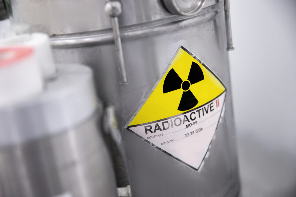
Additionally, Tc-99m widespread availability is a significant advantage. It is derived from Molybdenum-99 (Mo-99), which is produced in nuclear reactors and readily available in most hospitals and imaging centres. The isotope can be obtained from Mo-99 through a generator system, which can be easily installed on-site. This system allows for the continuous production of Tc-99m, ensuring a steady and reliable supply for daily diagnostic procedures.
The ease of preparation of Tc-99m labelled radiopharmaceuticals also contributes to its widespread use. Many Tc-99m compounds can be prepared quickly and used for a variety of imaging studies, ranging from cardiac and bone scans to renal and brain imaging. This versatility, combined with its safety and availability, makes Tc-99m an invaluable tool in modern medical diagnostics, providing clinicians with crucial insights into various medical conditions while ensuring patient safety.
Tc-99m radiopharmaceuticals
- Tc-99m Methylene Diphosphonate (MDP): Used for bone scans to detect bone metastasis, fractures, and infections.
- Tc-99m Sestamibi: Utilised in myocardial perfusion imaging for cardiac assessments, especially for detecting coronary artery disease.
- Tc-99m Tetrofosmin: Another agent for myocardial perfusion imaging, similar in use to Tc-99m Sestamibi.
- Tc-99m Pertechnetate: Used in thyroid scans to assess thyroid function and morphology.
- Tc-99m Mercaptoacetyltriglycine (MAG3): Employed in renal imaging to evaluate renal function and structure.
- Tc-99m Dimercaptosuccinic Acid (DMSA): Used in renal cortical imaging to detect and characterise renal cortical defects.
- Tc-99m Hexamethylpropyleneamine Oxime (HMPAO): Labeled white blood cells with this agent are used in infection and inflammation imaging.
- Tc-99m Exametazime: Used for cerebral perfusion imaging and also in leukocyte labelling for infection detection.
- Tc-99m Sulfur Colloid: Used in liver and spleen imaging and also in lymphoscintigraphy for sentinel node detection in cancer.
- Tc-99m Mebrofenin: Utilised in hepatobiliary imaging to assess gallbladder function and to diagnose bile duct obstruction.
These radiopharmaceuticals, each with their specific targeting capabilities, illustrate the versatility of Tc-99m in diagnosing a wide range of medical conditions.
The Impact of Advanced Imaging Technologies on Technetium-99m Applications
Technological advancements in medical imaging, particularly in gamma camera technology and hybrid systems like Single Photon Emission Computed Tomography/Computed Tomography (SPECT/CT), have significantly enhanced the applications of Technetium-99m in clinical diagnostics. These advancements have led to more precise and detailed images, thereby improving the accuracy and efficacy of diagnoses.
Modern gamma cameras, equipped with advanced detectors and sophisticated software, have improved the resolution and quality of Tc-99m images. These cameras are more sensitive and capable of producing clearer images, allowing for the detection of smaller lesions and finer anatomical details. This enhancement is crucial in early disease detection, particularly in oncology, cardiology, and neurology.
The integration of SPECT with CT in hybrid imaging systems has been a game-changer. SPECT/CT combines the functional imaging capability of SPECT with the detailed anatomical information provided by CT. This combination allows for precise localisation and characterisation of abnormalities detected with Tc-99m. For instance, in oncology, SPECT/CT can accurately pinpoint the location of metastases, improving staging and treatment planning.
Additionally, SPECT/CT provides a better understanding of complex anatomical regions, like the spine or joints, enhancing the diagnosis of orthopaedic and neurologic conditions. It also aids in differentiating between benign and malignant lesions, thereby improving patient management and reducing the need for invasive procedures.
These technological advancements not only enhance the diagnostic capabilities of Tc-99m but also contribute to more personalised and effective patient care. By providing detailed and accurate diagnostic information, these modern imaging techniques enable clinicians to tailor treatment strategies to individual patient needs, ultimately improving patient treatment outcomes and quality of life.
Conclusion
Technetium-99m radiopharmaceuticals are indispensable in modern medical imaging, offering high-quality images with relatively low risk, aiding in diagnosing, staging, and following various diseases, particularly in oncology, cardiology, and neurology. Their unique properties and the continuous development of new Tc-99m labelled compounds expand their potential in medical diagnostics.
Current clinical trials involving Technetium-99m radiopharmaceuticals include:
- Molecular Imaging of Prostate-Specific Membrane Antigen Using Labeled Technetium-99m BQ0413: This trial, with the identifier NCT06101927, is being conducted by the Tomsk National Research Medical Center of the Russian Academy of Sciences in collaboration with Uppsala University. It aims to evaluate the biological distribution of [99mTc]Tc-BQ0413 in patients with prostate cancer, focusing on its distribution in normal tissues and tumours, dosimetry, and safety after a single injection. The trial also seeks to compare SPECT imaging results using [99mTc]Tc-BQ0413 with CT, MRI, ultrasound, and immunohistochemical studies. This is a Phase 1 trial, enrolling 10 participants, and it started on October 19, 2023, with an estimated completion date of October 30, 2024.
- Molecular Imaging of Gastrin Releasing Peptide Receptors Using Labeled Technetium-99m DB8: This study, identified as NCT05940298, is also conducted by the Tomsk National Research Medical Center of the Russian Academy of Sciences. It investigates the distribution of 99mTc-DB8 in patients with prostate and breast cancer. Similar to the first trial, it assesses the distribution in normal tissues and tumours, dosimetry, and safety after a single injection. It also compares the SPECT imaging results with CT, MRI, ultrasound, and immunohistochemical studies. This Phase 1 trial involves 10 participants and started on July 3, 2023, with an expected completion date of February 1, 2024.
Disclaimer
The content provided in Technetium-99m Radiopharmaceuticals in Advanced Medical Imaging by Open Medscience is intended for informational and educational purposes only. It is not a substitute for professional medical advice, diagnosis, or treatment. The use of technetium-99m and other radiopharmaceuticals should only be conducted by qualified healthcare professionals within properly equipped medical facilities and in accordance with local regulations and safety guidelines.
While the article aims to present accurate and up-to-date information at the time of publication (12 November 2023), medical science and regulatory standards evolve, and newer findings may alter clinical practices. Open Medscience does not guarantee the completeness, accuracy, or applicability of the content to specific individual circumstances.
Readers should consult with appropriately qualified professionals before making any healthcare decisions based on this material. Neither Open Medscience nor the authors of this article accept any liability for any loss, damage, or injury resulting from the use or misuse of the information provided.
Use of technetium-99m radiopharmaceuticals involves exposure to ionising radiation, which carries risks. All such procedures must be justified and optimised under the supervision of licensed professionals, following national and international radiation safety standards.
home » blog » radiopharmaceuticals »