Introduction: A New Era in Vein Disease Diagnosis
The number of treatments for a variety of conditions continues to grow, but equally important to these treatments is the technology that allows practitioners to properly diagnose the conditions. It is now more common to diagnose patients with conditions related to circulation issues much earlier in the progression of the disease.
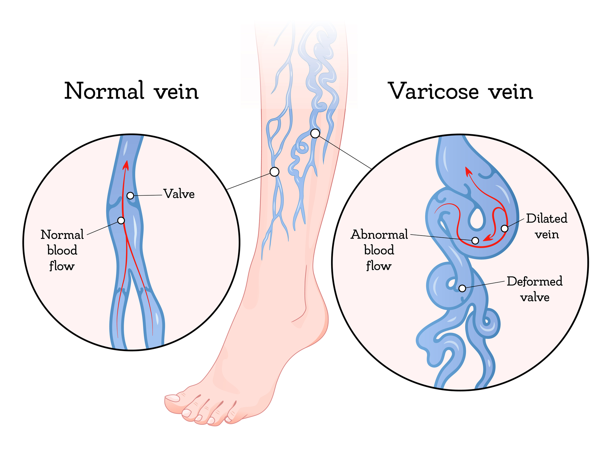
There are many conditions that compose vein disease, such as chronic venous insufficiency, deep vein thrombosis, and venous ulcers. Varicose veins and spider veins are also included in this disease category. However, the latter two conditions are not as life threatening as the others. The gradual onset of many of the conditions that comprise vein disease often results in patients mistaking symptoms of the disease as part of the aging process.
The progressive nature of many vein conditions, along with the insensitivity of many patients, poses a significant challenge to practitioners to timely and efficiently treat patients. Fortunately, technology has improved a clinician’s ability to accurately assess a patient’s vascular health. An accurate assessment of a patient’s vascular health will improve the patient’s quality of life and significantly alter and improve the treatment options available for a patient.
As technology continues to improve, the focus of vascular medicine will be on accurate assessment of a patient’s health. Patients experiencing symptoms such as leg swelling, heaviness, varicose veins, or persistent discomfort may benefit from seeking evaluation early and can receive care from experienced vein doctors in Daytona Beach who utilise advanced vascular imaging technologies to support timely diagnosis and personalised treatment planning.
Recognizing the Need for the Early Diagnosis of Vein Disease
When it comes to vein conditions that have a slow progression, the early symptoms of these often include noticeable fatigue and mild swelling in the legs. The presence of these symptoms may even be associated with aging by the patient. Accordingly, it is not unusual for patients to postpone the treatment of these symptoms.
The trouble is that venous disease may still worsen in the background even with manageable symptoms on the surface. Blood flow may still be affected and pressure on the veins still be problematic. Eventually significant and serious issues may develop.
Having the ability to catch disease processes on the decline may help uneven the course symptoms may take. Early diagnosis can be the difference between the treating options. Invasive procedures may be less warranted and ultimately create favorable outcomes.
The potential for early diagnosis increases with the advancement in imaging technology. Clinicians now have access to the information that could only be imagined in a physical exam.
Vascular Diagnostics Progression
The development of vein disease diagnosis was reliant upon the observation of the clinician and the description of the symptoms by the patient. Physicians would look for symptoms of vascular disease in the enlargement of the veins, skin changes, swelling and discomfort.
Presenting these symptoms, however, is only partial. Many problems of circulation occur and remain under the surface of the skin.
With advancement in vascular medicine, researchers and clinicians sought to develop diagnostic assessments that would allow further investigation of the venous system. This innovation spurred the development of advanced diagnostic imaging.
Specialists are now equipped with the technology to assess blood flow in real time. They can now appraise the health and function of venous valves and find disease with diagnostic imaging with great precision.
Evaluating Vein Pathology with Duplex Ultrasound Imaging
Duplex ultrasound imaging is unique compared with other imaging modalities used to diagnose vein pathology. Using a non-invasive means of assessing both anatomy and physiology, it employs conventional ultrasound imaging and Doppler technology.
Ultrasound is a means of generating images of structures, while Doppler technology provides a means of assessing flow. Utilizing both of these features gives insights of anatomy, physiology and pathology of veins and their valves.
One of the most notable features of duplex ultrasound is the identification of venous reflux. Unlike other imaging modalities, duplex ultrasound can diagnose venous reflux, the backward flow of blood due to valvular incompetence. Venous reflux is also the underlying cause of varicose veins and chronic venous insufficiency.
Because this imaging modality is painless, does not utilize ionizing radiation, and is cost-efficient, duplex ultrasound imaging is one of the mainstays of contemporary vascular diagnostics.
Imaging Advantage: Real Time Flow with Enhanced Diagnostic Accuracy.
A major advantage of the imaging technology of today is the ability of the clinician to visualize flow in real time. Gone are the days where a clinician has to rely on static representations of flow in order to assess the response of veins and other vascular structures to a variety of maneuvers.
Real time flow enhances the ability of the clinician to discover even the most minor of deviations to normal anatomy that may evolve in the presence of pathology. This also helps differentiate the many varied causes of leg complaints related to the presence of a circulatory disturbance.
Timely diagnosis creates the ability of the clinician to accurately design a plan to alleviate the pathology that creates the symptoms, rather than managing the symptoms associated with the circulatory disturbance.
Vein Mapping and Three-Dimensional Imaging
New imaging systems are not restricted to traditional, two-dimensional depictions of the vascular system. Advanced systems, such as three-dimensional imaging, enable clinicians to better visualize anatomy that contains complex structures.
Three-dimensional vein mapping systems can depict relationships among various blood vessels and help identify problem areas where circulation is not normal. These systems are particularly useful for the development of treatment plans to address complex venous problems. These problems often encompass a number of interconnected veins.
Three-dimensional modeling may have a growing impact on system imaging even beyond the health care industry for patient education and system diagnostics, as imaging software continues to develop.
When patients are shown the complex and detailed images of their vascular system, they often gain a greater understanding of the nature of their problem.
AI and Imaging for the Vascular System
Artificial Intelligence (AI) is starting to have an impact on every area of healthcare, and the field of vascular diagnostics is beginning to see the early application of this technology. Researchers are now looking to see how imaging systems that incorporate machine learning can help clinicians interpret imaging data more quickly and accurately.
Artificial Intelligence (AI) systems can easily analyze large amounts of flow imaging data, and in the process of image review, quickly identify data that contains abnormal circulation patterns. AI systems use Clinical Decision Support (CDS) to recommend a course of action. These systems are not a replacement for clinicians.
Automated imaging systems taken from this model may help to streamline the process of imaging for circulation as a diagnostic step in itself to provide a rapid assessment of the vascular system while better meeting the need of clinicians to provide a quality assessment of the vascular system.
AI systems may then serve a large number of support roles in vascular diagnostics and help design treatment plans.
The Contribution of Imaging to Personalized Medicine
Personalized medicine has to be one of the most breath-taking changes in modern healthcare. This technique focuses on altering the standardized treatment based on the patient’s characteristics and the results of the patient’s tests.
The use of advanced imaging plays a major role in this area. This technology focuses on the uniqueness of each patient’s anatomy and the circulation patterns of each patient. This diagnostic imaging also helps discover the various factors that contribute to each individual’s case of vein disease.
This approach also helps minimize the treatments that won’t help the patient’s case.
The Patient’s Journey with Advanced Diagnostics
Advanced imaging helps the patient in various ways. An example being the use of non-invasive procedures that help eliminate the discomfort the patient experiences.
Advanced imaging even helps the patient feel more included in the process. Providing the diagnostic tests and results in a way the patient can understand allows the patient to feel confident in the treatment they are about to receive.
The advanced imaging diagnostic tests even help the patient feel less uncertain about their condition as treatments are provided based on the results of the tests rather than the treatments being based on assumptions.
The diagnostics even helps the healthcare team feel more confident in the decisions they are making as they are based on tests rather than assumptions.
How Advanced Diagnostics Create Better Treatment
The use of advanced imaging helps the healthcare team feel confident in the treatment choices they are making. Creating advanced imaging technologies even allows the patient and healthcare team to feel confident in the treatment being provided.
With the help of cutting edge imaging, clinicians are able to locate unhealthy veins, assess the severity of a disease, and better quantify the efficacy of a given treatment over time. As a result, advanced imaging enhances the clinical decision-making process and optimally directs clinical interventions.
Patients with improved diagnostic accuracy often have a greater potential for symptom relief, better blood flow, and a greater prognosis with regard to their future health.
There is a strong correlation between the accuracy of a diagnosis and the successfulness of a treatment. However, the main reason that imaging is so important in modern vein care is due to the connection between the two.
Receiving Care
Since imaging technology is constantly advancing, patients benefit the most when expertise is combined with these tools. Skilled vascular specialists possess the expertise necessary to interpret clinical imaging and subsequently construct a viable treatment plan.
These practitioners employ cutting-edge imaging technology in order to assess the vascular health of the patient and develop targeted treatment in order to optimize the patient’s health.
The integration of modern imaging technology and specialized clinical practice is of utmost importance for the achievement of positive health outcomes.
Future of Detection in Vein Disease
The future of vascular imaging is highly promising due to rapid technological integration of imaging with AI, analytics, and even advanced therapies including personalized medicine. The efficacy of diagnostic imaging in locating vein disease will likely improve exponentially.
Researchers are developing new technology that may be able to detect vascular abnormalities that are currently beyond detection. Such technology would make it possible to conduct progressive vascular medicine care and act before the patient develops significant health concerns. Remote vascular medicine may be closer to a reality with the increased capability and integration of wearables that monitor health as they are used.
The integration of technologies into the clinic will improve the accuracy of diagnoses, the precision and personalization of treatment, and the quality of care patients receive.
The progress of diagnostics technologies that will make new therapeutic interventions possible and available will determine the level of care vein patients will receive.
Conclusion: Imaging at the Heart of the Modern Approach to Vein Care
The revolution in vascular diagnostics has impacted modern vein medicine dramatically. Contemporary vein diagnostics enables health practitioners to detect and treat vein disease with unparalleled precision.
Developments in duplex ultrasound and the evaluation of real-time blood flow, as well as 3D imaging and AI, will continue to innovate and improve the precision of modern diagnostics. These innovations will improve the timing of diagnoses, treatment, and consequent patient outcomes.
The future of modern vein care will include advanced imaging technologies as its foundation. The advanced imaging of the future will enable the integration of scientific advancements into the clinic and the practice of medicine. The future of technology in vascular diagnostics will deliver the promise of precision medicine to help patients at every segment of the vascular healthcare continuum.
Disclaimer: This article is intended for informational and educational purposes only and does not constitute medical advice, diagnosis, or treatment. Readers should consult a qualified healthcare professional regarding any medical concerns or before making decisions about their health. The views expressed are for general information and may not reflect the latest clinical guidelines or individual patient circumstances.


