The 2023 Nobel Chemistry Prize honours Bawendi, Brus, and Ekimov for quantum dots discovery, synthesis, advancing medical imaging, electronics, and solar energy.
Quantum Leap: The Trio of Bawendi, Brus, and Ekimov Honoured with 2023 Nobel Prize in Chemistry for Pioneering Quantum Dots Synthesis
Aleksey Ekimov first discovered Quantum Dots (QDs) in a glass matrix in 1981 whilst working in Saint Petersburg. This revolutionary discovery laid the foundation for the development and study of quantum dots, which since have found applications in various fields, including electronics, medicine, and renewable energy; this is due to their unique optical and electronic properties. Ekimov’s discovery marked the beginning of a new era in nanotechnology and quantum materials science. Consequently, the impact of quantum dots continues to grow as researchers explore new ways to harness their potential.
The foundation of Ekimov’s research on Quantum Dots led to the 2023 Nobel Prize being awarded to Aleksey Ekimov (Nanocrystals Technology Inc., New York, NY, USA), Louis Brus (Columbia University, New York, NY, USA) and Moungi Bawendi (Massachusetts Institute of Technology (MIT), Cambridge, MA, USA).
Aleksey Ekimov’s research managed to produce size-dependent quantum effects in coloured glass. The colour was generated from nanoparticles of copper chloride and proved that the particle size affected the colour of the glass due to quantum effects.
However, Louis Brus proved that size-dependent quantum effects in particles could float freely in a fluid while researching semiconductors at the AT&T Bell Laboratories in New Jersey. In two papers published in 1983 (A simple model for the ionization potential, electron affinity, and aqueous redox potentials of small semiconductor crystallites) and 1984 (Electron–electron and electron‐hole interactions in small semiconductor crystallites: The size dependence of the lowest excited electronic state).
Brus described QDs as small semiconductor crystallites. Finally, Moungi Bawendi produced the chemical production of quantum dots in almost perfect particles to be used in numerous applications.
Quantum dots, also known as artificial atoms, are semiconducting nanocrystals that exhibit unique optical and electronic properties, making them ideal candidates for medical imaging applications. These Quantum dots contain between 100 to 100,000 atoms and show strange properties as though they were comprised of a single atom.
Quantum dots now illuminate computer monitors and television screens based on QLED technology. They also add nuance to the light of some LED lamps, and biochemists and doctors use them to map biological tissue.
Quantum dots are thus bringing the most significant benefit to humankind. Researchers believe that in the future, they could contribute to flexible electronics, tiny sensors, thinner solar cells and encrypted quantum communication.
These nanoscale crystals can be synthesised using various methods, allowing control over their size, shape, and composition. Consequently, QDs have found potential use across multiple imaging techniques, including fluorescence, magnetic resonance imaging (MRI), and positron emission tomography (PET).
The advantages of using QDs in medical imaging include their tunable optical properties, high photostability, multifunctionality, and compatibility with multiple imaging modalities. However, challenges such as biocompatibility, toxicity, clearance, and stability must be addressed to realise the full potential of QDs in medical imaging.
Future prospects for this technology involve the continued development and optimisation of QDs for medical imaging applications, focusing on enhancing biocompatibility and targeted theranostic capabilities. QDs hold great promise as research progresses in revolutionising medical diagnostics, treatment monitoring, and personalised medicine.
Versatile Synthesis and Quantum Confinement-Driven Properties of Quantum Dots
Quantum dots (QDs) can be synthesised using various methods, including colloidal, hydrothermal, and sol-gel processes. Among these, the colloidal process is the most popular due to its versatility and ease of manipulating the size and shape of the QDs. This method involves precipitating QDs in a solution, allowing precise control over their size and composition to achieve desired optical properties.
The remarkable properties of QDs originate from quantum confinement effects. When semiconductor crystals are reduced to the nanoscale, electrons and holes become confined within a limited space, leading to discrete energy levels. As a result, QDs exhibit size-dependent emission and absorption spectra. These nanocrystals possess broad absorption spectra and narrow, tunable emission spectra, which can be adjusted by modifying their size and composition.
The unique combination of narrow emission spectra, high photostability, and quantum yield makes QDs particularly suitable for medical imaging applications. The ability to fine-tune the emission spectra allows for the simultaneous detection of multiple targets and the development of advanced imaging techniques.
Furthermore, their high photostability enables extended imaging sessions without significant degradation, while their impressive quantum yield ensures bright signals, even for low-abundance targets. Overall, QDs hold great promise for revolutionising the field of medical imaging, offering a versatile and powerful tool for researchers and clinicians alike.
Based on the information gathered, quantum dots (QDs) can be classified based on several criteria, including their material composition, structure, and properties, such as quantum confinement. Below is a table summarizing different types of quantum dots:
| Classification Criteria | Type of Quantum Dots | Description |
|---|---|---|
| Material Composition | Carbon QDs, Aluminium oxide QDs, Cellulose QDs, Ceramic QDs, Cobalt oxide QDs, Copper QDs, Gold QDs, Iron QDs, Iron oxide QDs, Iron-platinum QDs, Lipid QDs, Platinum QDs, Silver QDs, Titanium dioxide QDs | The material composition of QDs often determines their properties and applications. Different materials lead to QDs with different characteristics. |
| Structure | Core-Type QDs | These are basic types of quantum dots with a structure that exhibits quantum confinement effects, which means that the energy levels of the electrons in the QDs are quantized due to the small size of the crystals. |
| Quantum Confinement | – | Quantum confinement is a key property of QDs, which arises due to their small size, leading to quantized energy levels. The effect of quantum confinement on QDs has been studied theoretically. |
| Specific Applications | Donor QDs, Laterally gated QDs | These types of QDs are utilized in quantum computing. Donor QDs are created with a single dopant atom with a bound charge in a semiconductor, forming an intrinsically zero-dimensional system. Laterally gated QDs are a variation where gates are used to control the energies of the various quantum dot states. |
Applications of Quantum Dots in Medical Imaging
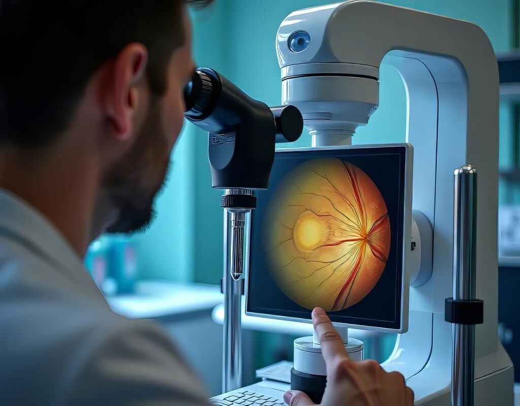
Optical Imaging
Fluorescence-based imaging is a prominent optical imaging technique, and quantum dots (QDs) have demonstrated considerable potential as fluorescent probes in this field. Their tunable emission spectra, high quantum yield, and photostability render them perfect candidates for multi-colour imaging, enabling the simultaneous detection of multiple targets with high sensitivity and specificity. The versatility of QDs allows for their use in a diverse range of fluorescence-based imaging techniques, expanding the possibilities for researchers and clinicians.
In fluorescence microscopy, QDs are superior to conventional organic dyes, offering enhanced photostability and brightness, contributing to improved image quality and reduced photobleaching. Additionally, their tunable emission spectra facilitate multi-colour imaging, allowing the study of complex cellular processes and interactions.
In vivo imaging using QDs provides real-time visualisation of biological processes within living organisms. Their excellent optical properties and surface functionalisation for targeted delivery make QDs valuable tools for studying molecular events, disease progression, and treatment response in preclinical models.
Flow cytometry is another application that has benefitted from QD technology. The use of QDs as fluorescent labels in this high-throughput technique allows for the rapid and precise analysis of multiple cellular markers simultaneously, improving the efficiency of cell sorting and characterisation.
QDs have revolutionised fluorescence-based imaging techniques by offering unparalleled optical properties, enabling simultaneous multi-target detection and expanding the capabilities of various imaging methods, including fluorescence microscopy, in vivo imaging, and flow cytometry.
Magnetic Resonance Imaging (MRI)
Quantum dots (QDs) are nanometer-scale semiconductor particles with unique properties due to their quantum confinement effects. These properties, including size-tunable fluorescence, have already been exploited for various applications in biological imaging, photovoltaics, and quantum computing. Recently, researchers have discovered that QDs can be modified to exhibit magnetic properties, making them suitable for use as contrast agents in magnetic resonance imaging (MRI).
By incorporating paramagnetic or superparamagnetic elements, such as gadolinium, manganese, or iron oxide, into the QD structure, these nanoparticles can generate contrast in T1- or T2-weighted images. T1-weighted images are sensitive to changes in longitudinal relaxation time, while T2-weighted images are sensitive to changes in transverse relaxation time. Magnetic QDs in the imaging area can significantly alter these relaxation times, enhancing image contrast.
QD-based MRI contrast agents offer several advantages over traditional contrast agents. Firstly, they possess higher relaxivities, allowing improved contrast enhancement at lower concentrations. This could potentially reduce the risk of side effects associated with contrast agents. Secondly, the magnetic properties of QDs can be tuned by adjusting their size, composition, and shape, enabling customisation for specific imaging requirements.
Moreover, QD-based contrast agents can be functionalised with biomolecules, such as peptides or antibodies, to facilitate targeted imaging of specific cellular or molecular targets. This targeted approach can improve diagnostic accuracy and enable early detection of diseases, such as cancer or neurodegenerative disorders.
QD-based contrast agents show great potential for revolutionising MRI by offering enhanced imaging capabilities, tunable magnetic properties, and targeted imaging. Further research is needed to optimise their design, ensure their biocompatibility, and evaluate their long-term safety for clinical use.
Positron Emission Tomography (PET)
Positron emission tomography (PET) is a highly sensitive nuclear imaging technique that provides quantitative information on biological processes at the molecular level. PET relies on detecting gamma rays emitted by radiolabelled tracers, typically small molecules or biologically relevant compounds. Radiolabelled quantum dots (QDs) have emerged as potential PET imaging agents due to their inherent advantages, such as high photostability, tunable emission spectra, and the ability to incorporate multiple radioisotopes.
One of the key benefits of using QDs in PET imaging is their high photostability, which allows for long-term imaging without significant signal degradation. Additionally, their tunable emission spectra enable the simultaneous detection of multiple QD-based tracers, providing a more comprehensive view of complex biological processes. Furthermore, the versatile surface chemistry of QDs allows for the conjugation of various radioisotopes, enhancing the signal-to-noise ratio and improving image quality.
By combining PET imaging with QD-based fluorescence imaging, researchers can achieve multi-modal imaging that provides complementary information on molecular and functional processes. PET imaging can offer high sensitivity and quantitative data on the distribution and kinetics of radiolabelled tracers. In contrast, QD-based fluorescence imaging can provide high-resolution, real-time visualisation of cellular and subcellular structures.
This multi-modal approach can facilitate a deeper understanding of biological processes in living organisms and improve the accuracy of disease diagnosis and monitoring. For example, the combined use of PET and QD-based fluorescence imaging can enhance the detection of cancerous lesions, enabling more precise localisation and characterisation of tumours and ultimately improving patient outcomes.
Theranostics
Theranostics is an emerging field that combines diagnostics and therapeutics, paving the way for personalised medicine. This innovative approach allows for the simultaneous detection, monitoring, and treatment of diseases, ultimately improving patient outcomes and reducing healthcare costs. Quantum dot (QD)-based theranostic agents have attracted considerable attention due to their unique properties, such as size-tunable fluorescence, high photostability, and versatile surface chemistry.
QDs can be functionalised with targeting moieties, such as antibodies or peptides, which bind them to bind to disease-associated biomarkers specifically. This targeted approach facilitates both precise imaging and therapy, increasing the accuracy of diagnosis and the efficiency of treatments. For instance, cancer cells often overexpress specific cell surface receptors. QDs conjugated with corresponding ligands can selectively target and accumulate in tumour tissues, allowing for enhanced imaging and targeted drug delivery.
Furthermore, QDs can act as photosensitisers in photodynamic therapy (PDT), a minimally invasive treatment that uses light to activate a photosensitiser, generating reactive oxygen species (ROS) that can destroy cancer cells or other pathological tissues. The tunable absorption and emission spectra of QDs make them ideal candidates for PDT, as they can be tailored to absorb light at specific wavelengths, facilitating the targeted production of ROS.
In addition to PDT, QDs can be employed as drug carriers, enabling the controlled release of therapeutic agents at the site of interest. By encapsulating drugs within the QD structure or conjugating them to the QD surface, researchers can develop stimuli-responsive drug delivery systems that release their cargo upon exposure to specific triggers, such as pH, temperature, or light changes.
| Property | Description |
| Material Composition | – Common materials include cadmium selenide (CdSe), cadmium telluride (CdTe), and indium arsenide (InAs), among others. |
| Size | – Typically range from 2 to 10 nanometers in diameter. |
| Colour (Emission Wavelength) | – The colour of emitted light can be tuned by changing the size and material composition of the QDs. Smaller QDs emit light at shorter wavelengths (blue), while larger QDs emit light at longer wavelengths (red). |
| Surface Modification | – To make QDs biocompatible and target-specific, their surface can be modified with various ligands, antibodies, peptides, or other biomolecules. |
| Optical Properties | – Bright, photostable fluorescence and broad absorption spectra with narrow emission spectra. |
| Applications in Medical Imaging | – Used in various imaging modalities, including fluorescence imaging, multiphoton imaging, computed tomography (CT), and magnetic resonance imaging (MRI). |
| Benefits | – High sensitivity, multiplexing capabilities, long-term stability. |
| Challenges | – Potential toxicity, regulatory approval, and long-term safety in biological systems. |
Advantages of Quantum Dots in Medical Imaging
The use of QDs in medical imaging offers several advantages compared to traditional imaging agents:
Tunable Optical Properties
Quantum dots (QDs) are nanometer-scale semiconductor particles with unique optical properties due to their quantum confinement effects. One of the most attractive features of QDs is the ability to easily control their size and composition during synthesis, allowing for tailoring their optical properties. This customisation enables QDs to have specific emission wavelengths, facilitating multi-colour imaging.
The emission wavelengths of QDs are directly related to their size, with smaller QDs emitting shorter wavelengths (blue light) and larger QDs emitting longer wavelengths (red light). This size-tunable fluorescence enables the production of QDs with a broad range of emission colours simply by adjusting their size during synthesis. Additionally, altering the composition of QDs by incorporating different semiconductor materials can further influence their optical properties and improve their performance in imaging applications.
Multi-colour imaging using QDs has numerous advantages, including the ability to simultaneously visualise multiple cellular or molecular targets within the same sample. This approach can provide a more comprehensive understanding of complex biological systems and interactions, essential for accurate diagnosis and targeted therapy. Furthermore, the high photostability and brightness of QDs allow for long-term imaging without significant signal degradation, enabling the tracking of biological processes over extended periods.
High Photostability and Brightness
Quantum dots (QDs) are an exceptional class of nanomaterials that have gained considerable attention in bioimaging due to their unique optical properties. One of the key advantages of QDs is their high photostability, which allows for prolonged imaging without significant photobleaching. Unlike conventional organic fluorophores, which tend to degrade and lose their fluorescence upon prolonged exposure to light, QDs can maintain their fluorescence over extended periods, making them ideal for long-term imaging applications.
In addition to their high photostability, QDs possess high quantum yield and brightness. Quantum yield measures a fluorophore’s efficiency in converting absorbed photons into emitted photons. The high quantum yield of QDs ensures that they emit many photons upon excitation, resulting in a bright signal. This characteristic makes QDs excellent candidates for detecting low-abundance targets, as they provide enhanced sensitivity and signal-to-noise ratio compared to traditional fluorophores.
These remarkable properties of QDs—high photostability, quantum yield, and brightness—have opened up new possibilities in the field of bioimaging. They enable the visualisation of dynamic biological processes over extended time periods, which is crucial for understanding complex cellular mechanisms and interactions. Furthermore, detecting low-abundance targets with high sensitivity can improve the accuracy of disease diagnosis, enabling early intervention and more effective treatment strategies. Overall, using QDs in bioimaging holds great potential for advancing our knowledge of biological systems and improving patient outcomes.
Multifunctionality
Quantum dots (QDs) possess unique properties that make them attractive candidates for various biomedical applications, including targeted imaging and therapy. One of the key features of QDs is their versatile surface chemistry, which allows them to be functionalised with various ligands or biomolecules, such as antibodies, peptides, or small molecules. This functionalisation enables QDs to target and bind to disease-associated biomarkers, resulting in targeted imaging and therapy.
The multifunctionality of QDs allows for the simultaneous detection and treatment of diseases, making them ideal theranostic agents. In theranostic applications, QDs can serve as imaging agents that provide real-time visualisation of disease processes and deliver therapeutic agents directly to the site of interest. This dual functionality can improve diagnostic accuracy, enable early intervention, and increase the efficiency of treatments, ultimately leading to better patient outcomes.
By combining the unique optical properties of QDs with their ability to be functionalised for targeted delivery, researchers can develop innovative theranostic platforms that hold great promise for personalised medicine. QD-based theranostic agents have the potential to revolutionise the diagnosis and treatment of various diseases, including cancer, neurodegenerative disorders, and cardiovascular diseases. Further research is needed to optimise the design and performance of these agents and ensure their long-term safety and biocompatibility for clinical use.
Multi-modal Imaging
Quantum dots (QDs) are versatile nanomaterials that have gained significant interest in the field of bioimaging due to their unique properties. They can be utilised in various imaging modalities, such as fluorescence, magnetic resonance imaging (MRI), and positron emission tomography (PET). By combining these modalities, researchers can obtain complementary information on molecular, functional, and anatomical processes, ultimately improving diagnostic accuracy and treatment monitoring.
In fluorescence imaging, QDs offer size-tunable emission spectra, high photostability, and brightness, enabling the visualisation of cellular structures and processes with high resolution and sensitivity. In MRI, QDs can be modified to exhibit magnetic properties by incorporating paramagnetic or superparamagnetic elements, making them suitable as contrast agents for enhanced imaging. In PET, radiolabelled QDs can provide quantitative information on biological processes at the molecular level.
The integration of QD-based imaging agents across multiple modalities can offer a more comprehensive view of complex biological systems. This multi-modal approach facilitates the simultaneous assessment of molecular targets, functional processes, and anatomical structures, providing valuable insights into disease progression and response to treatment. Furthermore, it can enhance the detection and classification of diseases, such as cancer or neurodegenerative disorders, enabling early intervention and more effective treatment strategies.
Challenges and Limitations of Quantum Dots
Despite their potential advantages, the use of quantum dots (QDs) in medical imaging poses numerous challenges and limitations that must be addressed to ensure their safe and effective application. The potential toxicity of QDs is a significant concern for their use in medical applications, as some QDs, particularly those containing heavy metals like cadmium, can be toxic to cells and organisms. This toxicity can lead to complications and adverse effects on human health if not properly mitigated.
Strategies to reduce the toxicity of QDs include the use of biocompatible coatings that can shield the toxic core and prevent interaction with biological systems. Additionally, researchers are working to develop heavy-metal-free QDs, such as those based on silicon or carbon, which exhibit lower toxicity while maintaining the desirable optical properties required for medical imaging applications.
Efficient clearance of QDs from the body is another critical aspect of their use as imaging agents. However, due to their size and surface properties, QDs often accumulate in organs such as the liver and spleen, leading to potential long-term complications. Further research is needed to optimise the biodistribution and clearance of QDs, including the development of strategies to modify their size, shape, and surface properties to improve renal clearance and minimise non-specific accumulation in organs.
The stability of QDs in biological environments is essential for their effective use in medical imaging. However, when exposed to biological fluids, QDs can aggregate or undergo chemical changes, altering their optical properties and consequently reducing their imaging capabilities. Researchers are developing robust coatings and surface functionalisation strategies to overcome these stability issues, protect QDs from degradation, and maintain their optical properties in complex biological environments.
Future Prospects
Quantum dots (QDs) have shown immense potential for medical imaging due to their unique optical properties, multifunctionality, and compatibility with various imaging modalities. These nanoscale semiconductors exhibit size-dependent tunable fluorescence emission, high quantum yield, and excellent photostability, making them particularly suitable for a variety of biomedical applications. Despite the challenges associated with their toxicity, clearance, and stability, ongoing research in this field is addressing these issues. It is paving the way for the development of safe and effective QD-based imaging agents.
In the future, we can expect the continued development and optimisation of QDs for medical imaging, emphasising biocompatibility and targeted theranostic applications. These theranostic agents will allow for simultaneous diagnosis and therapy, potentially revolutionising how diseases are treated. By developing QDs with specific targeting moieties, it will be possible to deliver imaging agents directly to the site of interest, such as tumours or inflammation, reducing off-target effects and improving diagnostic accuracy.
Moreover, combining QDs with other emerging nanomaterials, such as graphene and upconversion nanoparticles, may lead to the development of novel imaging agents with enhanced properties and functionalities. These hybrid nanomaterials could offer synergistic advantages, such as improved biocompatibility, multi-modal imaging capabilities, and increased targeting efficiency, which could further advance the field of medical imaging.
In addition to their potential use in diagnostics, QDs could be vital in treatment monitoring and personalised medicine. Real-time tracking of QD-based theranostics could enable clinicians to assess the effectiveness of therapies and adjust treatment plans accordingly, improving patient outcomes and reducing the likelihood of relapse.
Quantum dots hold great promise for advancing medical imaging, potentially revolutionising diagnostics, treatment monitoring, and personalised medicine. As research in this field progresses, we can expect the further development and refinement of QD-based imaging agents and their eventual translation to clinical applications. The integration of quantum dots into the medical imaging toolbox will undoubtedly contribute to more accurate diagnoses, better-informed treatment decisions, and, ultimately, improved patient care.
Disclaimer
The content presented in this article, “Nobel Prize Quantum Dots and their Applications in Medical Imaging”, is intended for informational and educational purposes only. While every effort has been made to ensure the accuracy and reliability of the information at the time of publication, Open MedScience does not warrant or guarantee its completeness or suitability for any particular purpose.
This article summarises scientific and medical developments relating to quantum dots and their potential applications in imaging and diagnostics. It is not intended as medical advice, nor should it be used as a substitute for professional healthcare guidance, diagnosis, or treatment. Readers should consult qualified medical or scientific professionals for specific advice regarding any medical condition or technological application.
Any mention of specific individuals, organisations, or products does not imply endorsement by Open MedScience. The article may contain references to experimental or emerging technologies that are subject to ongoing research and regulatory approval. As such, applications discussed may not yet be approved for clinical use or widely available in healthcare settings.
Open MedScience disclaims any responsibility for loss, injury, or damage incurred as a result of reliance on the information contained herein. Views expressed are those of the authors and do not necessarily reflect those of Open MedScience or its affiliates.
home » blog » optical imaging »