Medical optical imaging has entered a period of remarkable progress. Once confined to basic structural visualisation, it now supports advanced diagnostic decision-making, procedural guidance, remote patient monitoring, and early-stage disease detection. A combination of improved hardware, sophisticated computational methods, and greater integration with multimodal diagnostic workflows is steadily transforming how clinicians observe and interpret biological tissues. This article outlines the latest developments driving this transformation, with attention to areas experiencing the most significant momentum across research and clinical spheres.
Optical Coherence Tomography and the Rise of Wider, Faster, Deeper Imaging
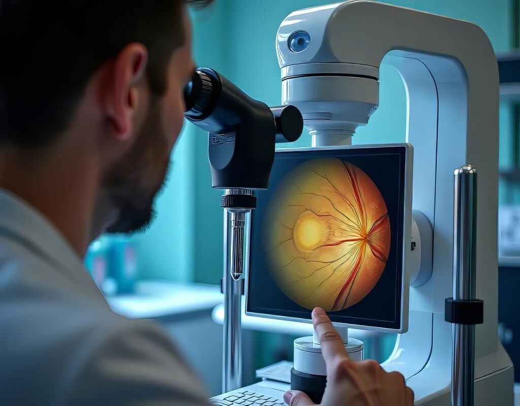
Optical Coherence Tomography (OCT) remains one of the most active and fast-moving areas in medical optical imaging. Originally developed for retinal assessment, the modality has grown far beyond its early use. Improvements in acquisition speed, image resolution, and depth penetration have expanded what can be seen, how quickly it can be captured, and how the images can be interpreted.
Swept-source OCT, in particular, has altered expectations about the field of view and clinical usability. By using longer wavelengths and tunable lasers, these systems capture images at greater depths and at higher speeds than traditional spectral-domain systems. This allows clinicians to observe peripheral regions of the retina that were once difficult to image effectively and to assess sub-surface structures with greater clarity.
At the same time, OCT angiography (OCTA) continues to grow in relevance. By detecting motion contrast from blood flow, OCTA provides a noninvasive map of the retinal vascular network. Researchers and clinicians now have access to vascular biomarkers that would previously have required invasive dye-based imaging. With commercial OCTA devices becoming more refined and faster, vascular assessment is moving into standard care pathways for diseases such as diabetic retinopathy, macular degeneration, and glaucoma.
Another emerging direction involves intraoperative OCT, which offers real-time visual guidance during surgery. Surgeons can assess tissue planes, membrane position, and changes in the vitreoretinal interface without interrupting the procedure. Many institutions have already incorporated microscope-integrated OCT, enabling continuous observation without workflow disruption.
There is also a growing market for compact, portable OCT devices intended for use outside specialist clinics. Some systems aim for technician-free deployment, particularly for basic screening applications. The opportunity to decentralise high-resolution imaging and expand access to earlier diagnosis is now becoming realistic.
Home-based Optical Imaging and the Move Towards Remote Care
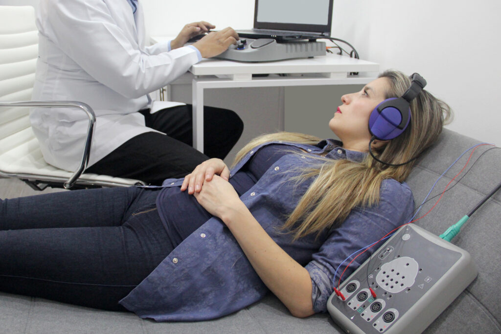
The growing interest in remote monitoring for chronic disease management has led to significant innovations in home-based optical imaging. Retinal conditions such as age-related macular degeneration and diabetic maculopathy require regular surveillance, often more frequently than patients can practically attend clinics. Home OCT devices are designed to address this challenge by enabling patients to capture retinal images themselves, which can be transmitted to clinics for analysis.
These systems often rely on artificial intelligence (AI) to assess image quality, detect relevant features, and flag abnormalities requiring clinical intervention. This alleviates the burden on clinical staff while ensuring consistent monitoring. Early studies indicate that patient-operated devices can achieve clinically acceptable image quality when supported by automated alignment and focusing systems.
The appeal of home optical imaging extends beyond ophthalmology. Portable optical devices are being explored for skin lesion monitoring, wound assessment, and even remote otolaryngology evaluations. The combination of compact hardware and cloud-based analysis tools is creating a framework in which diagnostic imaging is no longer tied to hospital-based devices. This trend has considerable potential within public health strategies, particularly where access or mobility is limited.
AI-Driven Optical Diagnostics and Insights Beyond Traditional Imaging
The influence of artificial intelligence in optical imaging continues to grow at a rapid pace. Algorithms are increasingly integrated into both research and clinical optical systems, improving accuracy, reducing interpretation time, and enabling new forms of diagnostics.
One of the most promising areas involves the use of multimodal datasets. OCT, OCTA, colour fundus photography, and adaptive optics imaging can now be combined to create a richer and more nuanced picture of tissue health. AI models that ingest these combined datasets are capable of detecting subtle structural and vascular changes that may be overlooked by humans.
Another important development is the exploration of systemic disease markers within retinal imaging. Several recent studies suggest that microscopic retinal features correlate with cardiovascular risk, neurodegeneration, cognitive decline, and even some metabolic conditions. Machine learning models trained on large retinal datasets appear to uncover patterns that do not directly relate to traditional ophthalmic disease but instead provide insight into whole-body physiology.
This work is still preliminary in many domains, and questions remain regarding generalisability, bias, and robustness. Nevertheless, the possibility of using optical imaging as a window into broader systemic health is gaining serious attention.
AI also plays a role in advancing acquisition and reconstruction techniques. Models are increasingly used for denoising, motion correction, artefact suppression, and enhanced visualisation. For low-resource imaging settings, these improvements are particularly valuable, as they help deliver usable images from equipment that may not otherwise reach the performance of high-end clinical devices.
Quantum Optical Imaging: Moving Beyond Classic Photon-Based Detection
Quantum imaging techniques represent one of the most forward-looking developments in the field. Although still in early research stages, the potential impact is significant. These techniques use quantum correlations, entangled photon pairs, and various quantum-state manipulations to achieve imaging performance beyond that of classical light sources.
Key benefits include improved signal-to-noise ratios, greater phase sensitivity, and the potential for lower light-dose imaging. Lower doses are particularly relevant in medical contexts where tissue exposure must be minimised.
Quantum illumination and ghost imaging have drawn notable attention. In ghost imaging, an image is reconstructed from photons that do not directly interact with the object, while correlated photons follow a different path. This phenomenon has intriguing implications for imaging delicate biological structures.
Another promising technique is the use of squeezed light to reduce quantum noise. By altering the quantum fluctuations of a light field, it becomes possible to exceed the classical shot-noise limit, thereby improving measurement precision.
Practical medical applications remain some distance away, as current devices are complex and delicate. However, as optical hardware becomes more stable and miniaturised, clinical use may eventually become feasible.
Computational Optical Imaging: Integrating Algorithms with Hardware
Computational imaging has become central to optical system innovation. Rather than relying solely on optics to capture images, researchers now design hardware and algorithms in tandem, optimising the entire imaging pipeline.
One promising approach uses differentiable imaging techniques, where both the optical system and the reconstruction algorithm are treated as optimisable components within a single mathematical framework. This allows researchers to fine-tune light propagation, sensor response, and algorithmic reconstruction simultaneously.
This form of co-design is particularly useful for deep-tissue imaging and low-signal environments. It enables targeted enhancement of specific structures or contrasts that are challenging to visualise with conventional optics alone.
Another area of progress is the use of neural rendering and physics-informed models to reconstruct three-dimensional structures from limited optical information. Such systems reduce the need for specialised or expensive hardware while improving image utility.
Computational optical imaging is also being applied to dynamic tissues, where motion increases complexity. Algorithms are now being developed that reconstruct volumetric sequences with minimal artefacts, supporting real-time surgical imaging and in vivo studies of microvascular flow.
Optical Imaging in Interventional and Surgical Practice
Optical imaging is no longer restricted to diagnostic rooms. Increasingly, it integrates directly into operative and interventional workflows. Surgeons across ophthalmology, oncology, and cardiology are now leveraging optical systems to improve safety, precision, and outcomes.
In ophthalmic surgery, microscope-integrated OCT provides continuous visual feedback during procedures such as vitrectomy, membrane removal, and macular hole repair. Surgeons can assess the completeness of membrane peeling, confirm tissue plane separation, and adjust techniques immediately. This reduces uncertainty and improves the likelihood of successful outcomes.
Another area undergoing rapid progress is the integration of fluorescence imaging into surgical oncology. Optical agents such as Pegulicianine help surgeons detect residual tumour tissue during resection. By illuminating cancerous regions with high contrast, this technology supports more complete tumour removal and reduces the likelihood of secondary surgeries.
Cardiology is also benefiting from optical innovation. Intravascular OCT is increasingly used during angioplasty to assess plaque composition, guide stent placement, and confirm optimal deployment. AI-enabled systems are now emerging that interpret OCT vessel images in real time, offering procedural guidance and automated lesion assessment.
The advantage of optical imaging in these contexts is the high spatial resolution and immediacy it provides. Procedures that once relied on indirect visual cues or lower-resolution modalities can now be performed with the support of detailed internal imaging at the point of intervention.
Expanding Access Through Portability and Lower-Cost Devices
Another important trend is the growing emphasis on portability, accessibility, and reduced equipment costs. Smaller devices are becoming more robust, and improvements in battery efficiency, optics, and sensor technology are decreasing the cost of entry for clinics and community health services.
Handheld fundus cameras, compact OCT units, and low-cost fluorescence systems are all contributing to greater access in regions with limited imaging infrastructure. These devices are often supported by AI-based interpretation tools, which reduce reliance on specialist readers.
This shift supports broader screening initiatives and earlier detection of disease. It also aligns with sustainability goals by reducing the need for large, energy-intensive equipment and extensive clinical space.
Challenges That Still Need to Be Addressed
Although the progress in medical optical imaging is impressive, several challenges remain. One major issue is the need for standardisation across devices, protocols, and data formats. Without consistent imaging practices, it is difficult to compare results across studies or ensure the reliability of AI models.
Data diversity is another pressing concern. AI models trained on specific populations or imaging platforms may not perform well elsewhere. Ensuring that datasets represent varied demographics, devices, and clinical settings is vital for widespread clinical adoption.
The regulatory pathway for advanced optical systems, especially those using AI or quantum methods, also requires careful consideration. Ensuring safety, repeatability, and reliability is crucial before deployment in clinical practice.
Cost remains an obstacle for some emerging technologies, particularly those involving sophisticated lasers or quantum light sources. Long-term viability will depend on improvements in manufacturing and reductions in component expense.
A Glimpse Into the Future of Optical Imaging
The developments discussed here suggest that optical imaging is moving toward a future defined by integration, portability, intelligence, and new physics-based capabilities. Instead of acting as a stand-alone modality, optical imaging will increasingly complement other imaging techniques, contributing fine-scale detail in multimodal diagnostic workflows.
Remote and home-based imaging is likely to expand, supported by AI systems capable of triage and early detection. Surgical and interventional environments will continue to adopt optical modalities for guidance and improved outcomes. Research-grade innovations such as quantum imaging may eventually reach clinical maturity, offering new levels of sensitivity and precision.
For healthcare professionals, researchers, and medical device developers, optical imaging is a rapidly evolving field full of opportunity. Continued progress will rely on collaborative engineering, strong clinical partnerships, and careful attention to validation and safety. As these technologies mature, the impact on patient care is set to be substantial.
Disclaimer
This article is intended for informational purposes only and does not constitute medical advice, diagnosis, or treatment. The technologies, research developments, and clinical applications discussed are subject to ongoing study and regulatory review. Clinical adoption may vary by region, device, and healthcare provider, and not all methods described are approved for routine use. Readers should not rely on this content as a substitute for professional medical guidance, nor should it be used to inform clinical decisions without consulting qualified healthcare practitioners. Open MedScience accepts no responsibility for actions taken based on the information presented, and all clinical or research decisions should be made in accordance with current standards, regulatory requirements, and institutional policies.
home » blog » optical imaging »