Breakthroughs in Positron Emission Tomography Imaging have significantly enhanced imaging resolution and accuracy, expanded clinical applications, and integrated AI, revolutionising the diagnosis and treatment of various diseases.
Advancements in PET Technology
Positron Emission Tomography (PET) is a sophisticated imaging technology utilised extensively in the medical field to observe metabolic processes in the body. It involves the use of radioactive tracers, typically introduced into the body by injection, inhalation, or ingestion. These tracers emit positrons that interact with electrons in the body, producing gamma rays that are then detected by the PET scanner to create detailed images.
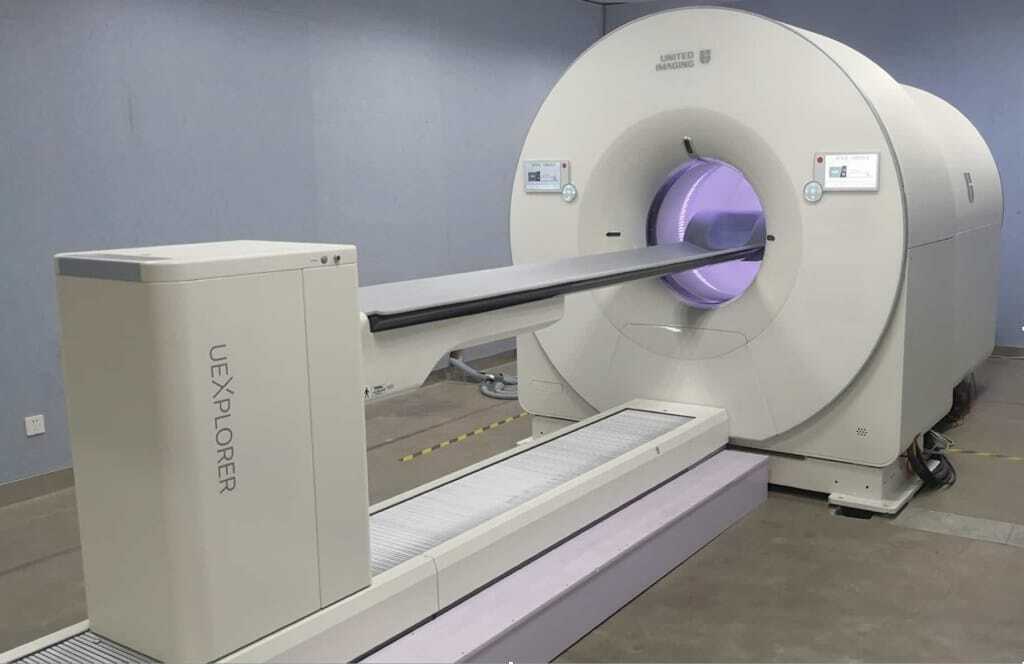
Recent advancements in PET technology have significantly improved the resolution of PET scanners. Modern PET scanners now feature improved spatial resolution, allowing for the detection of smaller lesions and more precise imaging of metabolic processes. Detector technology advancements, such as silicon photomultipliers (SiPMs), have achieved this enhancement. These detectors offer better timing resolution and higher sensitivity, leading to clearer and more accurate images.
Time-of-Flight (TOF) PET is a significant development that enhances image quality by incorporating the precise measurement of the time it takes for gamma rays to travel from the point of annihilation to the detectors. By accurately determining the difference in arrival times of the two gamma rays, TOF PET improves the signal-to-noise ratio, resulting in higher image resolution and better lesion detectability. This technique is particularly beneficial in oncology, where detecting small and early-stage tumours is crucial.
The shift from analogue to digital PET technology represents a significant advancement. Digital PET systems utilise digital detectors and advanced data processing algorithms to produce high-quality images with improved contrast and resolution. These systems also offer faster acquisition times and reduced radiation exposure for patients. Digital PET is particularly advantageous in dynamic imaging, where rapid changes in tracer distribution need to be captured accurately.
Novel Tracers and Applications
The development of novel radiotracers has expanded the applications of PET beyond traditional oncology. Researchers continuously explore new tracers targeting specific molecular pathways and physiological processes. For instance, tracers targeting amyloid plaques and tau protein are being used in the early diagnosis of Alzheimer’s disease. Similarly, tracers targeting dopamine receptors are utilised in the study of neurodegenerative diseases such as Parkinson’s disease.
The integration of PET with Magnetic Resonance Imaging (MRI) has led to the development of PET/MRI hybrid systems. These systems combine the metabolic imaging capabilities of PET with the superior soft tissue contrast of MRI, providing comprehensive anatomical and functional information in a single scan. PET/MRI is particularly valuable in neurology, as it offers insights into brain function and structure, and in oncology, where it aids in precise tumour localisation and characterisation.
Theranostics, a combination of therapy and diagnostics, is an emerging field that leverages PET imaging for both diagnostic and therapeutic purposes. Radiolabelled compounds used in PET can be designed to target specific disease markers, allowing for personalised treatment strategies. For instance, in prostate cancer, PET imaging with tracers targeting prostate-specific membrane antigen (PSMA) can be used for both diagnosis and to guide targeted radionuclide therapy, improving treatment efficacy and reducing side effects.
Advancements in Clinical Applications
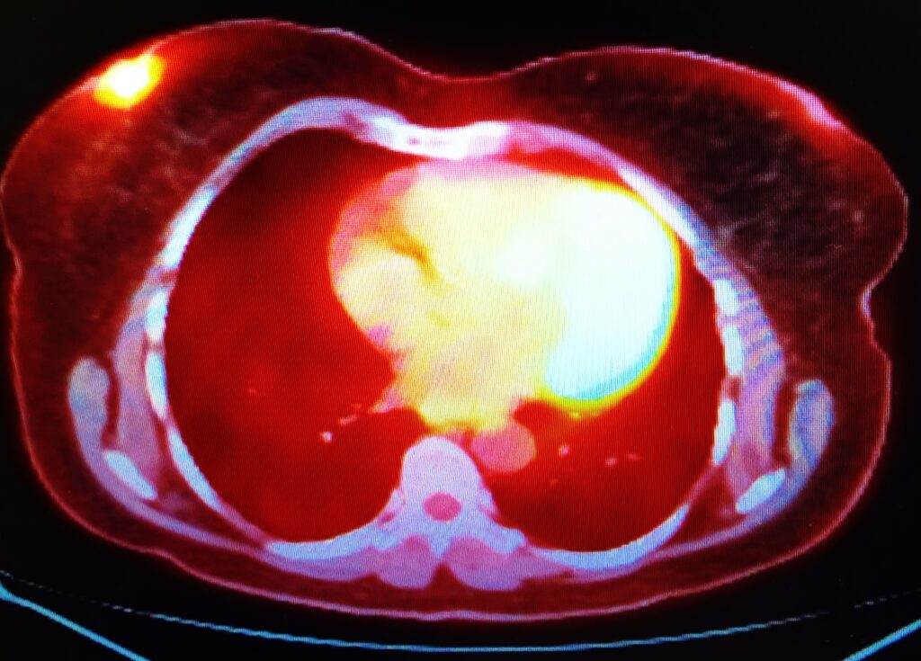
PET has long been a cornerstone in oncology for tumour detection, staging, and treatment monitoring. Recent advancements have further solidified its role in cancer care. Improved tracers and imaging techniques now allow for the detection of smaller tumours and metastases, leading to earlier and more accurate diagnoses. Additionally, PET imaging is increasingly used to assess treatment response, helping oncologists tailor therapies to individual patients and adjust treatment plans based on real-time metabolic changes in tumours.
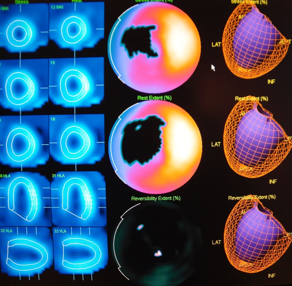
In cardiology, PET is utilised to assess myocardial perfusion and viability, helping to identify areas of the heart muscle that may benefit from revascularisation. Recent developments include the use of new tracers such as 18F-flurpiridaz, which provides high-quality images of myocardial blood flow. PET imaging is also being explored for its potential to detect early stages of atherosclerosis and monitor the efficacy of therapies aimed at reducing cardiovascular risk.
Neurological applications of PET have expanded with the development of tracers targeting specific brain pathologies. For example, PET imaging with amyloid and tau tracers is revolutionising the early diagnosis of Alzheimer’s disease, allowing for timely intervention and better management of the disease. PET is also used in epilepsy to identify epileptogenic foci, aiding in surgical planning. Furthermore, research is ongoing to develop tracers for other neurological conditions, such as multiple sclerosis and traumatic brain injury.
Integration with Artificial Intelligence
Artificial intelligence (AI) is increasingly being integrated into Positron Emission Tomography Imaging to enhance image reconstruction and interpretation. Machine learning algorithms can process vast amounts of data to improve image quality, reduce noise, and correct motion artefacts. AI-based reconstruction techniques have shown promise in producing high-quality images with lower radiation doses, making PET imaging safer and more accessible for patients.
AI algorithms are also being developed to assist in detecting and characterising diseases. These algorithms can analyse PET images to identify patterns and anomalies that may be indicative of specific conditions. For example, AI can help in the early detection of cancer by identifying subtle changes in metabolic activity that human observers may miss. In neurology, AI can aid in the classification of different types of dementia based on PET imaging data.
The integration of AI with PET imaging holds great potential for predictive analytics and personalised medicine. By analysing PET data along with other clinical information, AI can help predict disease progression and response to treatment. This predictive capability can inform personalised treatment plans, allowing for more targeted and effective therapies. In oncology, for instance, AI can help determine which patients are likely to benefit from specific treatments based on their PET imaging profiles.
Regulatory and Safety Considerations
While PET imaging involves the use of radioactive tracers, advancements in technology have helped reduce radiation exposure to patients. The development of high-sensitivity detectors and improved imaging protocols allow for lower doses of radiotracers without compromising image quality. Additionally, digital PET systems and AI-enhanced reconstruction techniques contribute to further dose reduction, making PET imaging safer for patients, particularly in paediatric and longitudinal studies.
Standardisation and quality control are essential for ensuring the accuracy and reliability of PET imaging. Efforts are being made to establish standardised imaging protocols and guidelines for the use of new tracers and technologies. International collaborations and initiatives, such as the European Association of Nuclear Medicine (EANM) guidelines, play a crucial role in promoting best practices and ensuring consistency across different PET imaging centres.
Rapid advancements in PET technology and the development of new radiotracers pose ethical and regulatory challenges. Critical considerations include ensuring patient safety, obtaining informed consent, and addressing privacy concerns related to the use of AI in PET imaging. Regulatory bodies are working to establish frameworks for the safe and ethical use of PET imaging technologies, balancing the need for innovation with the protection of patient rights.
Future Directions and Research
Research is ongoing to develop next-generation PET scanners with even higher sensitivity and resolution. Advances in detector technology, such as the use of novel scintillators and photodetectors, are expected to enhance the performance of PET systems further. Additionally, efforts are being made to design more compact and portable PET scanners, which could expand the use of PET imaging in various clinical settings, including remote and underserved areas.
The discovery and development of novel radiotracers continue to be a focus of research. Scientists are exploring new tracers that can target a broader range of biological processes and disease markers. For example, radiotracers targeting immune cells are being developed to study the immune response in cancer and autoimmune diseases. Additionally, tracers for imaging inflammation and infection are being investigated, which could have significant implications for diagnosing and managing infectious diseases.
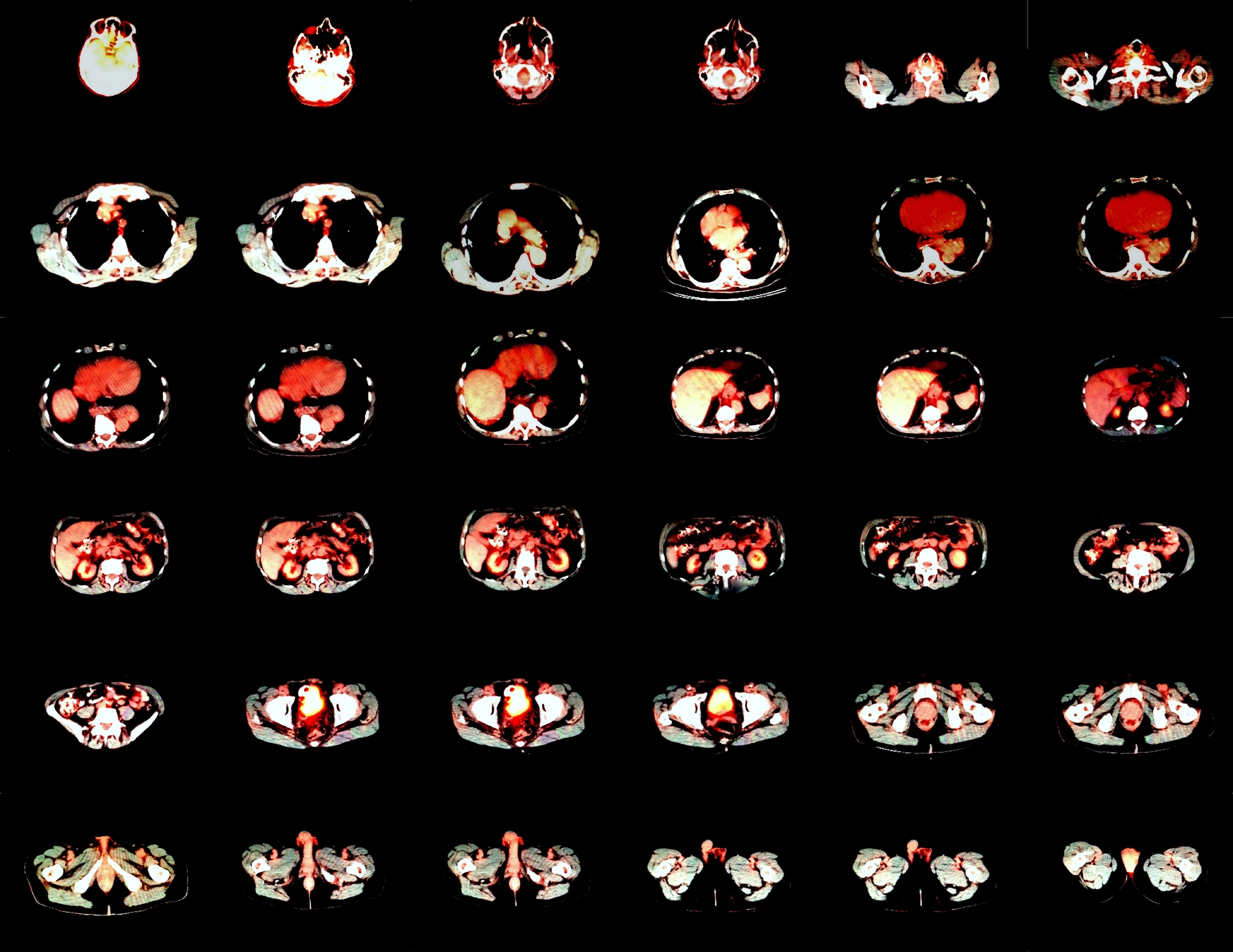
The integration of PET with other imaging modalities, such as MRI, CT, and optical imaging, is an area of active research. Multimodal imaging systems that combine the strengths of different modalities can provide comprehensive information about the structure, function, and metabolism of tissues. This approach has the potential to improve diagnostic accuracy and guide treatment decisions more effectively. For instance, PET/CT combines the metabolic information from PET with the anatomical detail from CT, offering a powerful tool for cancer diagnosis and staging.
PET imaging is poised to play a crucial role in the era of precision medicine. By providing detailed insights into the molecular and metabolic characteristics of diseases, PET can help tailor treatments to individual patients. Research is focused on identifying biomarkers that can predict treatment response and monitoring the effects of therapies in real-time. This approach has the potential to improve patient outcomes and reduce healthcare costs by ensuring that treatments are targeted and effective.
Conclusion
Positron Emission Tomography (PET) has undergone significant advancements in recent years, enhancing its role in medical imaging and expanding its applications across various fields. Improved scanner resolution, novel radiotracers’ development, and AI integration are driving PET technology’s evolution. These advancements improve diagnostic accuracy and pave the way for personalised medicine and innovative therapeutic strategies. As research continues and new technologies emerge, PET is expected to play an increasingly vital role in diagnosing, treating, and managing a wide range of diseases.
Disclaimer
The content presented in this article, “Breakthroughs in Positron Emission Tomography Imaging”, is intended for informational and educational purposes only. While every effort has been made to ensure the accuracy and reliability of the information provided, Open Medscience does not guarantee the completeness, timeliness, or suitability of the content for any specific purpose. This article is not intended to substitute professional medical advice, diagnosis, or treatment. Readers should consult qualified healthcare professionals regarding any medical condition or decisions related to diagnosis and therapy.
Any mention of specific technologies, radiotracers, companies, or products is not intended as an endorsement. Opinions expressed are those of the authors and do not necessarily reflect the views of Open Medscience or its affiliates.
Open Medscience assumes no responsibility or liability for any loss or damage resulting from the use of the information in this article. Use of this content is at the reader’s own discretion and risk.
home » blog » nuclear medicine imaging »