Neurology is not always defined by a single diagnosis; it is an ongoing clinical process. The procedure begins by gathering complex physiological metrics and continues into the formulation of long-term care plans.
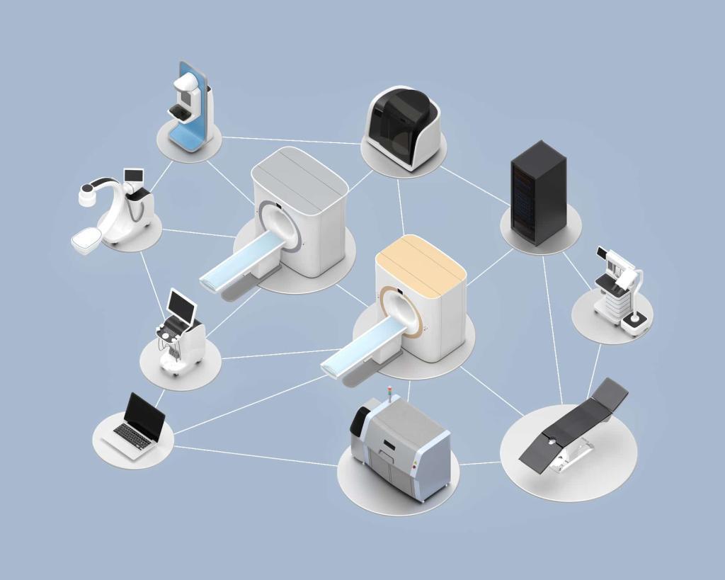
The primary challenge for modern practices is not merely collecting information; it is ensuring the transmission of that information to the necessary point of care. Information frequently remains isolated within silos. Medical imaging may reside in a radiology system while the neurologist attempts to synthesise a comprehensive patient history within the examination room.
To deliver the standard of care required, these clinical disconnections must be resolved. Clinical efficacy requires a seamless transition from diagnostics to results, where radiology and neurology systems operate in synchronisation. This integration enables the transition from analysing brain scans to making clinical decisions.
Phase 1: Radiology Imaging and Diagnostic Data Capture
The process initiates with radiology, where underlying neurological activity is captured through specialised imaging. Contemporary neuro-diagnostics provide a deeper view of the Central Nervous System (CNS). This process extends beyond anatomy to facilitate an understanding of the function and metabolism of the human brain.
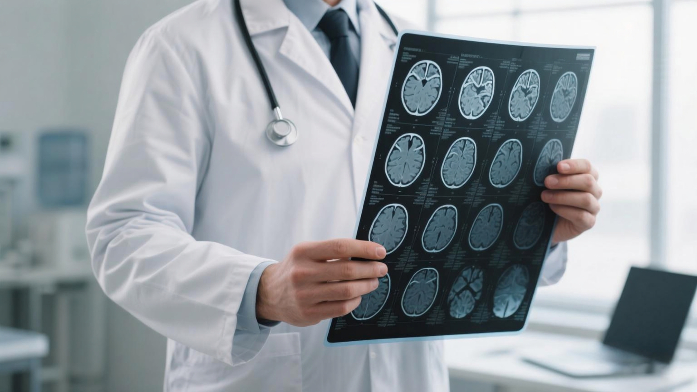
When neurologists establish a comprehensive assessment of a patient’s condition, imaging is a primary component. However, standard scans often require additional diagnostic depth. Functional Magnetic Resonance Imaging (fMRI) addresses this requirement. By detecting time-varying changes in brain metabolism associated with regional blood flow, fMRI allows specialists to map neural activity.
Whether planning a surgical intervention or assessing how a tumour impacts critical brain tissues, this insight supports informed clinical decisions.
Even with MRI, the diagnostic process is not exhaustive. While an MRI identifies structural location, nuclear medicine evaluates functional and metabolic activity.
By using radiopharmaceuticals to study brain metabolism, clinicians can identify subtle changes in the central nervous system before they manifest in standard scans. This facilitates early detection for conditions such as Alzheimer’s or Parkinson’s. At this stage, detailed diagnostic metrics exist within a radiology system and require connection to the broader clinical context.
Phase 2: Data Integration Between Radiology Systems and Neurology EHRs
This is a critical juncture in patient management. A scan is of no utility if the specialist cannot access it during a consultation. The technical infrastructure allows information to move from the capture site (Radiology) to the decision site (Neurology).
In a modern configuration, this does not involve printing reports or utilising facsimile transmissions. The focus remains on interoperability, supported by imaging and diagnostic review tools. When analysing how integrated systems manage this, specific integration technologies are utilised:
- DICOM Imaging Support: Many Neurology EHRs provide direct access to full-resolution DICOM images within the patient record, rather than limiting clinicians to static summaries. This allows specialists to review scan slices while documenting findings.
- e-Labs and Imaging Integrations: Systems utilise integrated diagnostic interfaces to synchronise results. When the radiology lab clears a result, it is electronically received and filed into the patient record. This removes manual entry and ensures the neurologist has the most recent results prior to the patient consultation.
- Seamless Data Exchange: Through established protocols, patient information moves consistently across systems. This ensures that previously captured CNS metrics remain accessible, allowing imaging insights to be incorporated into clinical evaluation and care planning.
Phase 3: Neurology Care Delivery and Long-Term Management
Once the information has been successfully transferred and resides within the neurology EHR software, the focus shifts. The emphasis transitions from initial diagnostic results to executing long-term management of chronic neurological conditions.
Neurology is inherently longitudinal. Conditions such as epilepsy, multiple sclerosis, and stroke recovery require ongoing monitoring and structured follow-up over many years. At this stage, specialised systems facilitate the translation of initial imaging and diagnostic metrics into continuous care planning.
Structured SOAP Assessments: Each clinical encounter is typically documented in a structured and consistent manner. Neurology-specific templates support SOAP (Subjective, Objective, Assessment, Plan) documentation, often pre-aligned with common neurological presentations. This helps ensure that findings from imaging modalities such as fMRI or nuclear medicine are accurately reflected in clinical notes.
Treatment Plan Tracking: Determining medication efficacy requires treatment plan tracking. Tools within neurology EHRs allow providers to monitor disease progression over time and compare historical results with current symptoms. If a scan from a previous period established a baseline, the EHR helps track the intervention’s effectiveness against that baseline.
Patient Engagement via Portals: Patient understanding and access to their own health data are important for long-term care adherence. Secure patient portals integrated within EHRs allow individuals to view imaging reports and laboratory results and manage routine tasks such as prescription refills and appointment scheduling. This supports consistent engagement throughout long-term neurological care.
Multi-Provider Collaboration: Neurological care typically involves a multidisciplinary team that includes neurosurgeons, physical therapists, and primary care physicians. Collaboration tools help ensure that all providers are working from a unified and up-to-date treatment plan. This shared access to synchronised clinical information supports more coordinated decision-making, grounded in consistent diagnostic data.
Aligning Clinical Imaging Data with Neurology EHR Systems
When analysing the full transition from diagnostics to results, the key factor is not just imaging technology or clinical expertise in isolation, but the synchronisation between them. Medical science generates metrics, while clinical software enables those metrics to be utilised effectively in care delivery.
By connecting high-quality medical imaging with day-to-day clinical documentation, we reduce the risk of information being lost between systems. Advanced imaging modalities such as fMRI and nuclear medicine techniques (e.g., PET or SPECT) provide deeper diagnostic insights, which can then be integrated into a neurology EHR through capabilities like DICOM support and treatment plan tracking.
This creates a more continuous and structured flow from diagnosis to long-term care, ensuring that the information captured in radiology meaningfully contributes to accurate, ongoing clinical decision-making.
Disclaimer: This article is for general informational and educational purposes only and does not constitute medical advice, diagnosis, treatment, or professional clinical guidance. The content should not be used as a substitute for advice from a qualified healthcare professional. While Open MedScience aims to provide accurate and current information, no guarantee is made regarding completeness, accuracy, or applicability. Open MedScience accepts no liability for any loss or outcome resulting from reliance on this content.
home » blog » medical technologies »