Recombinant antibodies (rAbs) are artificially produced monoclonal antibodies (mAbs). Unlike mAbs, which are produced using hybridoma-based techniques, rAbs don’t rely on animals. Instead, these antibodies are produced via genetic engineering techniques that use DNA sequence coding for the antibody.
So, recombinant antibodies can be produced from any species, provided there are appropriate oligonucleotide primers or hybridization probes available. This modern approach provides researchers the ability to control, replicate and modify antibodies with high precision.
rAbs are nowadays incredibly valuable for diagnostics, therapeutics and basic research. According to ScienceDirect, these antibodies and their fragments now represent over 30% of all biological proteins undergoing clinical trials for disease diagnosis and therapy.
But how are recombinant antibody sequences obtained? Let’s discuss the same.
The Genetic Structure of Antibodies
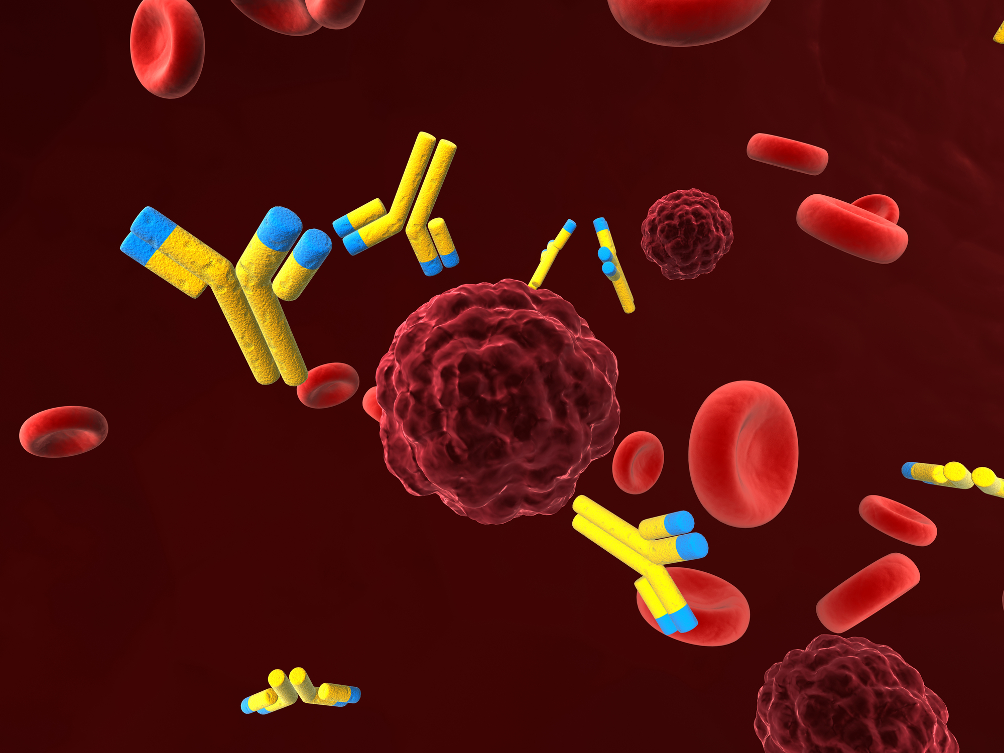
Before we go ahead, it is crucial to understand the genetic structure of antibodies. Antibodies are Y-shaped proteins produced by our body’s immune system. Its primary function is to identify and neutralize foreign substances such as bacteria and viruses.
Each antibody consists of 2 heavy chains and 2 light chains. The tips of the “Y” form are known as the antigen-binding site—this is unique for each antibody. The genes encoding these chains determine the structure and function of the antibody.
When it comes to recombinant antibody production, it is the genes that scientists look for to identify, isolate and reproduce antibodies.
How are Recombinant Antibody Sequences Obtained?
Step 1: Isolating the Antibody-Producing Cells
The first step in obtaining recombinant antibody sequences is to identify and isolate B cells (the immune cells that naturally produce antibodies) from a human or animal that has been exposed to a specific antigen.
This could be an immunized animal or human, spleen or lymph nodes, or bone marrow. From these samples, B cells or plasma cells are selected because they contain the antibody genes of interest.
Step 2: Extracting mRNA from B Cells
Once the B cells are isolated, scientists extract messenger RNA (mRNA) from them. mRNA is the molecule that carries genetic instructions from DNA to the cell’s protein-making machinery. The mRNA specifically encodes the variable regions of the antibody’s heavy and light chains—the parts that bind to antigens.
Note: In some cases, single B-cells sorting using flow cytometry is done to ensure high specificity.
Step 3: Reverse Transcription and Amplifying Antibody Genes via PCR
In the next step, extracted mRNA is reverse-transcribed into complementary DNA (cDNA) using the enzyme reverse transcriptase. After this, the specific segments of DNA are amplified using polymerase chain reaction (PCR).
Researchers use primers that match conserved regions of immunoglobulin genes to amplify:
- VH (Variable region of heavy chain)
- VL (Variable region of light chain)
The amplified DNA contains the genetic blueprint for the antibody’s binding region. This DNA can then be cloned into suitable expression vectors for further study and production.
Step 4: Cloning into Expression Vectors
The amplified VH and VL gene segments are inserted into plasmids or other vectors. They serve as carriers to deliver the genes into host cells. Researchers can clone these genes in two formats:
- ScFV (Single-chain variable fragment): VH and VL domains are linked by a short peptide.
- Fab (Fragment antigen-binding): Includes a constant region along with VH and VL.
These vectors also contain regulatory elements, such as:
- Promoters
- Signal peptides
- Selectable markers
These are introduced into the host cells via transfection to produce the antibody protein.
Step 5: Expression in Host Systems
To introduce the vectors in the host system, various expression systems are used, such as:
- Bacteria (e.g., E. coli)
- Yeast (e.g., Pichia pastoris)
- Mammalian cells (e.g., CHO or HEK293 cells)
The expression system is chosen based on the goal. For instance, for cost-effective and quick results, bacteria are used, whereas to produce complex proteins, like glycosylated proteins.
However, mammalian cells are used if the researchers’ primary goal is to produce therapeutic antibodies. Once the vectors are introduced, the host cells translate the inserted DNA and produce the recombinant antibodies.
Step 6: Antibody Purification and Screening
Once expressed, the antibodies are purified from the culture medium using various techniques:
- Protein A/G affinity chromatography
- Size exclusion chromatography
- Ion exchange chromatography
After purification, the antibodies undergo screening for affinity, specificity, and function. This is usually done using high-throughput screening methods like ELISA, flow cytometry, or surface plasmon resonance (SPR).
Step 7: Sequence Validation and Optimization
After selection, the DNA sequences of the VH and VL regions are fully sequenced to confirm their identity.
Additional sequence optimization may be performed to:
- Improve binding affinity
- Reduce immunogenicity
- Enhance expression yields
Tools such as codon optimization and humanization (replacing animal sequences with human counterparts) are commonly used, especially for therapeutic use.
The Bottom Line
Recombinant antibody sequencing is a powerful technique that has transformed the way scientists develop and produce antibodies. By isolating the genetic code that creates antibodies and inserting it into expression systems like bacteria or mammalian cells, researchers can produce precise, consistent, and scalable antibodies for a wide variety of uses.
Disclaimer
The content provided in this article is intended for informational and educational purposes only. Open Medscience does not provide medical, diagnostic, or therapeutic advice. While we strive to ensure the accuracy and timeliness of the information presented, it is not a substitute for professional scientific or medical consultation.
All information relating to recombinant antibody sequencing reflects general research practices and may not apply to every specific case or application. Readers are encouraged to consult peer-reviewed literature and qualified professionals before making decisions based on the content of this article.
Open Medscience shall not be held responsible for any direct, indirect, or consequential loss or damage arising from the use or interpretation of the information provided.
home » blog » medicine »