This article explores the latest advancements in Magnetic Resonance Imaging (MRI), a noninvasive diagnostic tool pivotal in medical imaging. We explore the cutting-edge techniques that have revolutionised MRI’s capabilities alongside the development of novel contrast agents that enhance image quality and diagnostic accuracy. Through detailed sections, the text illuminates the principles behind MRI, the strides made in improving the technology, the introduction of state-of-the-art contrast agents, and their impact on patient care.
Introduction to Magnetic Resonance Imaging (MRI)
Magnetic Resonance Imaging (MRI) represents a revolutionary advancement in medical diagnostics. It provides an unparalleled view into the human body’s internal structures without ionising radiation, a notable distinction from traditional imaging techniques like X-rays and CT scans. The foundational principle of MRI is rooted in the physics of hydrogen protons, which are abundant in the body due to the high water content of human tissue.
At the heart of MRI technology is the manipulation of these hydrogen protons using a potent magnetic field. Under normal conditions, the protons within the body spin on their axes in a random and disordered manner. However, when placed within an MRI scanner’s strong magnetic field, these protons align parallel to the direction of the field. The degree of uniformity in their alignment is a critical factor in the MRI process, as it determines the strength of the signal that can be obtained.
The next stage involves the application of a specific type of energy known as radiofrequency (RF) current. This energy is carefully calibrated to match the resonance frequency of the hydrogen protons in the magnetic field. When the RF current is applied, it temporarily disrupts the aligned state of the protons, causing them to absorb energy and move into a higher energy state. Upon removal of the RF current, the protons gradually return to their original aligned state, a process known as relaxation. During relaxation, the protons release the absorbed energy in the form of RF signals.
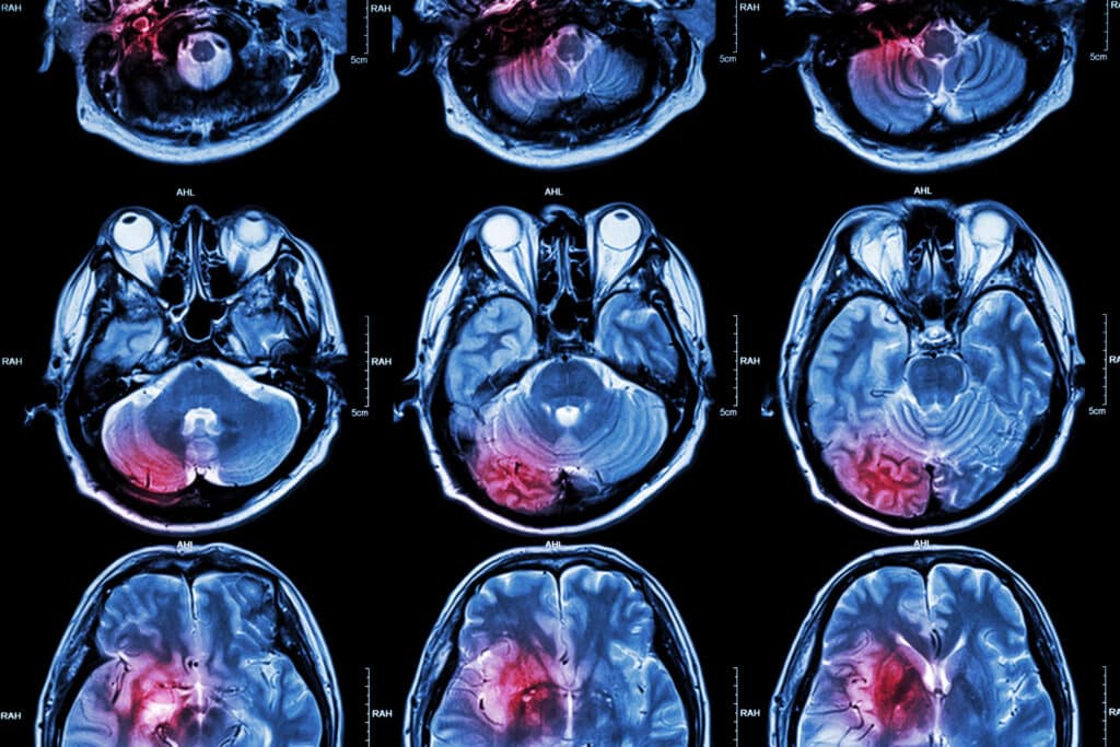
Receivers within the MRI machine then capture these emitted signals. The intensity and characteristics of these signals vary depending on the type of tissue and its environment, allowing for a detailed contrast between different tissues. Advanced computer algorithms process these signals to construct high-resolution images that depict the body’s internal structures in remarkable detail. This capability to differentiate between various types of tissues is one of the most significant advantages of MRI, making it especially useful for imaging the brain, spinal cord, muscles, and joints and detecting tumours and other abnormalities.
MRI is noninvasive in nature, and its avoidance of ionising radiation is a critical benefit. This makes it a safer option for repeated imaging and for populations sensitive to radiation, such as pregnant women and children. Additionally, MRI provides a level of detail that is often unachievable with other imaging methods, offering an invaluable tool for diagnosis, treatment planning, and monitoring of various medical conditions.
Although it demonstrates numerous advantages, MRI is not without limitations. The process can be time-consuming, requiring patients to remain still for extended periods inside the machine, which can be challenging for some individuals. Moreover, using strong magnetic fields means that MRI is unsuitable for patients with certain types of metal implants or devices within their bodies.
MRI is a cornerstone in medical diagnostics, offering a powerful, noninvasive method for visualising the body’s internal structures with exceptional clarity and detail. Its reliance on the unique properties of hydrogen protons and the sophisticated interplay of magnetic fields and radiofrequency energy enables healthcare professionals to diagnose and monitor a wide range of conditions with unprecedented precision. As technology advances, the applications and capabilities of MRI continue to expand, solidifying its role as an indispensable tool in modern medicine.
Technological Advancements in MRI
Recent years have witnessed remarkable technological advancements in Magnetic Resonance Imaging (MRI), revolutionising its application in medical diagnostics. These innovations have been driven by the quest to enhance image clarity, reduce scan times, and improve patient comfort, thereby providing more detailed insights into the human body with greater efficiency and less inconvenience.
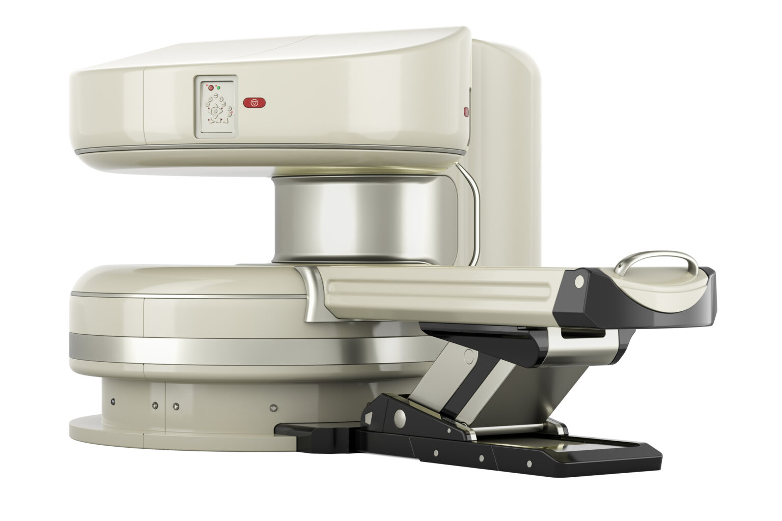
One of the most significant advancements in MRI technology is the development of high-field MRI scanners. These scanners are equipped with stronger magnetic fields, typically measured in teslas (T). While traditional MRI machines operate at 1.5T or 3T, high-field scanners use 7T or higher fields. The stronger magnetic field creates a greater alignment of hydrogen protons in the body, which, in turn, produces a stronger signal and, ultimately, images of higher resolution. This enhancement in image clarity is particularly beneficial for identifying and diagnosing small or subtle pathological changes within the body, offering a clearer picture of patients’ conditions.
Moreover, high-field MRI scanners have led to quicker acquisition times. The increased signal strength allows for faster imaging sequences, reducing the overall time a patient spends in the scanner. This reduction in scan time improves patient comfort by lessening the need for prolonged stillness and increases the throughput of MRI procedures, enabling healthcare facilities to serve more patients.
Parallel imaging techniques represent another leap forward in MRI technology. This method uses multiple radiofrequency (RF) coils to simultaneously receive signals from different body parts. By doing so, parallel imaging can significantly reduce scan times and improve image quality. It works by gathering data from multiple channels at once, allowing for quicker image reconstruction. This technique is particularly useful in dynamic studies, such as those of the heart or joints in motion, as it can capture high-quality images of moving organs with reduced motion artefacts.
The introduction of 3D imaging has further elevated the potential of MRI. Unlike traditional 2D imaging, which captures slices of the body one at a time, 3D imaging acquires volumetric data in a single scan. This comprehensive data collection facilitates the generation of highly detailed images that can be viewed from multiple angles and reconstructed into various planes. The ability to manipulate these 3D images enhances the visualisation of complex anatomical structures, aiding in more accurate diagnoses and better planning for surgical interventions.
Additionally, advancements in software and computing power have dramatically improved the processing and analysis of MRI data. Artificial intelligence (AI) and machine learning algorithms are increasingly being integrated into MRI systems, automating image segmentation and abnormality identification tasks. These technologies streamline the diagnostic process and enhance the precision and reliability of MRI interpretations.
These technological advancements in MRI over recent years have significantly improved the diagnostic capabilities of this imaging modality. High-field MRI scanners, parallel imaging techniques, and 3D imaging, combined with AI and advanced computing integration, have collectively enhanced image quality, reduced scan times, and improved patient comfort. These innovations continue to push the boundaries of medical imaging, offering deeper insights into the human body and facilitating more accurate and efficient diagnoses.
The Role of Contrast Agents in MRI: Enhancing Diagnostic Precision
In the sphere of Magnetic Resonance Imaging (MRI), contrast agents play an instrumental role in amplifying the visibility and distinction of internal organs, blood vessels, and tissues. These agents, often based on gadolinium or iron particles, are administered to patients during an MRI scan to modify the magnetic properties of water molecules in the vicinity. This alteration significantly enhances the contrast between different bodily tissues in the images produced, enabling more precise diagnoses.
Contrast agents are not universally required for all MRI scans. The specific details and objectives of the examination determine their use. For instance, when the aim is to discern fine details within soft tissues, detect tumours, or assess blood vessels, the introduction of a contrast agent can markedly improve the quality and diagnostic utility of the images obtained. This enhanced delineation aids medical professionals in identifying abnormalities, such as cancerous lesions or areas of inflammation, with greater accuracy and confidence.
Gadolinium-based contrast agents (GBCAs) are among the most commonly used in MRI procedures. Gadolinium is a rare earth metal with strong paramagnetic properties, making it ideal for altering the magnetic resonance properties of water molecules. When gadolinium is chelated, or bound, to a molecule that ensures its safety for human use, it can be injected into the body without harm. Upon administration, GBCAs circulate through the bloodstream and interact with surrounding tissues, creating a distinct contrast that highlights differences in tissue composition and vascularity on the MRI images.
Even though the use of GBCAs is not without concerns. Nephrogenic systemic fibrosis (NSF) is a rare but serious condition associated with the use of certain GBCAs in patients with severe renal impairment. This has led to rigorous guidelines regarding the use of GBCAs, particularly in patients with known kidney issues. Moreover, recent studies have raised questions about the long-term deposition of gadolinium in the brain and other tissues. These concerns have spurred the development of new GBCAs with improved safety profiles and the investigation of alternative contrast agents, such as those based on iron oxide, which offer different magnetic properties and may be safer for certain patient groups.
Iron-based contrast agents, while less commonly used than GBCAs, provide an alternative mechanism for enhancing MRI contrast, particularly in the liver and lymph nodes. The body’s macrophages take up these agents and can highlight areas of inflammation or disease in tissues where these immune cells are concentrated.
The development and refinement of MRI contrast agents is an ongoing field of research driven by the pursuit of safer, more effective compounds that can provide clearer, more informative images. Novel agents targeting specific types of cells or molecules are also under investigation. These agents promise a future where MRI can visualise anatomy with high clarity and provide molecular-level information, paving the way for highly personalised diagnostics and treatment planning.
These contrast agents are vital to the MRI toolkit, significantly enhancing the modality’s diagnostic capabilities. Their development reflects a balance between maximising diagnostic utility and minimising risks, illustrating medical imaging technology’s complexities and continual evolution.
Innovative MRI Contrast Agents
The landscape of Magnetic Resonance Imaging (MRI) diagnostics is undergoing a remarkable transformation, thanks to the advent of innovative MRI contrast agents. These novel agents represent a significant leap forward in the precision and safety of MRI scans, providing clearer and more detailed images of the human body. This evolution is particularly crucial in enhancing the diagnostic accuracy for various conditions, including complex pathologies where traditional imaging techniques may fall short.
Contrast agents play a pivotal role in MRI by improving the visibility of internal structures. They work by altering the magnetic properties of nearby water molecules, thereby enhancing the contrast between different tissues. This differentiation is vital for accurately identifying and assessing disease. Among the groundbreaking developments in this area are tissue-specific and pathology-specific agents, which have opened new doors for targeted diagnostics.
Hepatobiliary contrast agents are a prime example of this innovation. Designed specifically for liver imaging, these agents are absorbed by the liver cells and excreted into the bile, providing a clear view of liver anatomy and function. This specificity allows for a more detailed examination of liver lesions, differentiating benign from malignant growths with greater accuracy. Such precision is indispensable in the early detection and treatment planning of liver diseases, including liver cancer, cirrhosis, and hepatitis.
Another significant advancement is the introduction of macrocyclic contrast agents. The structure of these agents provides a more stable chelate, which means that the gadolinium ion (a heavy metal used in many contrast agents for its magnetic properties) is less likely to be released into the body. This structural stability significantly reduces the risk of gadolinium deposition diseases, such as nephrogenic systemic fibrosis (NSF), a rare but serious condition associated with the use of gadolinium-based contrast agents in patients with severe kidney dysfunction. The development of macrocyclic agents represents a major stride towards safer MRI procedures, minimising the potential for adverse effects while maintaining the high-quality imaging necessary for accurate diagnostics.
The exploration and development of innovative contrast agents are driven by the need for more effective and safer diagnostic tools. Researchers are continually seeking agents that can provide enhanced specificity to certain types of tissues or pathological changes. This includes the potential for agents that can cross the blood-brain barrier for improved central nervous system imaging, agents targeted at specific cancer cells for early detection and monitoring of tumours, and agents that can illuminate cardiovascular diseases with unprecedented clarity.
The implications of these innovations extend far beyond improved diagnostic capabilities. They are poised to revolutionise patient care, enabling earlier detection of diseases, more precise treatment planning, and monitoring of treatment efficacy. Moreover, the emphasis on safety through developments like macrocyclic agents addresses a critical concern in medical imaging, ensuring that the benefits of MRI can be accessed by a broader patient population, including those with existing health concerns that may have previously precluded the use of contrast agents.
The development of novel MRI contrast agents marks a significant advancement in medical imaging. By offering greater specificity, enhanced imaging quality, and improved safety profiles, these innovations are setting a new standard in diagnostic precision. As research continues to push the boundaries of what is possible with MRI technology, the future holds the promise of even more targeted and safer contrast agents, heralding a new era in patient care and disease management.
Safety and Efficacy of New Contrast Agents
The expansion of contrast agents in Magnetic Resonance Imaging (MRI) diagnostics has significantly enhanced the capacity to visualise and diagnose a vast array of conditions with unprecedented clarity and detail. However, as the use of these agents becomes more widespread, their safety and efficacy have come under intense scrutiny. Recent advancements in the formulation and application of MRI contrast agents have been particularly focused on addressing concerns related to nephrogenic systemic fibrosis (NSF) and gadolinium retention, which have been associated with older generations of gadolinium-based contrast agents (GBCAs).
Nephrogenic systemic fibrosis is a rare but serious condition that has been linked to the use of certain gadolinium-based contrast agents in patients with severe kidney impairment. NSF can lead to thickening and hardening of the skin, joint immobility, and even life-threatening complications. The recognition of this risk led to stringent guidelines on the use of GBCAs, particularly in patients with renal dysfunction. In response to these concerns, the development of new contrast agents has been oriented towards improving safety profiles, notably through the creation of more stable gadolinium chelates that reduce the risk of free gadolinium release into the body.
Macrocyclic agents represent a significant advancement in this regard. Their chemical structure ensures that the gadolinium ion is tightly enclosed, minimising the risk of gadolinium release and subsequent retention in the body. Studies have shown that these newer agents have a much lower association with NSF, even in patients with compromised kidney function, thereby expanding the applicability of contrast-enhanced MRI scans to a broader patient demographic.
Furthermore, the issue of gadolinium retention has been another focal point of safety considerations. Gadolinium ions from some contrast agents can be deposited in the brain and other tissues, leading to concerns about potential long-term effects. The development of agents with lower retention rates has been a priority, with research demonstrating that macrocyclic and newer linear agents exhibit significantly less gadolinium deposition than earlier formulations. This development is crucial in ensuring that the benefits of enhanced diagnostic imaging do not come with unacceptable risks to patient health.
The efficacy of these new contrast agents also plays a critical role in their safety profile. Agents that provide clearer, more distinct imaging results can reduce the need for repeat scans and the associated exposure to gadolinium, further mitigating risk. Additionally, the advent of tissue-specific contrast agents allows for more targeted imaging, enhancing the diagnostic process’s efficacy and efficiency.
Regulatory bodies and the medical community have strongly emphasised the rigorous evaluation of the safety and efficacy of new contrast agents. This involves extensive clinical trials and post-marketing surveillance to monitor for adverse effects and ensure that these agents meet the highest standards of patient safety. The ongoing assessment of contrast agents is dynamic, adapting to new evidence and technological advancements to safeguard patient health.
The safety and efficacy of new MRI contrast agents are paramount to their development and application. The move towards agents with improved safety profiles, including lower toxicity and reduced retention rates, addresses the critical concerns associated with their use. These advancements not only enhance the diagnostic capabilities of MRI but also ensure that this vital imaging tool can be used safely across a wider spectrum of the patient population, underscoring the commitment to patient care and safety in the field of radiology.
As the array of contrast agents expands, their safety and efficacy become paramount. Recent studies have underscored the importance of using highly safe agents, especially concerning nephrogenic systemic fibrosis (NSF) and gadolinium retention. The latest generation of contrast agents demonstrates lower toxicity and reduced retention rates, effectively addressing previous safety concerns.
Future Directions in MRI Technology and Contrast Agents
As we gaze into the future of Magnetic Resonance Imaging (MRI), it’s clear that the promise of groundbreaking innovations and transformative advancements illuminates the horizon. The relentless pursuit of efficiency, precision, and safety is steering the development of MRI technology and contrast agents towards a new era of medical diagnostics that envisions a more personalised and patient-centred approach to healthcare.
One of the most exciting frontiers in MRI research is the development of highly efficient and targeted contrast agents. Scientists are exploring the potential of biodegradable contrast agents, which can be broken down and eliminated by the body naturally, thereby reducing the risk of long-term retention and associated complications. This innovation would mark a significant step forward in enhancing the safety profile of contrast agents, making MRI scans safer for a broader range of patients, including those with renal impairments or allergies to traditional contrast materials.
Furthermore, the advent of molecular imaging capabilities in MRI contrast agents opens up new vistas for diagnostics. These agents are designed to target specific cellular or molecular markers, providing insights into the anatomy and the biochemical and physiological processes occurring within the body. This level of detail could revolutionise the early detection and monitoring of diseases, allowing for more precise and effective treatment plans.
Technological advancements are not limited to contrast agents alone. The field of MRI is also witnessing the integration of cutting-edge technologies such as Artificial Intelligence (AI) and machine learning algorithms. These tools promise to significantly enhance the speed and accuracy of image analysis and interpretation, reducing scan times and improving the overall patient experience. AI algorithms can learn to recognise patterns and anomalies in imaging data, assisting radiologists in diagnosing conditions more quickly and with greater confidence. Furthermore, these technologies have the potential to automate and optimise various aspects of the MRI process, from patient positioning to scan parameter adjustments, ensuring optimal image quality with minimal human intervention.
The exploration of hyperpolarised agents presents another exciting avenue for MRI technology. These agents can increase the signal strength of certain molecules in the body, providing a much clearer and detailed view of metabolic processes. This could be particularly beneficial in the field of cancer diagnostics and treatment monitoring, offering new insights into tumour metabolism and response to therapy.
In summary, the future of Magnetic Resonance Imaging (MRI) is productive, with the promise of innovation and advancement. The development of advanced contrast agents and the integration of AI and machine learning into MRI technology are set to revolutionise the field, enhancing imaging quality and efficiency while ensuring greater patient safety and comfort. These advancements promise a future where diagnostics are more accurate and tailored to individual needs and highlight the ongoing commitment to improving patient outcomes through precision in diagnosis and treatment planning. As MRI technology continues to evolve, its role in modern medicine becomes ever more invaluable, reaffirming its position at the forefront of diagnostic imaging.
Disclaimer
The information presented in this article is intended for general educational and informational purposes only. It is not intended to substitute professional medical advice, diagnosis, or treatment. Readers should consult qualified healthcare professionals for any medical concerns or decisions related to MRI procedures or the use of contrast agents.
While Open Medscience strives to ensure the accuracy and reliability of the information provided, the content reflects the knowledge available at the time of publication and may not include the most recent developments in MRI technology or contrast agent research. Open Medscience and its contributors do not accept any liability for any loss, injury, or damage incurred as a result of the use or reliance on the information provided herein.
The article may mention products, technologies, or procedures that are still under research or may not be available or approved in all regions. Inclusion of such information does not constitute endorsement or recommendation.