Summary: This article explores how medical image quality is influenced by equipment, imaging methods, and variables chosen by the operator. It examines factors like contrast, blur, noise, artefacts, and distortion, while also discussing strategies to meet diagnostic imaging standards, achieve radiological clarity, utilise optimised imaging parameters, apply image contrast enhancement, focus on minimising imaging artefacts, and emphasise reducing image distortion.
Keywords: diagnostic imaging standards; radiological clarity; optimised imaging parameters; image contrast enhancement; minimising imaging artefacts; reducing image distortion.
Understanding Medical Image Quality
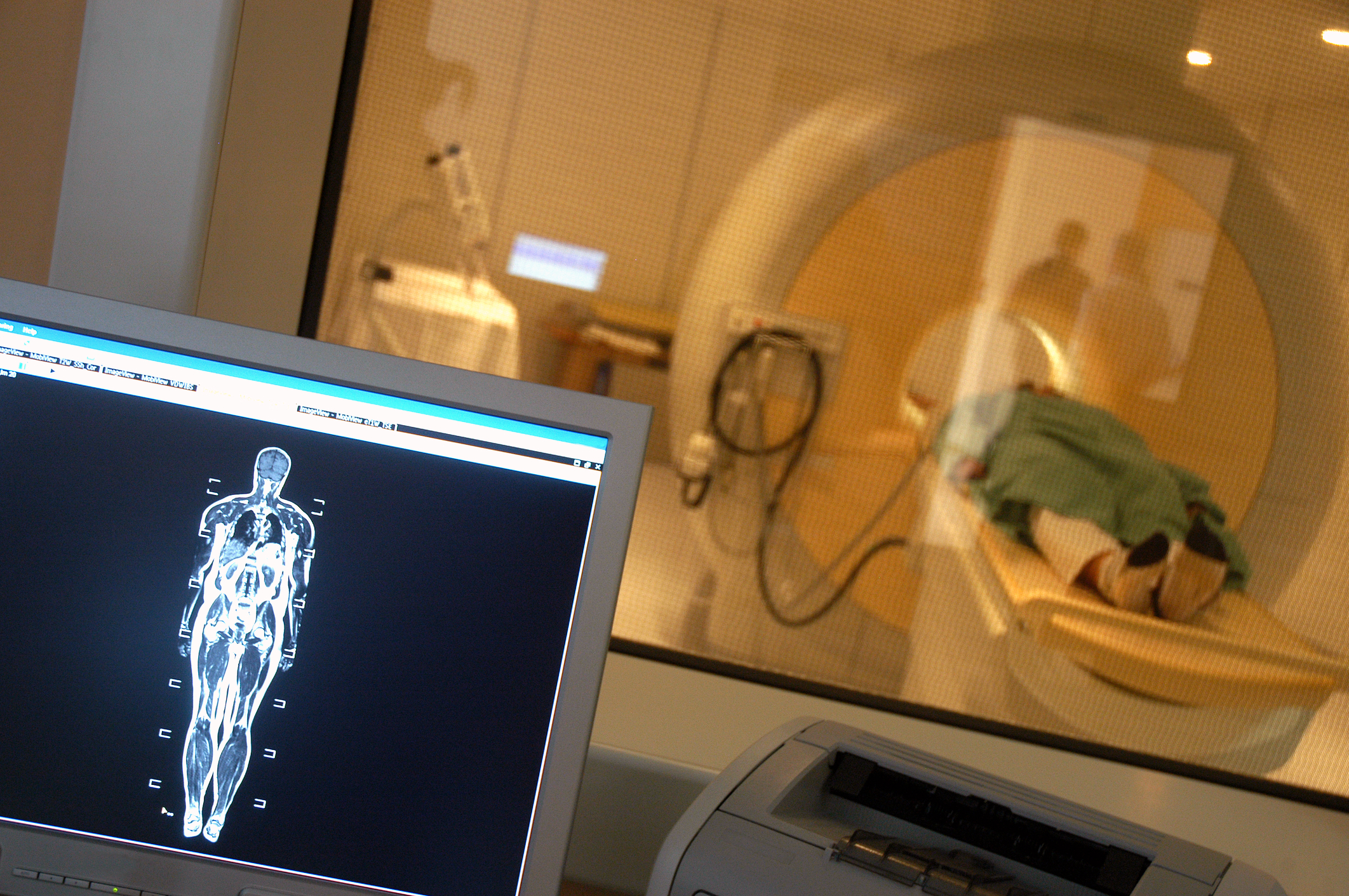
In the field of healthcare, the role of medical image quality cannot be overstated. High-quality medical imaging underpins accurate diagnosis, effective treatment, and ongoing patient monitoring. From routine X-rays to advanced MRI scans and CT imaging, the subtle differences in image quality can mean the difference between early detection and delayed intervention. By adhering to diagnostic imaging standards, healthcare professionals can ensure that the images they produce meet the highest clinical benchmarks. To achieve this, a careful balance of equipment selection, imaging methodology, and the manipulation of optimised imaging parameters is required.
At its core, producing excellent medical images involves understanding how factors like image contrast enhancement, radiological clarity, and the control of imaging variables affect overall picture fidelity. There are key considerations such as minimising imaging artefacts and reducing image distortion that can dramatically improve diagnostic confidence. As we shall see, every aspect of the imaging process—from machine calibration to operator technique—contributes to the resultant medical image quality.
In clinical practice, medical image quality refers to the degree to which an image accurately represents the anatomy or pathology of interest. It is not merely a subjective notion; rather, it is defined by measurable parameters that determine whether the image is fit for diagnostic purposes. By achieving consistent radiological clarity, professionals ensure that every subtle structure is visible. Additionally, adhering to diagnostic imaging standards is essential, as these guidelines help maintain consistency and ensure images are reliably interpreted across different facilities and practitioners.
Core Principles of Image Formation
The foundation of medical image quality lies in understanding how the image is formed. Equipment selection, such as the choice of X-ray tubes, MRI magnets, or ultrasound transducers, plays a pivotal role. However, an operator’s skill in applying optimised imaging parameters—like exposure time, voltage, and current—can significantly enhance the final result. High-quality images are those that deliver the best balance between contrast, spatial resolution, signal-to-noise ratio, and geometric accuracy. Through careful calibration and methodical testing, operators work towards maintaining consistent diagnostic imaging standards that guide every imaging decision.
The Role of Equipment and Technology
Modern medical imaging depends on cutting-edge technology, such as advanced MRI scanners, high-resolution CT machines, and specialised detectors that enable image contrast enhancement and improved radiological clarity. Such equipment must be regularly maintained and updated to ensure optimal performance. By continually refining imaging protocols and employing optimised imaging parameters, healthcare facilities can enhance medical image quality. Technological advancements also play a crucial part in minimising imaging artefacts and reducing image distortion—critical steps in producing reliable and reproducible diagnostic images.
Key Factors Affecting Medical Image Quality
Several key factors influence medical image quality, including contrast, blur, noise, artefacts, and distortion. Each of these components must be carefully managed to meet diagnostic imaging standards and achieve exemplary radiological clarity.
Contrast and Image Contrast Enhancement
Contrast is the difference in brightness between various areas of an image. Adequate contrast is vital for identifying subtle abnormalities, especially when looking for small lesions or early-stage pathologies. Through various techniques, including the use of contrast agents in modalities like MRI and CT, operators can achieve effective image contrast enhancement. By adjusting parameters such as beam intensity, energy levels, and the type of contrast media, radiologists can obtain images that highlight specific tissues, ensuring that medical image quality remains uncompromised. Through repeated calibration and analysis, the right degree of contrast can be established, adhering to diagnostic imaging standards and promoting radiological clarity.
Blur and Radiological Clarity
Blur occurs when the boundaries between structures become indistinct, making it harder to pinpoint specific details. Achieving optimal radiological clarity involves minimising motion blur through patient cooperation, shorter exposure times, and advanced stabilisation techniques. Equipment capabilities and optimised imaging parameters also influence blur; for instance, fine-tuning focal spot size or sampling intervals can sharpen the final image. By focusing on radiological clarity, healthcare professionals ensure that the images reveal accurate anatomical details, thus enhancing medical image quality.
Noise and Minimising Imaging Artefacts
Noise refers to random variations in pixel intensity, often resulting from low signal strength or technical limitations. Excessive noise can obscure important details and detract from medical image quality. To address this, many facilities incorporate noise reduction algorithms, improved detector materials, or better shielding techniques. Hand in hand with noise control is the necessity of minimising imaging artefacts. Artefacts can arise from patient movement, foreign objects, or operator error. Systematic approaches—such as ensuring consistent patient positioning, using proper scanning protocols, and selecting optimised imaging parameters—help control these issues. Through careful management of noise and minimising imaging artefacts, professionals maintain compliance with diagnostic imaging standards and achieve the desired radiological clarity.
Distortion and Reducing Image Distortion
Distortion refers to geometric inaccuracies that appear when an image does not represent the true shape, size, or position of anatomical structures. By focusing on reducing image distortion, radiologists and technologists can ensure that measurements derived from images are reliable. Adjusting scanner calibration, improving magnet homogeneity in MRI, or refining beam geometry in CT scans can all reduce distortion. Accurate and consistent positioning of the patient is also pivotal. Maintaining medical image quality requires ongoing vigilance and adaptation, as reducing image distortion directly impacts the utility of the image in clinical decision-making.
Achieving Diagnostic Imaging Standards
One of the overarching goals of all imaging departments is to meet and surpass diagnostic imaging standards. These standards are set by professional bodies and regulatory agencies to ensure that every image produced can confidently guide patient care. Emphasising medical image quality means consistently achieving the benchmarks that define excellence in clinical imaging. Incorporating optimised imaging parameters, maintaining radiological clarity, and ensuring image contrast enhancement are key steps towards meeting these standards.
Selecting Optimised Imaging Parameters
The selection of optimised imaging parameters is integral to achieving both diagnostic imaging standards and radiological clarity. Parameters such as exposure time, tube current, voltage, and detector settings must be meticulously chosen. By adjusting these parameters, operators improve the balance between contrast and noise, ensure adequate spatial resolution, and prevent excessive patient radiation dose. Through this careful calibration, the imaging team ensures that each scan meets the criteria of medical image quality, while also integrating minimising imaging artefacts and reducing image distortion into the standard operating procedures.
Strategies for Consistent and Reliable Results
Maintaining medical image quality over time involves not just meeting diagnostic imaging standards but surpassing them. Ensuring consistent, high-quality results requires a culture of continuous improvement and ongoing training. By frequently reviewing protocols, refining optimised imaging parameters, and adopting novel techniques for image contrast enhancement, the imaging team demonstrates their commitment to excellence.
Training Operators and Technologists
Human expertise lies at the heart of producing exceptional medical image quality. Skilled technologists and radiographers must know how to harness the capabilities of their equipment fully. This means understanding how to achieve radiological clarity, how to apply appropriate exposure settings, and how to implement optimised imaging parameters for each patient scenario. Regular training sessions, workshops, and credentialing programmes help staff remain abreast of current best practices. Additionally, promoting knowledge about minimising imaging artefacts and reducing image distortion ensures that each member of the team contributes to consistent, top-tier results.
Continuous Quality Improvement
Establishing a quality assurance framework ensures that high standards are not just attained but maintained. Routine equipment checks, audits against diagnostic imaging standards, and the use of test phantoms verify that equipment performance remains stable. These routines help detect issues early, allowing for timely interventions that preserve medical image quality. Periodic reviews of imaging protocols and the adaptation of optimised imaging parameters keep pace with technological advances and emerging clinical requirements. Moreover, continuous quality improvement initiatives focus on enhancing radiological clarity, facilitating image contrast enhancement, and consistently minimising imaging artefacts and reducing image distortion.
The Importance of Patient-Centred Imaging
Central to the concept of medical image quality is patient welfare. Good image quality leads to accurate diagnoses, less invasive treatment plans, and potentially better outcomes. By upholding diagnostic imaging standards, medical teams ensure that each patient’s imaging experience leads to clinically valuable information. Good practices, such as minimising imaging artefacts caused by patient movement, can also help reduce the number of repeat scans, lowering radiation exposure and cost.
High-quality images also facilitate better communication among multidisciplinary teams. When images adhere to diagnostic imaging standards and exhibit excellent radiological clarity, surgeons, oncologists, and other specialists can make informed decisions more rapidly. In this context, achieving medical image quality is not merely a technical goal but a patient-centred endeavour that supports safer, more effective healthcare delivery.
Advancements in Imaging Technology
Modern imaging systems increasingly incorporate artificial intelligence (AI) and machine learning algorithms to improve medical image quality. These technologies can automatically identify and correct common issues, thereby minimising imaging artefacts and reducing image distortion. AI-driven tools can suggest optimised imaging parameters based on patient-specific characteristics, ensuring consistent adherence to diagnostic imaging standards.
Meanwhile, the development of better hardware, detectors, and image reconstruction techniques continues to boost radiological clarity. Enhanced MRI coils, advanced CT detector materials, and state-of-the-art image processing software all contribute to better image contrast enhancement. These innovations allow for more accurate diagnoses, more targeted treatments, and overall improved patient care.
Practical Tips for Operators
For operators and technologists, achieving and maintaining medical image quality is a daily pursuit. Below are some practical tips:
- Know Your Equipment: Understanding your imaging system’s capabilities and limitations is fundamental. Familiarity with control settings and routine maintenance ensures consistent radiological clarity and adherence to diagnostic imaging standards.
- Select Appropriate Parameters: Adjust exposure settings, voltage, and detector gains to ensure optimised imaging parameters. Experimenting with small changes can lead to significant improvements in medical image quality.
- Use Contrast Agents Wisely: When seeking image contrast enhancement, follow established protocols and patient safety guidelines. Proper use of contrast can greatly improve the visibility of anatomical structures.
- Train Continuously: Stay updated on best practices. Regular training on new imaging techniques, patient positioning, and methods for minimising imaging artefacts and reducing image distortion helps maintain top-quality outputs.
- Collaborate with Colleagues: Discussing difficult cases with peers and consulting radiologists about image quality issues helps identify areas for improvement. Collaboration fosters an environment where diagnostic imaging standards are consistently met.
Overcoming Common Challenges
Challenges in maintaining medical image quality arise from both technological and human factors. Problems with outdated equipment, inadequate maintenance, or non-adherence to optimised imaging parameters can lead to compromised image quality. Similarly, patient cooperation issues—such as difficulty remaining still—can introduce motion artefacts.
Addressing these challenges involves a combination of preventive maintenance, regular equipment upgrades, comprehensive training, and patient communication. Encouraging patient comfort and providing clear instructions can make a difference in minimising imaging artefacts. Similarly, using specialised coils, upgrading software, and applying corrective algorithms in MRI or CT scanning can support reducing image distortion. Through diligence and ongoing improvement efforts, these hurdles can be effectively overcome, preserving both radiological clarity and compliance with diagnostic imaging standards.
The Future of Medical Imaging
Looking ahead, medical imaging technology is poised to become even more patient-centred, efficient, and accurate. As AI-driven protocols become more commonplace, systems will automatically fine-tune optimised imaging parameters, enhancing medical image quality without increasing operator workload. There will be an even greater focus on advanced techniques for image contrast enhancement, enabling doctors to detect minute abnormalities earlier than ever before.
Research into novel detector materials, new imaging modalities, and advanced image reconstruction algorithms continues to push the boundaries of what can be achieved. With these improvements, healthcare professionals can expect even greater radiological clarity, more robust strategies for minimising imaging artefacts, and innovative solutions for reducing image distortion. As these technologies mature, adherence to diagnostic imaging standards will remain a cornerstone, ensuring that the benefits translate into tangible clinical outcomes.
Conclusion
The pursuit of high-quality medical images is at the heart of modern healthcare, guiding critical diagnostic decisions and shaping patient journeys. Attaining superior medical image quality relies on multiple interwoven factors: the meticulous selection of equipment, the application of optimised imaging parameters, rigorous adherence to diagnostic imaging standards, and continuous efforts towards image contrast enhancement, radiological clarity, minimising imaging artefacts, and reducing image distortion.
From the fundamental principles of image formation to the practical tips that operators can employ, every aspect of the imaging process matters. As technology advances, these improvements will continue to refine imaging capabilities, enabling earlier disease detection, personalised treatment planning, and better patient outcomes. The future of medical imaging is one of precision, innovation, and, most importantly, high-quality images that serve the needs of patients and clinicians alike.
Q & A – Medical Image Quality
Q: How can I ensure that my images meet diagnostic imaging standards?
A: Consistent training, routine equipment maintenance, and careful selection of optimised imaging parameters help maintain medical image quality, ensuring that your images meet established standards and offer dependable radiological clarity.
Q: What practical steps can I take to achieve image contrast enhancement?
A: To achieve image contrast enhancement, select the right contrast agent for the modality, follow established scanning protocols, and carefully adjust imaging parameters. These steps will improve medical image quality and highlight subtle pathologies.
Q: How do I focus on minimising imaging artefacts in everyday practice?
A: Minimising imaging artefacts involves careful patient positioning, ensuring patients remain still, and performing regular equipment checks. Adhering to established diagnostic imaging standards and using optimised imaging parameters ensures more consistent, artefact-free images.
Q: What strategies can help in reducing image distortion effectively?
A: Reducing image distortion includes calibrating equipment regularly, maintaining magnet homogeneity in MRI, and utilising corrective algorithms. By doing so, you enhance radiological clarity, support accurate diagnoses, and maintain medical image quality.
Q: How important are optimised imaging parameters in meeting patient expectations?
A: Optimised imaging parameters reduce the need for repeat scans and help produce images that accurately represent the patient’s anatomy. This leads to quicker, more confident diagnoses, ultimately improving patient experience and outcomes.
Disclaimer
The content of this article, Mastering Medical Imaging Quality: Tools, Techniques, and Outcomes, is intended for informational and educational purposes only. It does not constitute professional medical advice, diagnosis, or treatment. While efforts have been made to ensure the accuracy and relevance of the information provided, Open MedScience makes no guarantees regarding its completeness or suitability for clinical decision-making. Medical professionals should consult appropriate clinical guidelines and exercise their own professional judgement in practice. Open MedScience is not liable for any outcomes resulting from the application of information contained in this article.


