
Transforming Health: How Medical Imaging is Shaping the Future of Wellbeing
 By
By
Mental health imaging technologies enable precise diagnostics, enhance treatment planning, and improve understanding of neurological conditions.
Breast cancer remains one of the most commonly diagnosed cancers in women across the globe. Therefore, early detection is crucial in improving treatment outcomes and enhancing the patient’s quality of life. In recent years, Positron Emission Mammography (PEM) has emerged as an innovative diagnostic tool for breast cancer, providing more accurate and precise results than traditional imaging techniques.
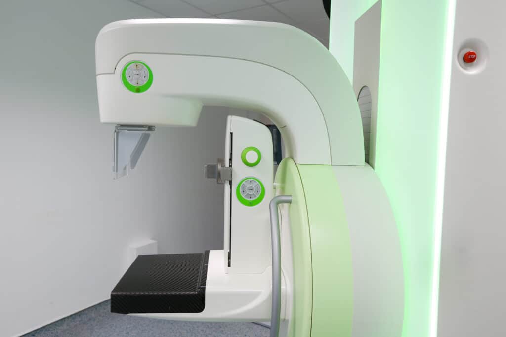
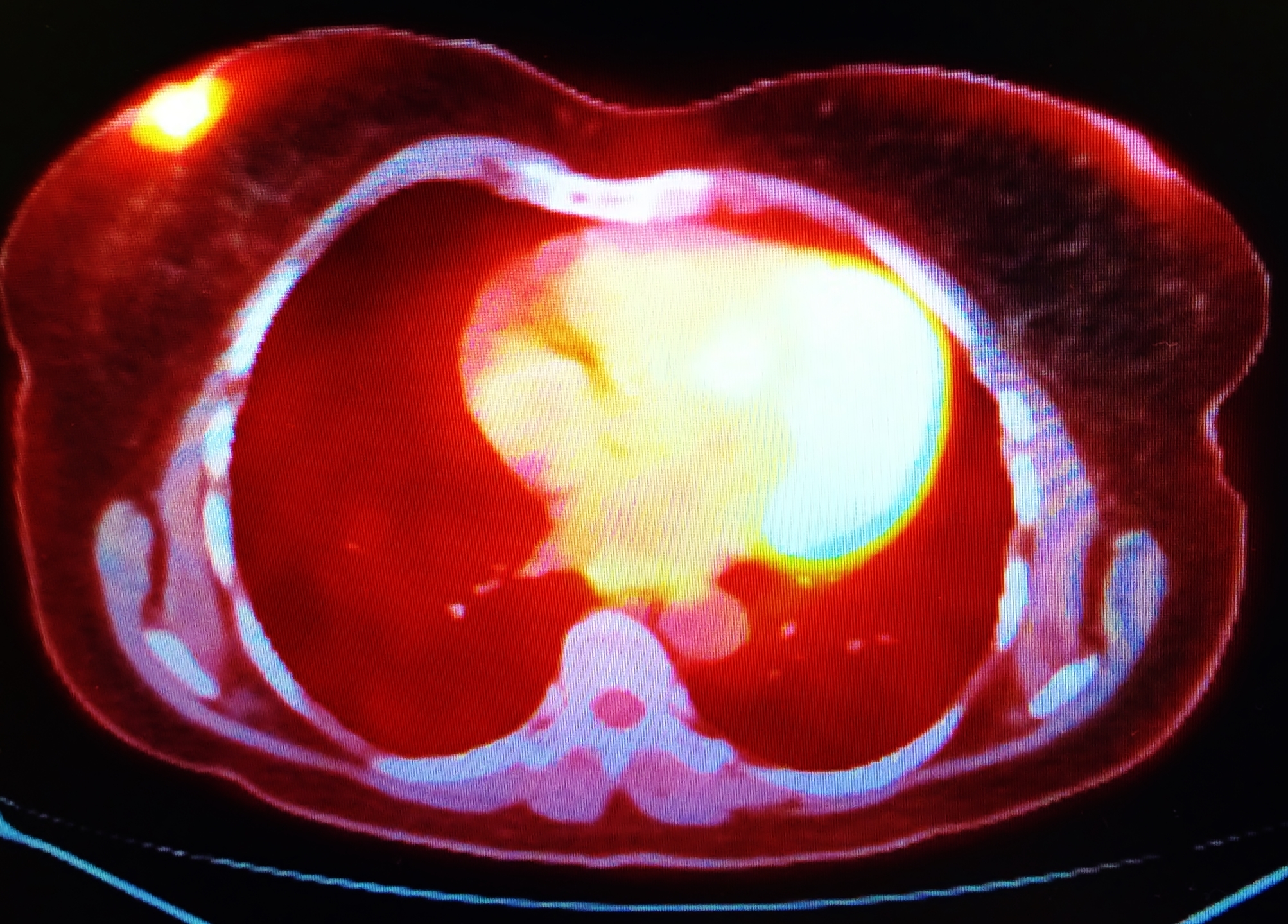
Positron Emission Mammography (PEM) is a nuclear medicine imaging technique that uses a small amount of radioactive material and a specialised camera to produce detailed, high-resolution images of breast tissue. By visualising the metabolic activity of cells, PEM can help doctors identify malignant tumours and differentiate them from benign growths.
PEM involves injecting a small amount of radioactive material, usually a radiolabeled glucose analogue called fluorodeoxyglucose (FDG), into the patient’s bloodstream. This radioactive tracer accumulates in areas with high metabolic activity, such as cancer cells, which consume more glucose than normal cells. The PEM camera then detects the gamma rays emitted by the tracer and produces a three-dimensional image of the breast tissue, highlighting areas with high tracer uptake.
Advantages of PEM
Despite its advantages, PEM is not without limitations. It is not recommended as a primary screening tool for the general population due to its relatively high cost and limited availability. Additionally, PEM may not detect very small or slow-growing tumours, which may not take up enough FDG for detection.
home »
By
Mental health imaging technologies enable precise diagnostics, enhance treatment planning, and improve understanding of neurological conditions.
 By
By
Mammography, using low-dose X-rays, is essential for detecting early-stage breast cancer, improving treatment outcomes, and reducing mortality rates.
 By
By
The diagnostic breast imaging tool Positron Emission Mammography uses short-lived positron isotopes to detect breast cancer.