Mammography
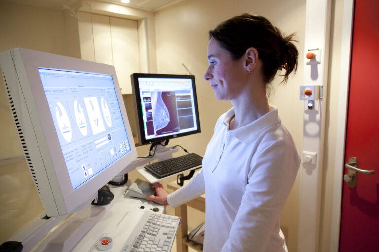
Mammography (Mastography) is a specialised medical imaging that uses a low-dose x-ray (30 kVp) system to see inside the breast. A mammography examination, called a mammogram, aims to detect the early stages of breast cancer by detecting characteristic masses or microcalcifications. An X-ray (radiograph) is a non-invasive procedure that helps diagnose and treat medical conditions.
Therefore, imaging with X-rays enables a picture of the human body by using a dose of ionising radiation. Wilhelm Conrad Roentgen discovered X-rays in 1895 by working with a cathode-ray tube: he observed a fluorescent glow of crystals near the tube. Since then, X-rays have become central to medical imaging, especially with advances in mammography, including breast tomosynthesis, digital mammography and computer-aided detection. Other imaging modalities which assist mastography include ultrasound, ductography, positron emission mammography (PEM) and magnetic resonance imaging (MRI).
In addition to this ductography is used to evaluate patients exhibiting areola and nipple problems. However, over four decades, mammography has historically been the gold standard for breast cancer screening tools. Alternative screening tools are becoming more prevalent, especially digital mammography, which allows images of the breasts to be depicted in a digital format compared to traditional mammography on film.
Thermography is a non-invasive procedure which does not emit radiation and is based on infrared technology to detect inflammation in breast tissue. Also, thermography can be performed during pregnancy as dense breast tissue is a limitation of mastography. Another non-radioactive technique used to assist breast screening programmes for pregnant women is ultrasound. This non-invasive diagnostic tool can detect breast cancer and microcalcifications at rates comparable to mammography.
Furthermore, the cumulative effect of routine mammography screening may increase the risk of radiation-induced breast cancer. In addition, women carriers of the BRCA gene may be at higher risk from the effects of radiation. Studies have shown the effect of diagnostic chest X-rays on breast cancer and found that medical radiation exposure increases the risk of developing breast cancer.
You are here:
home » mammography


