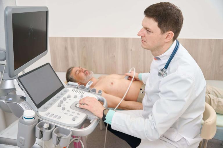
Automated Breast Ultrasonography
Automated Breast Ultrasonography (ABUS) is an innovative imaging procedure that has emerged as a valuable tool in breast cancer detection and evaluation. Utilizing ultrasound technology, ABUS automatically scans the breast, creating a 3-dimensional image of the breast tissue. This comprehensive view enables the reformatting of images in axial, sagittal, and coronal planes. The generated coronal plane, in particular, has been shown to enhance diagnostic accuracy, offering a significant advantage over traditional 2-dimensional ultrasound imaging.
Breast cancer is the most widespread cancer among women worldwide, and early detection is critical in improving patient outcomes. However, dense breast tissue, characterized by a high proportion of glandular and connective tissue compared to fat, can pose a significant challenge to traditional imaging techniques such as mammography. This is because dense tissue appears white on mammograms, and distinguishing between healthy tissue and potential tumours is difficult. ABUS, however, offers a valuable alternative for detecting breast disease, especially in women with dense breasts.
The high-resolution 3-dimensional images produced by ABUS offer a more detailed view of the breast tissue, enabling radiologists to better differentiate between benign and malignant masses. The coronal plane, in particular, allows for the visualization of the entire breast in a single view, leading to improved detection of lesions that may be obscured in other imaging planes.
In addition to its utility in early breast cancer detection, ABUS plays a critical role in evaluating breast cancer staging. Accurate staging is essential for determining each patient’s most appropriate treatment plan. By providing a comprehensive view of the breast tissue, ABUS can help determine the extent of the disease and identify the presence of any additional tumours that may not have been detected using other imaging modalities.
Moreover, ABUS has proven valuable in evaluating tumour response to neoadjuvant chemotherapy. This type of chemotherapy is administered before surgery to shrink tumours and increase the likelihood of successful surgical removal. By monitoring changes in tumour size and morphology through ABUS, healthcare professionals can assess the effectiveness of neoadjuvant chemotherapy and make informed decisions about subsequent treatment strategies.
home » automated breast ultrasonography

 By
By