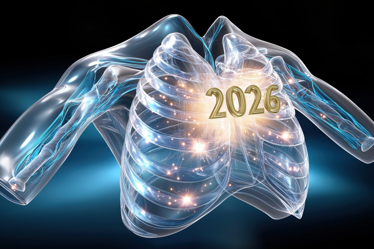
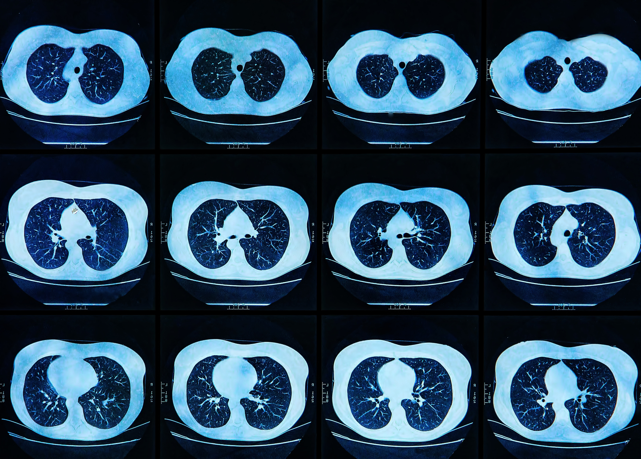
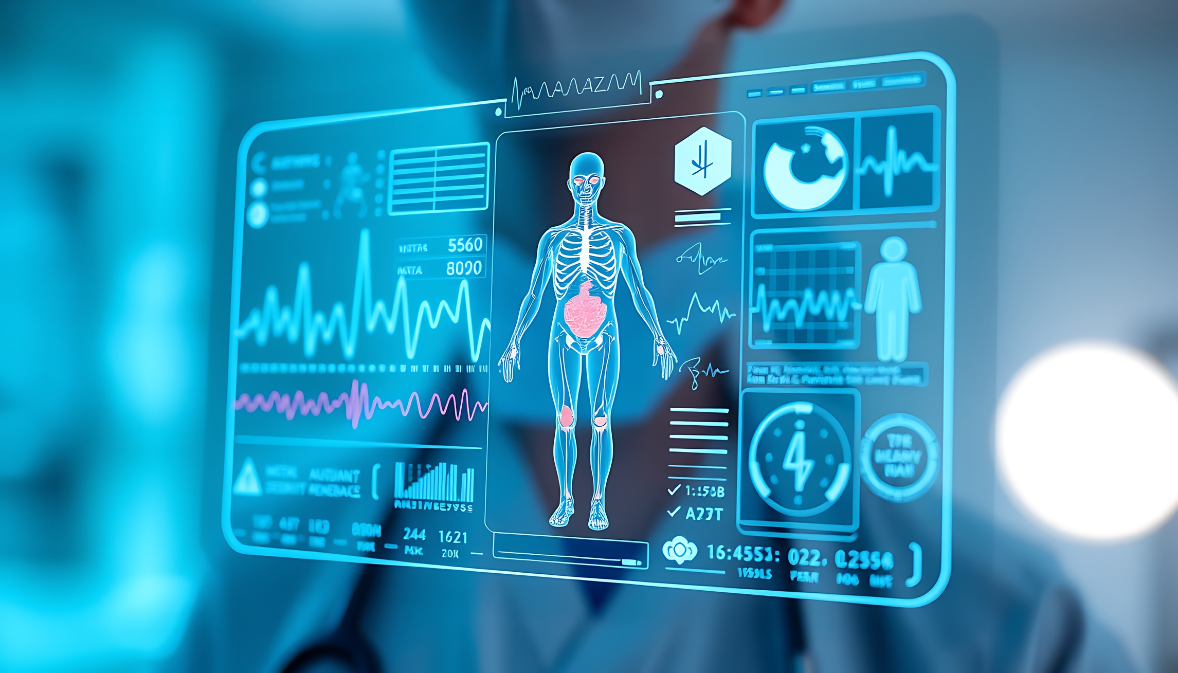
Medical Imaging in 2026: Smarter Scanners, Portable Diagnostics, and a Shift Towards Predictive Care
 By
By
Discover how medical imaging in 2026 is evolving through innovation, accessibility, and AI-driven decision support for healthcare.
By
Discover how medical imaging in 2026 is evolving through innovation, accessibility, and AI-driven decision support for healthcare.
 By
By
Discover how the integration into clinical practice is reshaping medtech with AI advancements and new manufacturing techniques.
 By
By
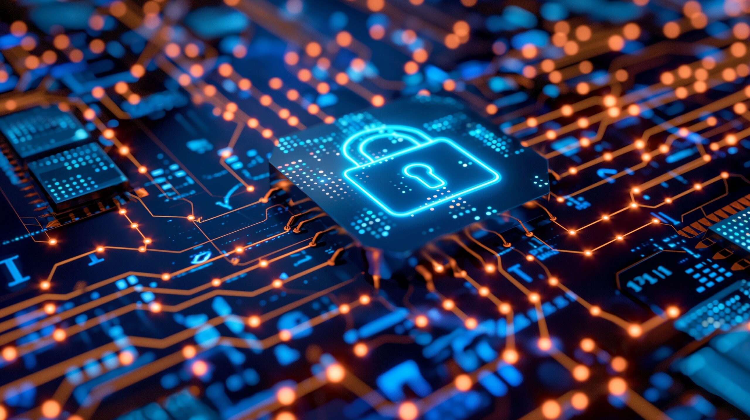
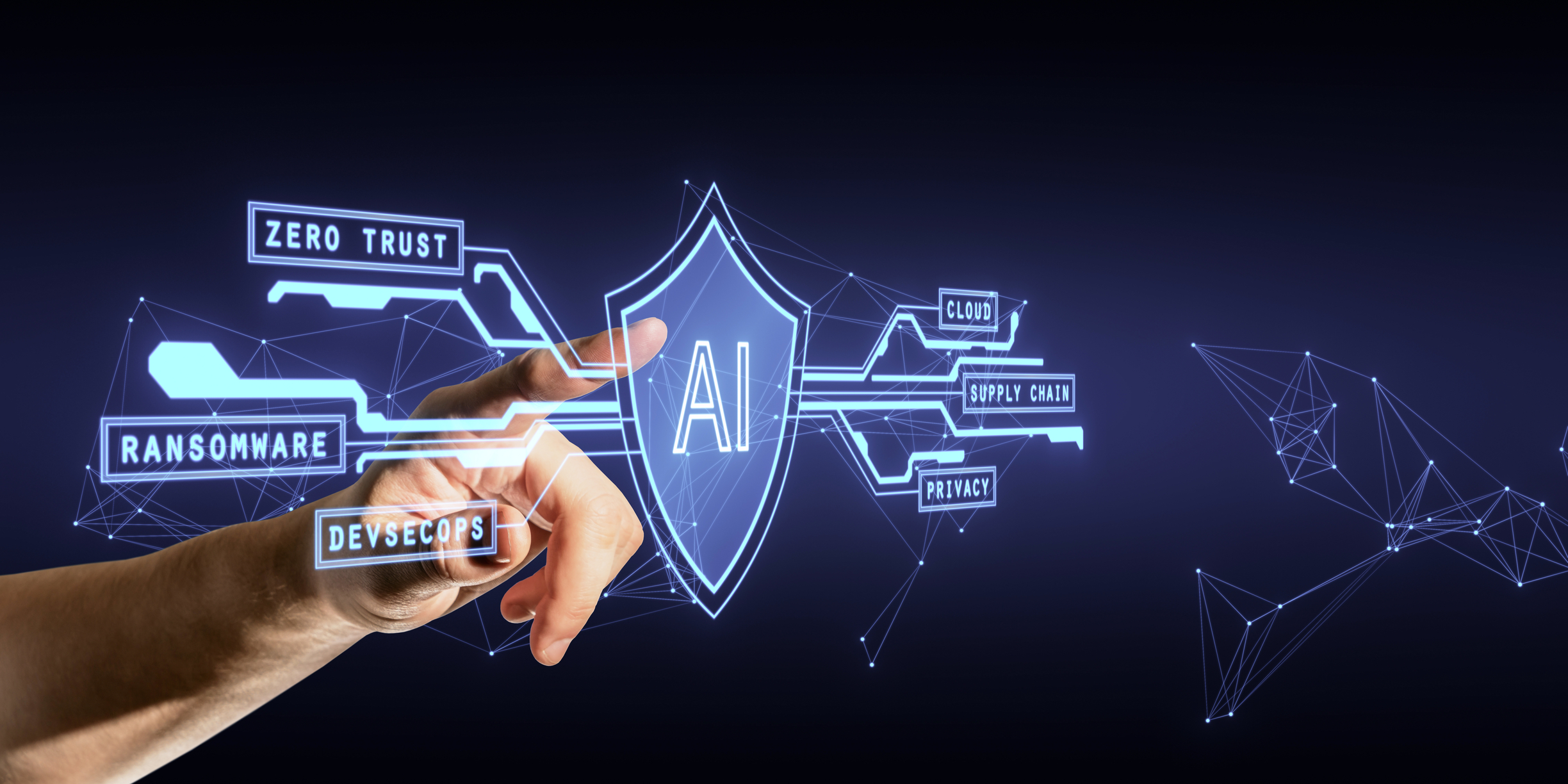
Learn how cybersecurity for medical imaging in 2026 addresses challenges with advanced encryption and vendor governance strategies.
 By
By
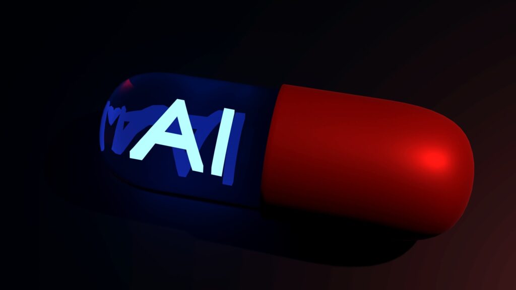
Discover the advantages of AI-powered radiology in modern healthcare, improving early detection and treatment success rates.
 By
By
Discover how AI-powered low-field MRI is transforming lung imaging, enhancing accessibility and affordability for better healthcare.
 By
By
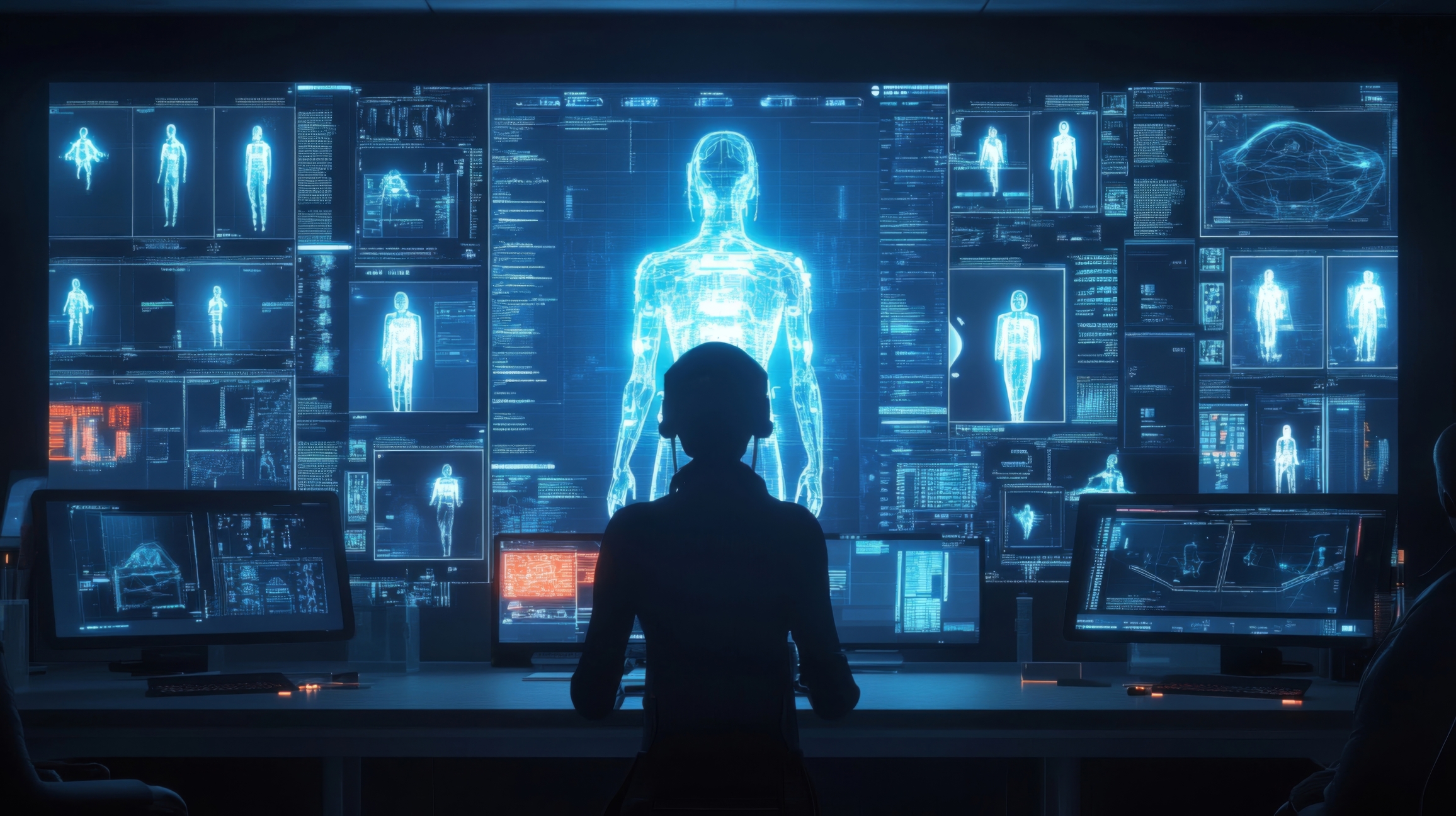
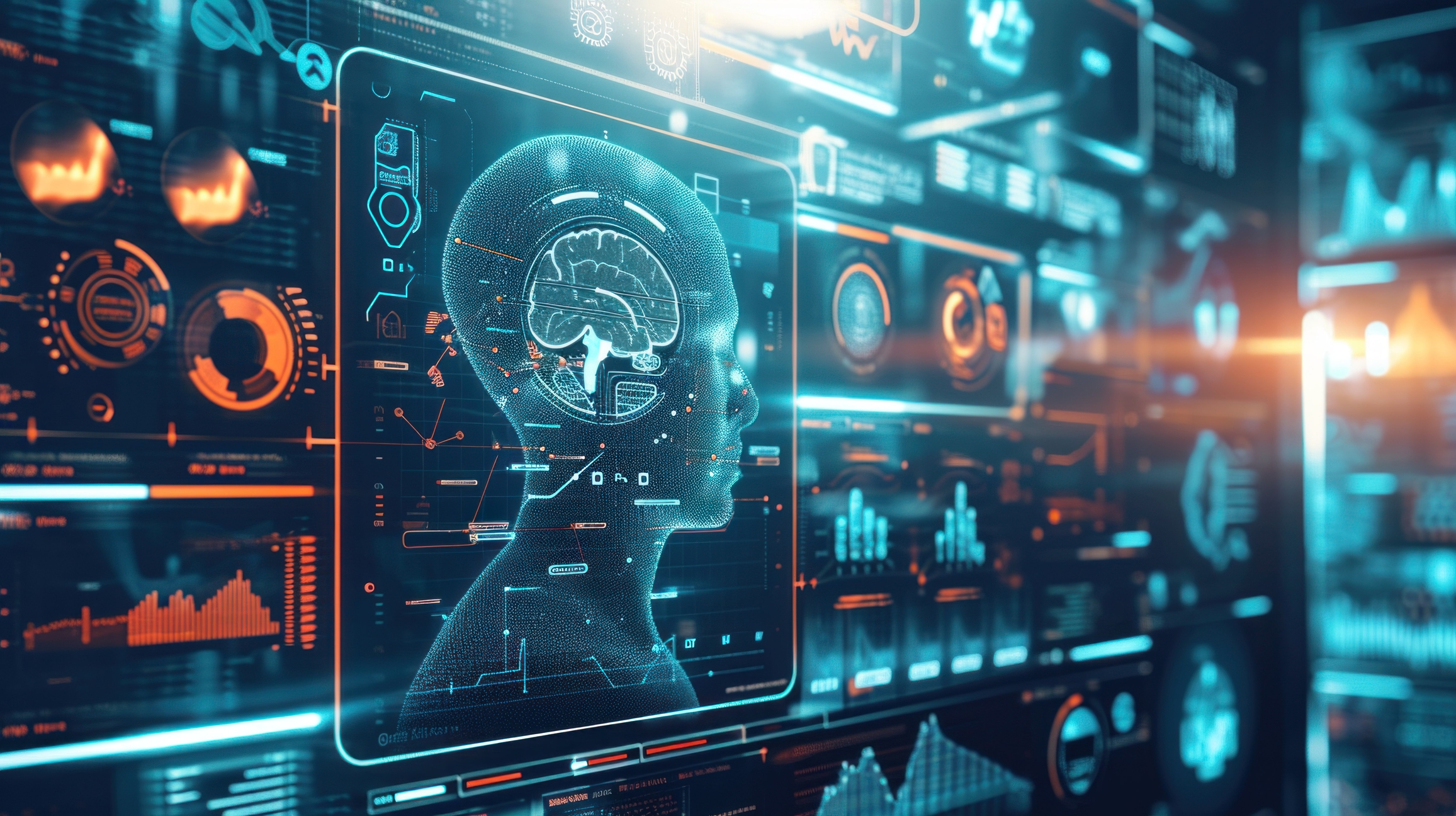
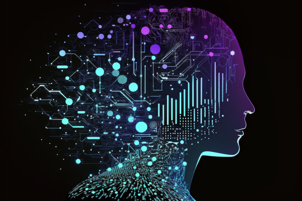
Discover how artificial intelligence in medical imaging is transforming clinical diagnosis and improving healthcare outcomes.
 By
By
Discover how AI-driven electronic health records transform patient data management and support cybersecurity in healthcare.
 By
By
Explore AI-enhanced cybersecurity in pharma and learn how it protects healthcare data while ensuring compliance and security.
 By
By
Understand the reasons behind addiction relapse: uncover hidden triggers and how technology assists in predicting relapses.
 By
By
Discover AI tools mental health professionals are using to cut down on paperwork and focus more on their patients’ needs.
 By
By
Discover how mental health AI tools streamline workflows and expand access to care while facing ethical challenges.
 By
By
Learn about the role of AI medical physics in enhancing accuracy and personalisation in modern patient care and healthcare strategies.
 By
By
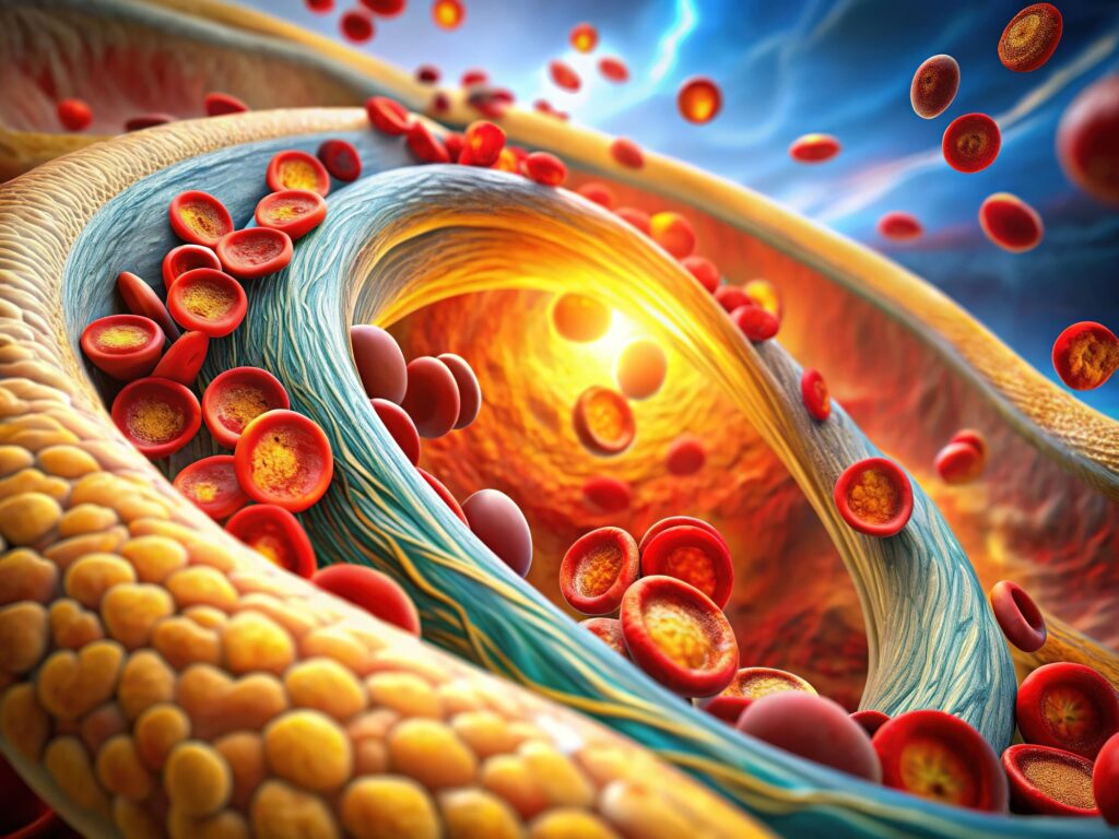
Understand the role of MRI carotid inflammation as a PET-equivalent method for detecting plaque inflammation in strokes.
 By
By
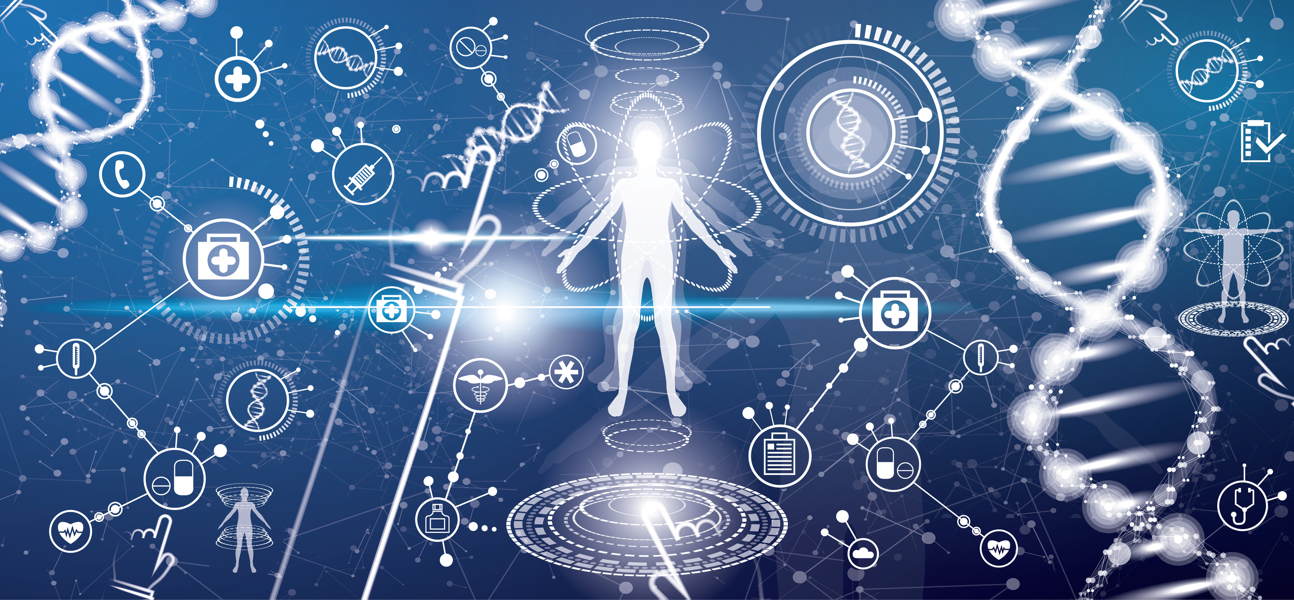
Explore the role of medical imaging and healthcare technology in enhancing patient care and advancing diagnostic sciences.
 By
By
Understand the importance of standardisation in radiomics and artificial intelligence for effective oncologic imaging.
 By
By
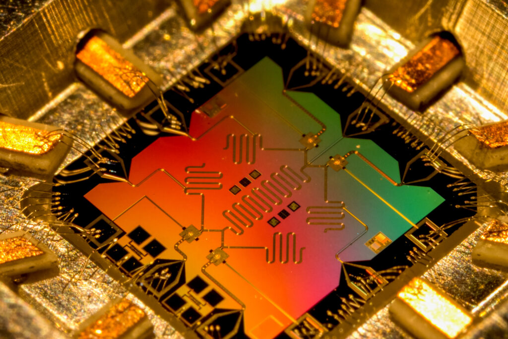
Uncover the significance of the Google Willow quantum chip, a leap in processor technology that outperforms classical supercomputers.
 By
By
Find out how quantum computing in medical imaging is transforming diagnostics with enhanced algorithms and AI integration.
 By
By
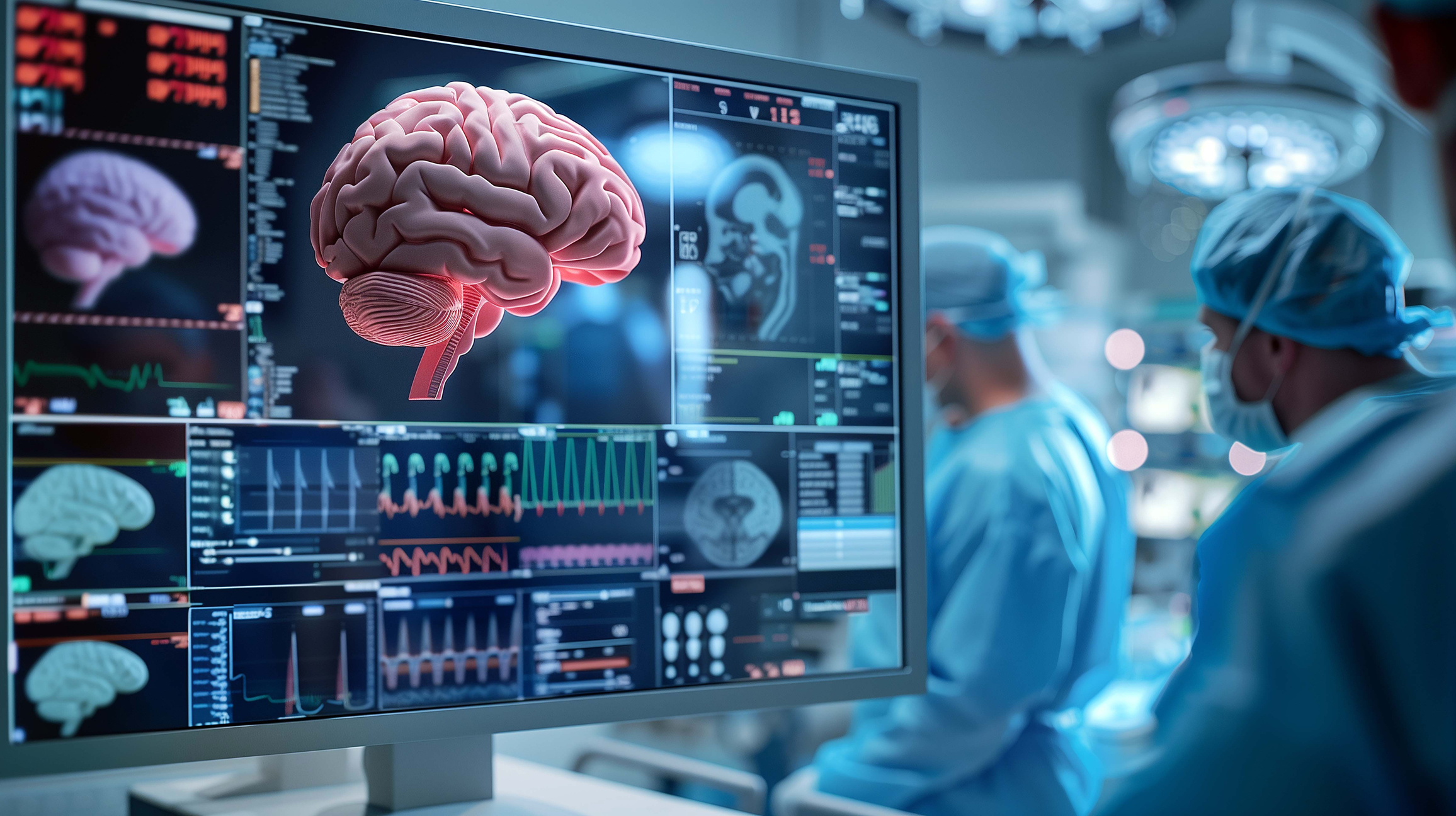
Uncover the role of AI intracranial haemorrhage detection in revolutionising emergency medicine and its clinical implications.
 By
By
Find out how generative AI antibiotics are paving the way for new solutions to modern infections and antimicrobial resistance.
 By
By
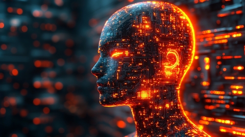
Explore the role of AI in medical imaging systems and how it enhances diagnostic accuracy while improving patient safety.
 By
By
Discover the role of AI cybersecurity in medical imaging and how it protects against threats while maintaining data integrity.
 By
By
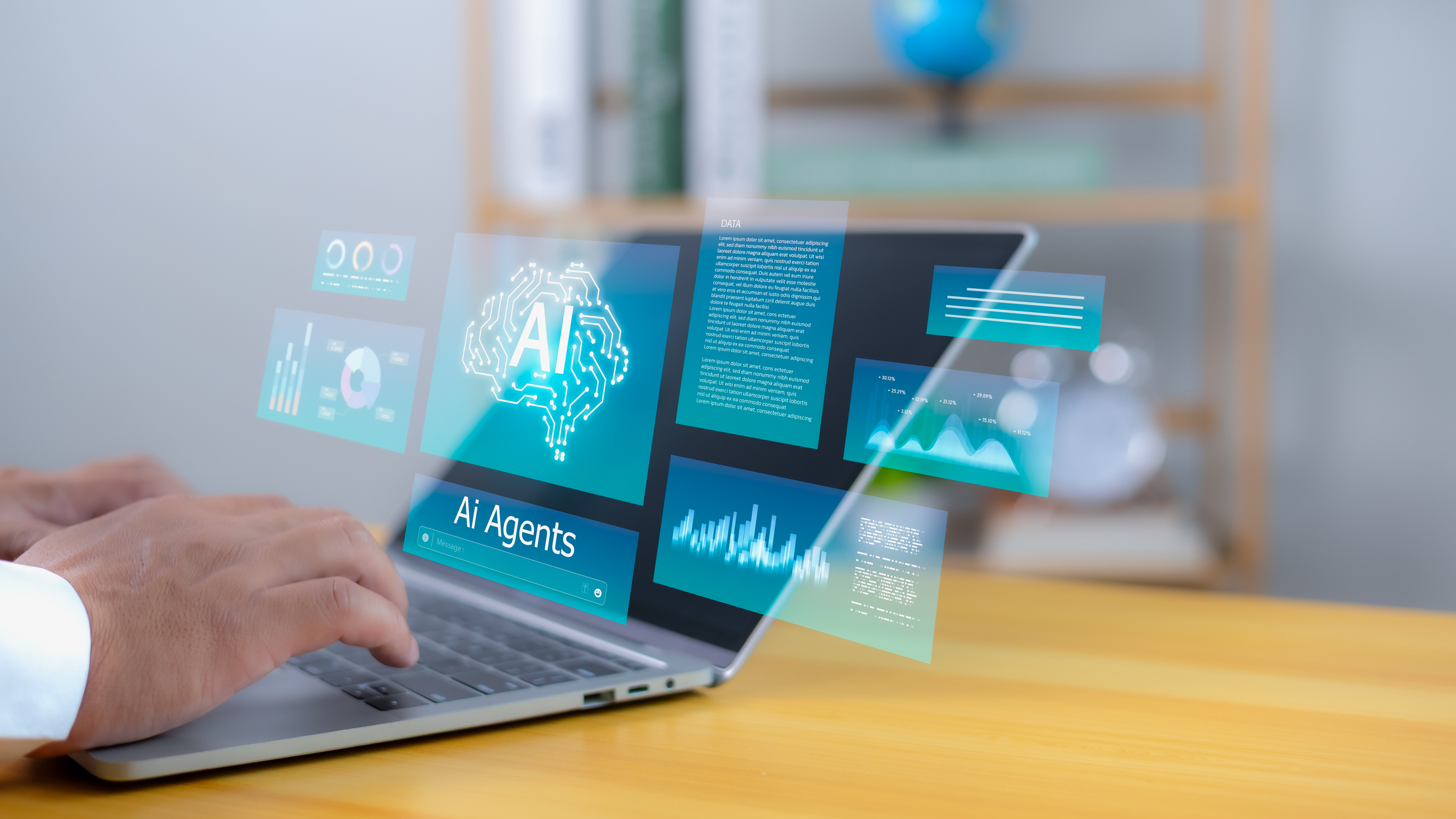
Discover the benefits of chat agents in medical imaging, aiding radiologists in scan interpretation and report generation.