
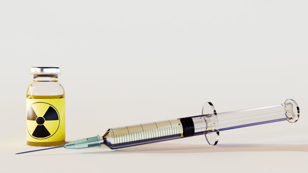
Medical Documentation After a Traumatic Brain Injury From a Road Collision
 By
By
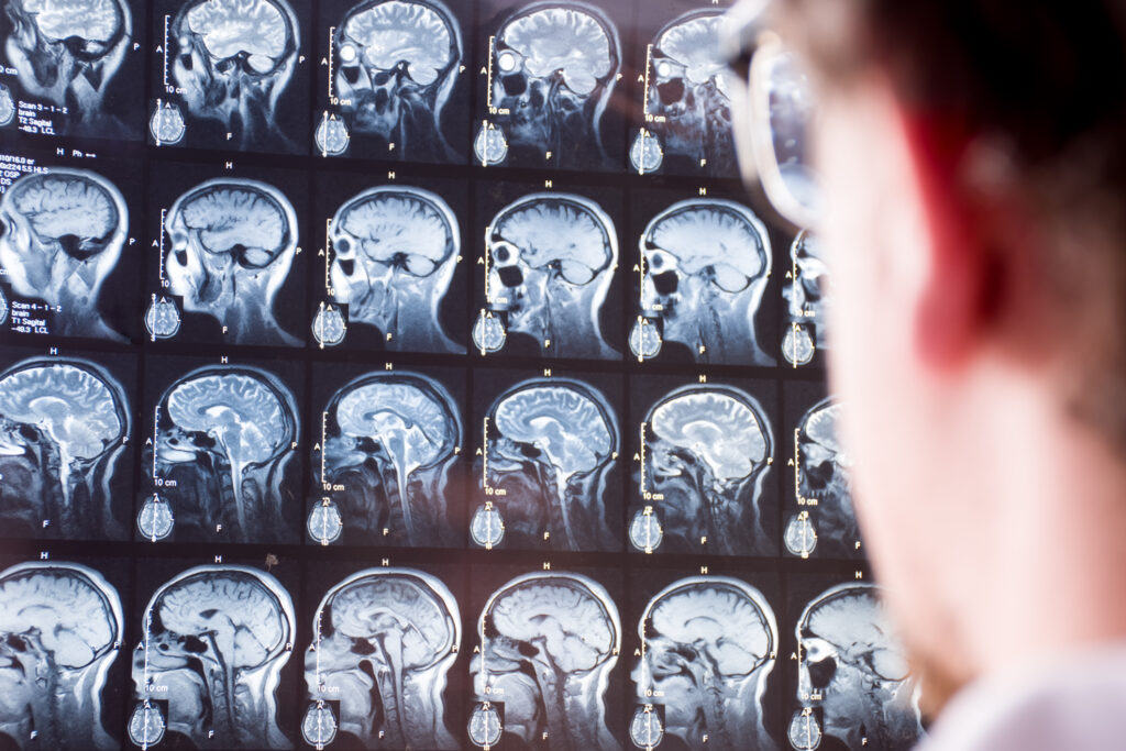
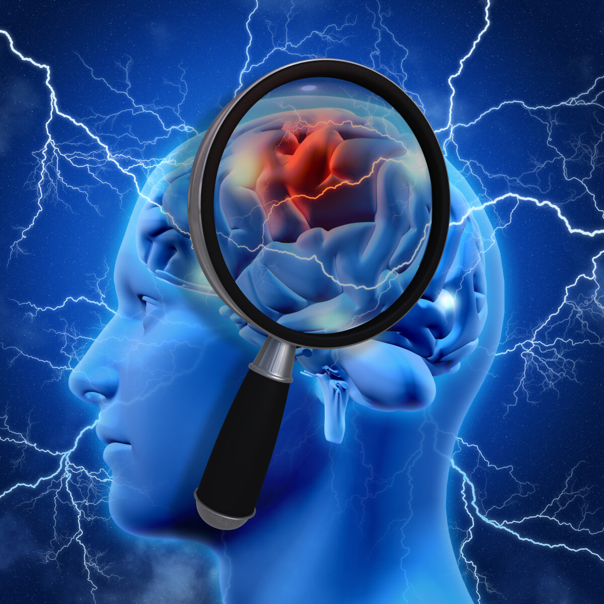
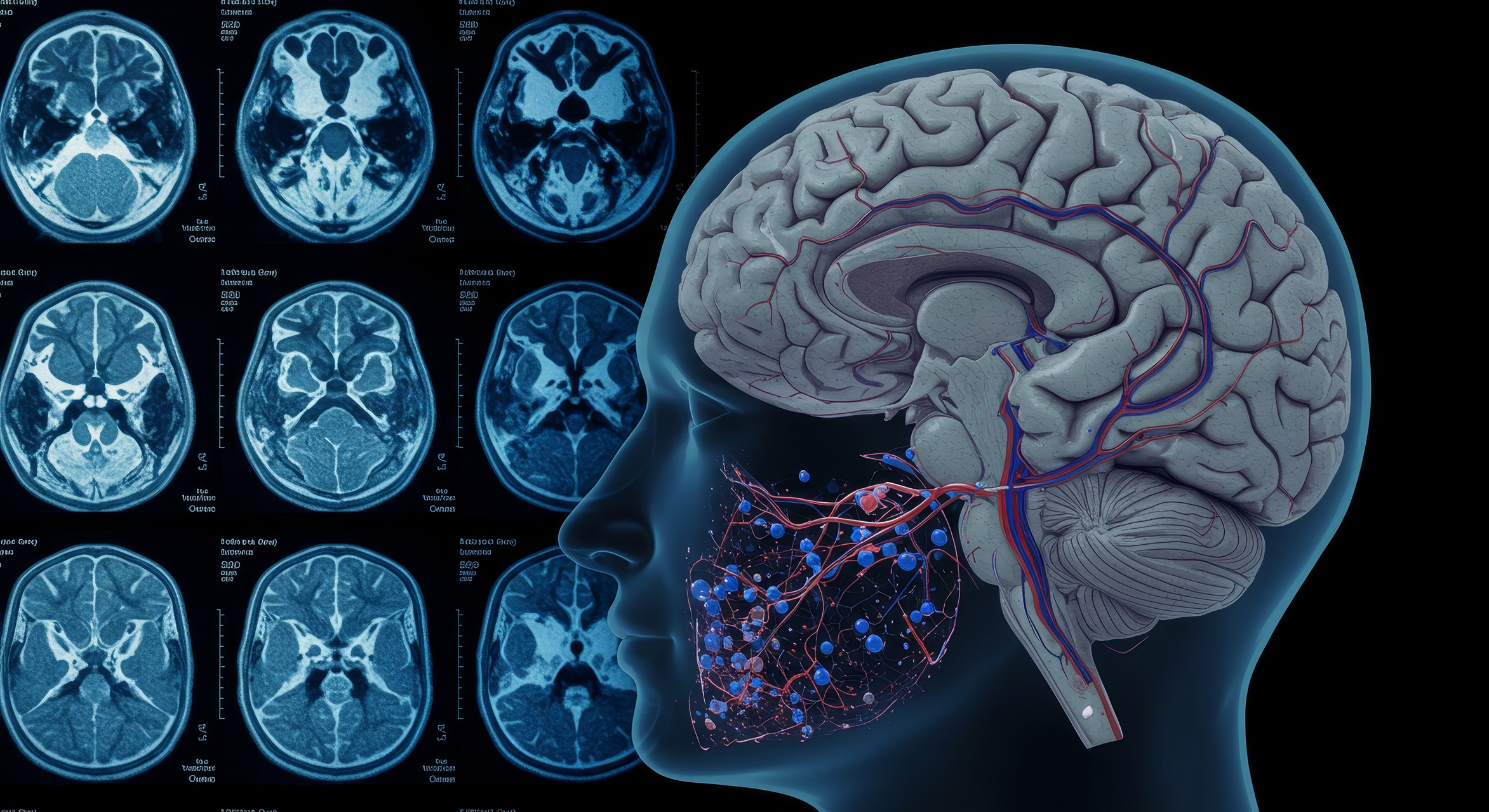
Recognizing Traumatic Brain Injury is vital. Learn about symptoms that may emerge days after a road accident.
By
Recognizing Traumatic Brain Injury is vital. Learn about symptoms that may emerge days after a road accident.
 By
By
Learn how functional MRI technology is used to assess brain injury. Understand the complexities of mTBI symptoms and recovery.
 By
By
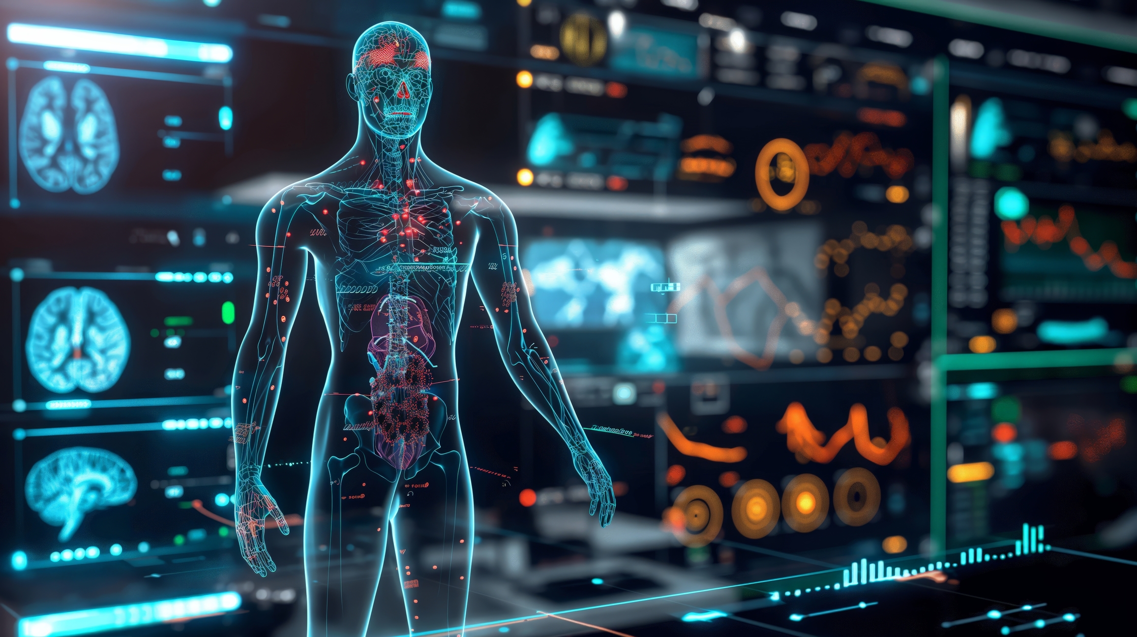
Explore the role of AI in medical imaging systems and how it enhances diagnostic accuracy while improving patient safety.
 By
By
Learn about the UK Biobank’s pioneering imaging study that has delivered unique insights from 100,000 participant scans.
 By
By
Radiology in drug addiction treatment provides essential tools for understanding addiction and crafting personalised therapies.
 By
By
Learn the impact of Imaging in Addiction Therapy, addressing challenges and offering insights into effective rehabilitation methods.
 By
By
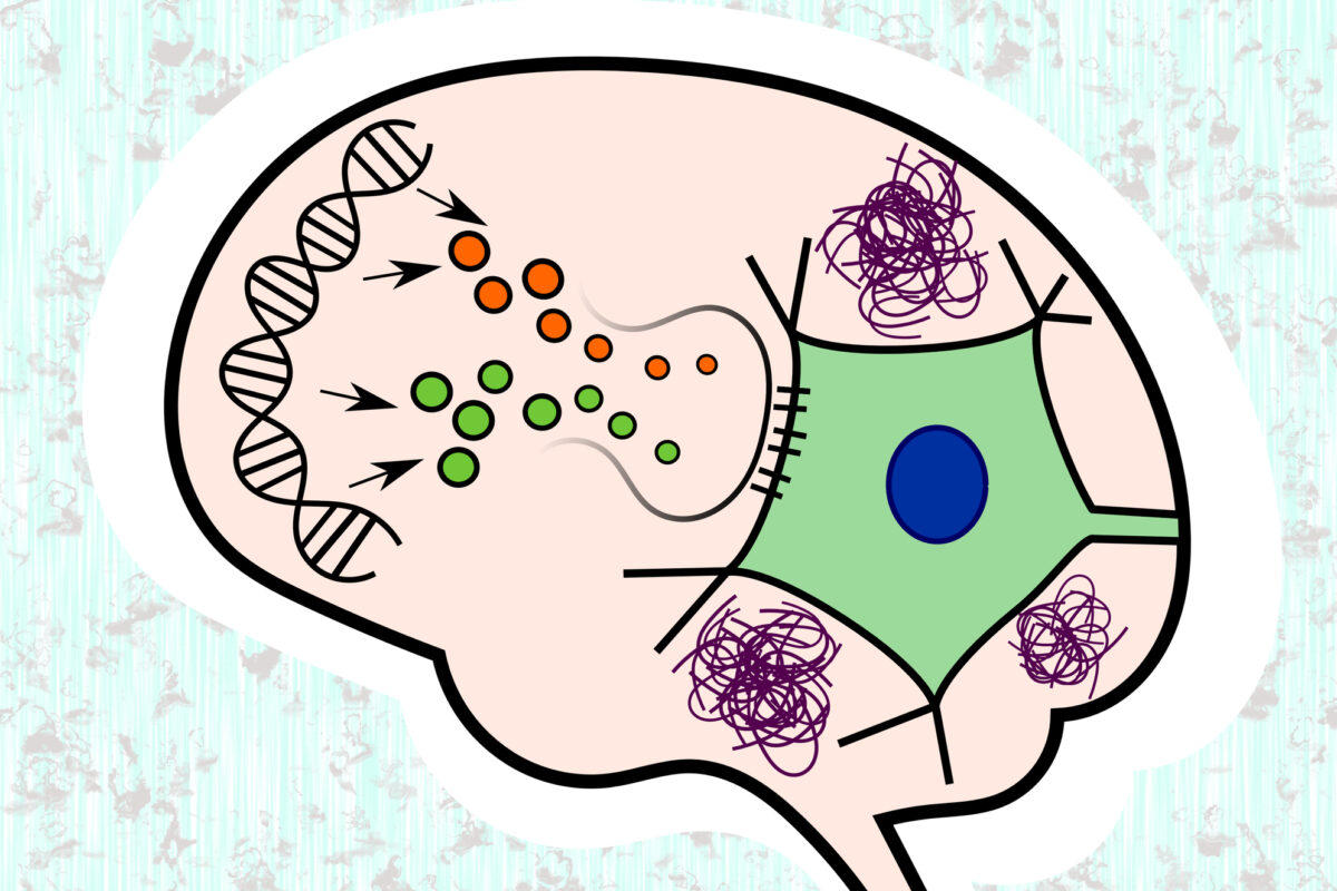
Memory loss clinical trials are crucial for understanding Alzheimer’s. Find out how they help shape effective interventions.
 By
By
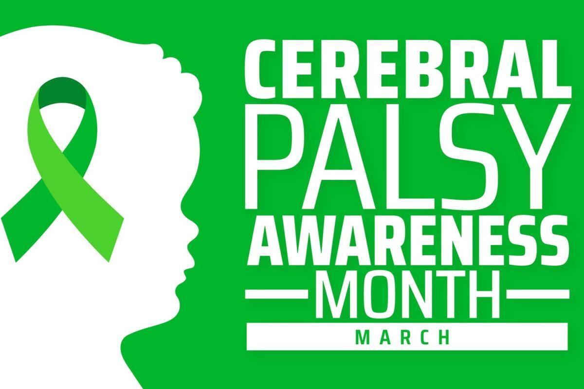
Cerebral Palsy advancement in stem cell therapy and AI-driven rehabilitation offers new hope for patients worldwide.
 By
By
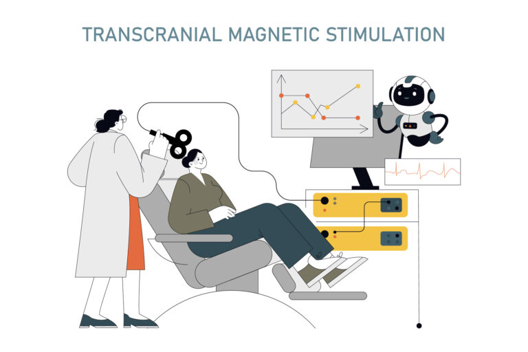
TMS for PTSD treatment provides a medication-free solution, using magnetic pulses to support brain healing effectively.
 By
By
Occupational Therapy Autism Benefits include improving communication, enhancing sensory processing, developing motor skills, and promoting independence.
 By
By
Mental health imaging technologies enable precise diagnostics, enhance treatment planning, and improve understanding of neurological conditions.
 By
By
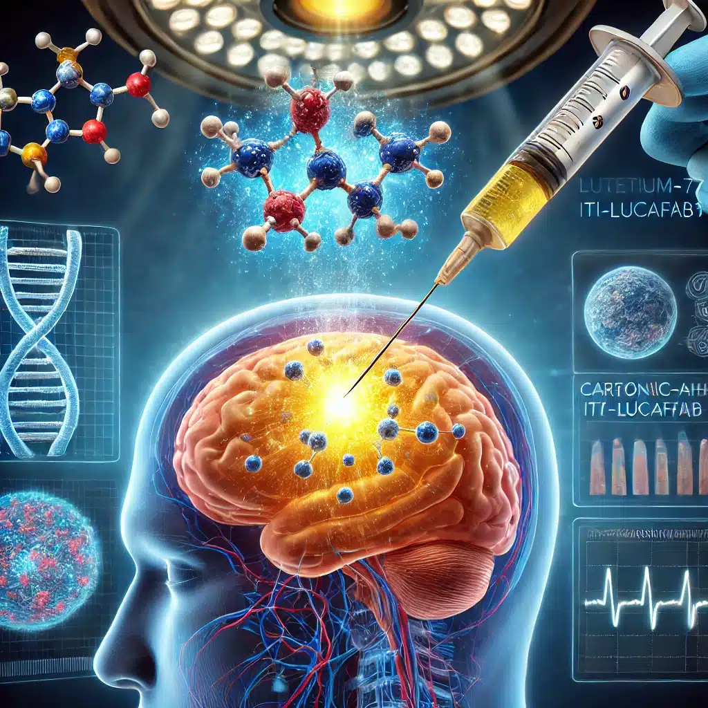
Targeting glioblastoma with Lutetium-177 ITM-31 offers precise intracavitary treatment, reducing recurrence by specifically addressing residual cancer cells post-surgery.
 By
By
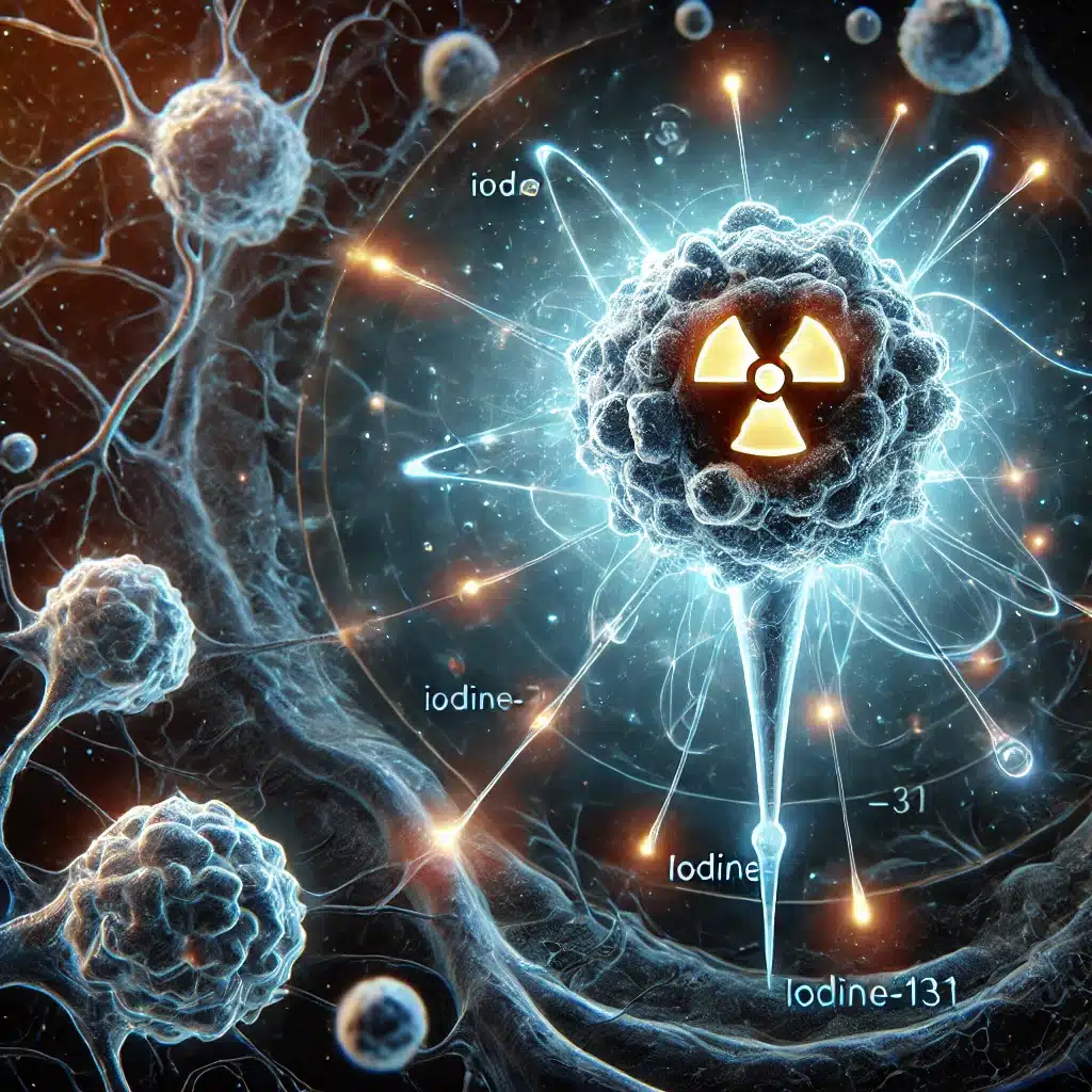
Iodine-131 Omburtamab offers targeted radiation therapy, significantly improving survival in neuroblastoma patients with CNS and leptomeningeal metastasis.
 By
By
Iodine-131 naxitamab (¹³¹I-3F8) targets GD2-expressing cancers, offering precise radioimmunotherapy for neuroblastoma, melanoma, and small cell lung carcinoma.
 By
By
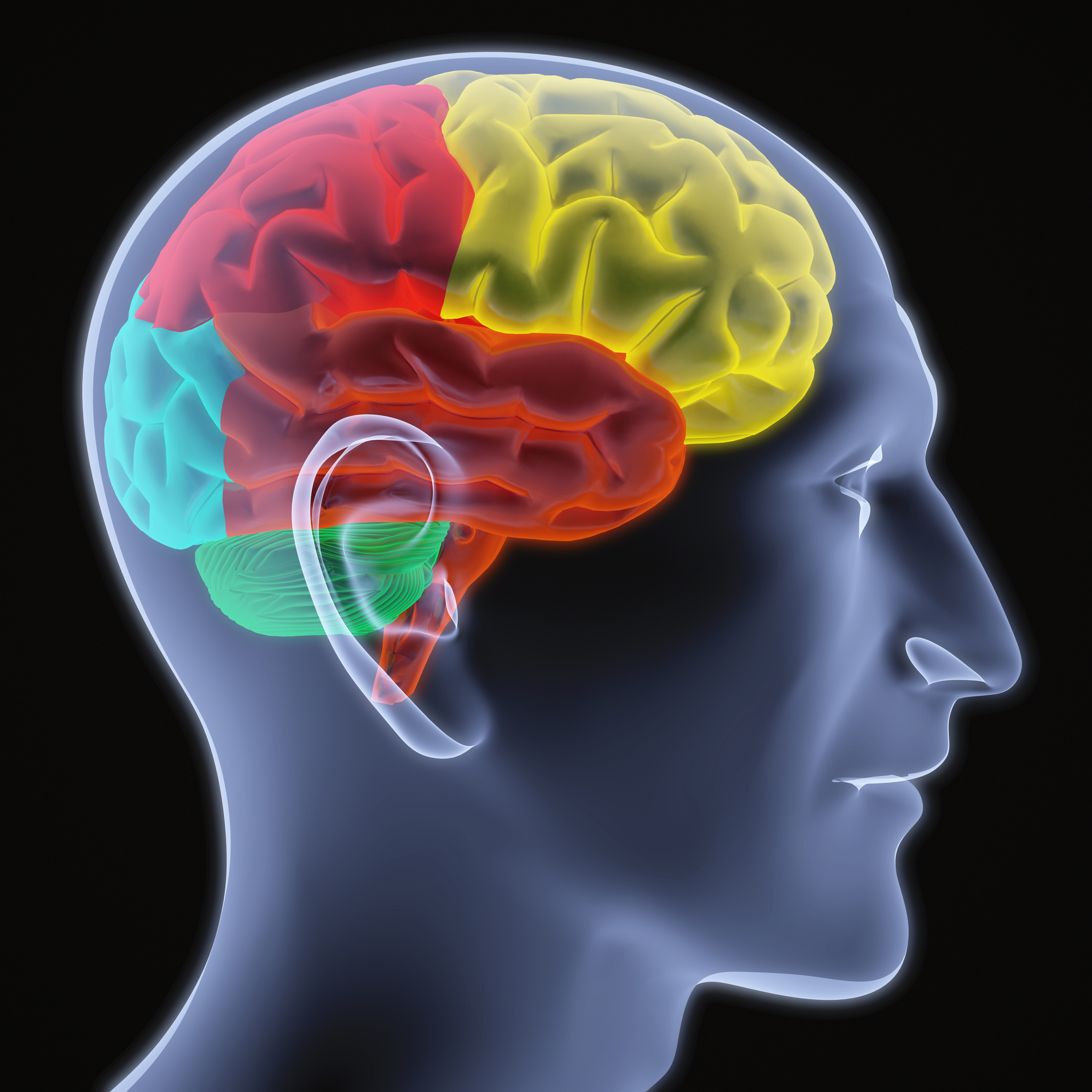
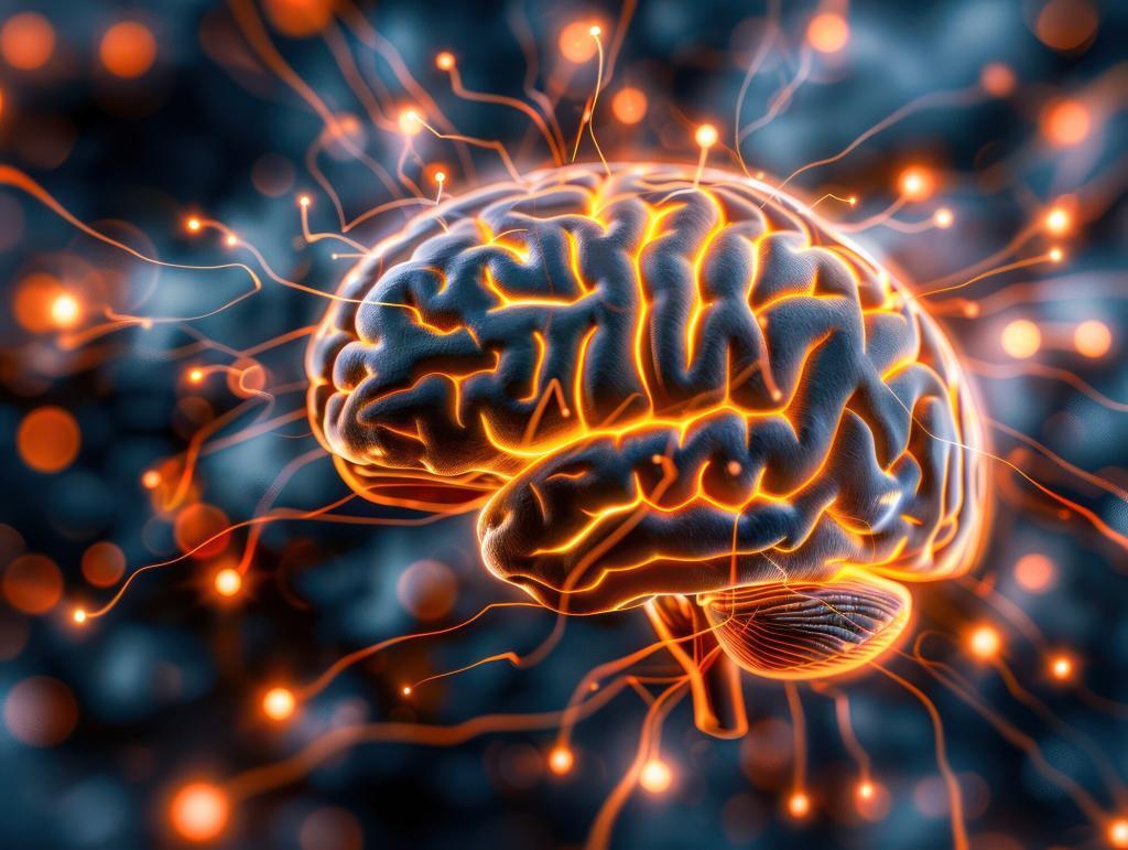
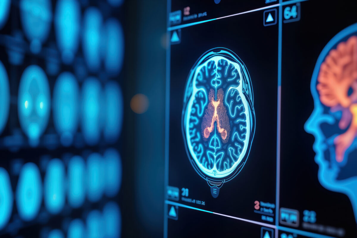
Neurological diagnostics have been transformed by advanced imaging techniques, enhancing accuracy in identifying brain disorders.
 By
By
Xenon-133 is pivotal for imaging lungs and assessing brain flow, enhancing diagnoses in modern medicine.
 By
By
Technetium-99m sodium pertechnetate is pivotal for brain, thyroid, cardiac imaging, and cancer detection in nuclear medicine.
By
Technetium-99m pentetate revolutionises brain and kidney imaging by providing crucial non-invasive insights into their function.
 By
By
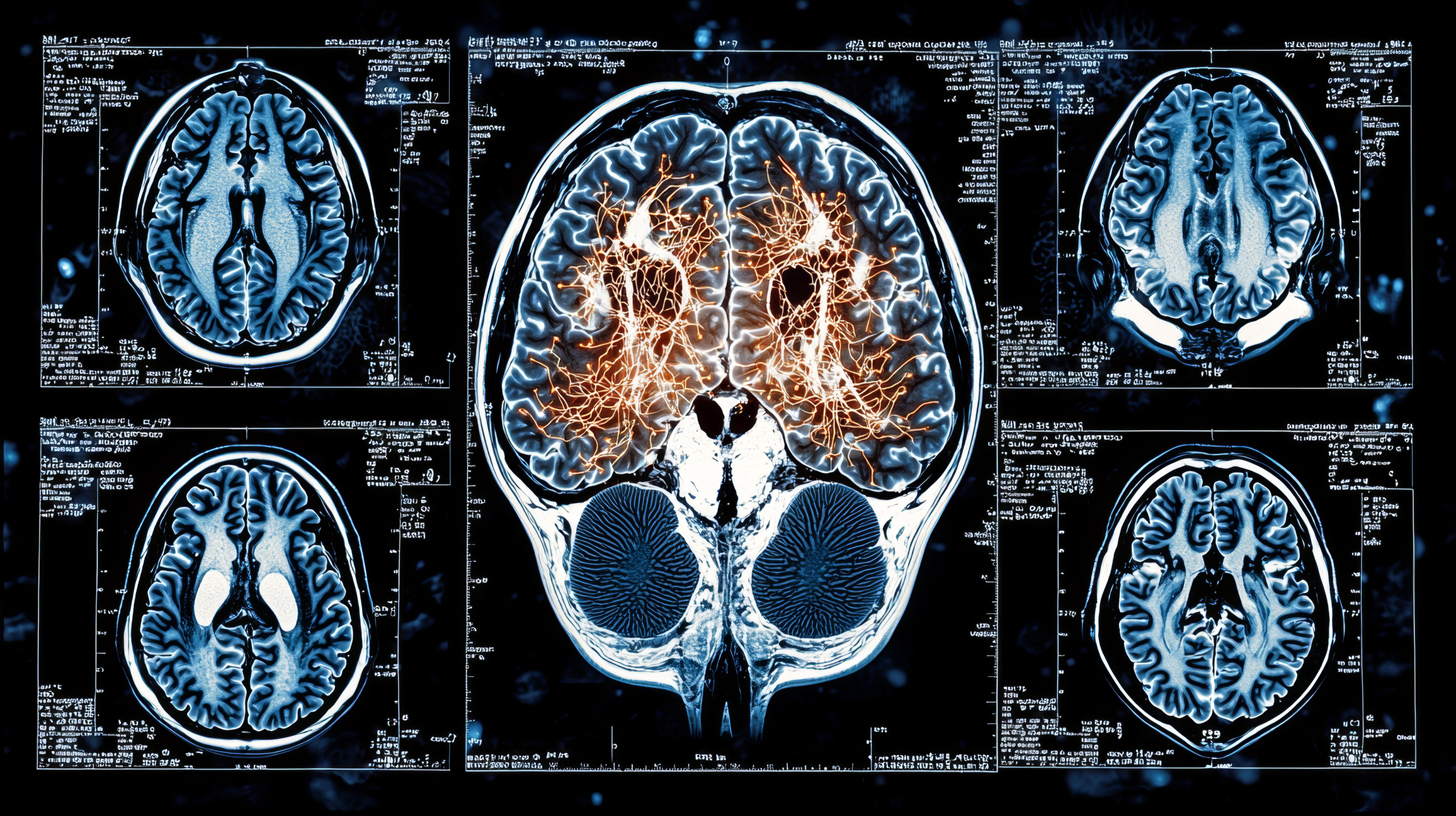
A useful brain imaging technique uses functional magnetic resonance imaging to analyse metabolic changes such as blood oxygenation.
 By
By
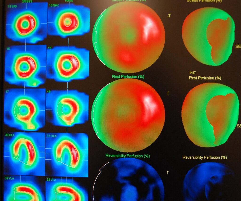
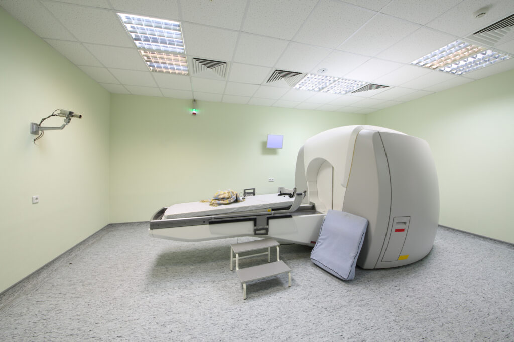
SPECT imaging is used to obtain a myocardial perfusion scan (SPECT scan) to investigate the function of the heart muscle.
 By
By
Stereotactic radiosurgery is a non-surgical procedure that utilises gamma radiation for the management of brain tumours amongst others.