SPECT-CT
SPECT-CT (Single Photon Emission Computed Tomography-Computed Tomography) is an advanced imaging technique that merges the functional imaging capabilities of SPECT with the high-resolution anatomical imaging of CT. SPECT-CT has transformed medical imaging by providing more accurate and comprehensive diagnostic information, improving patient outcomes. This hybrid modality benefits various medical fields, including oncology, cardiology, neurology, and orthopaedics. This article will explore SPECT-CT technology’s principles, applications, and advantages.
Principles of SPECT-CT
SPECT-CT is a dual-modality imaging technique that combines the principles of SPECT and CT scans. SPECT is a nuclear medical imaging technique that detects gamma rays from radiotracers injected into a patient’s body. These radiotracers accumulate in specific organs or tissues based on their biochemical properties, allowing for functional assessment of the targeted areas. SPECT generates 3D images of the radiotracer distribution within the body, providing valuable insight into the physiological processes at work.
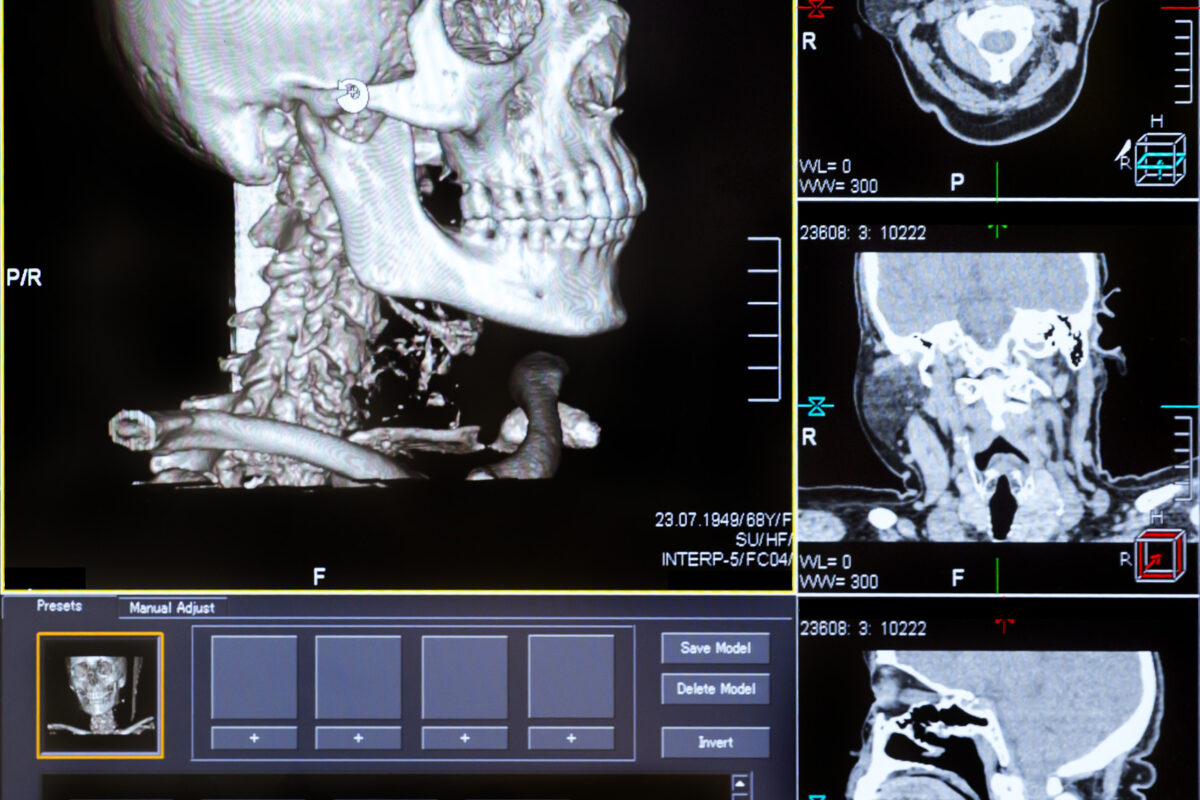
Conversely, CT is an X-ray-based imaging technique that generates detailed, high-resolution 3D images of the body’s internal structures. During a CT scan, X-rays are emitted from a rotating source and captured by detectors on the opposite side of the patient. This generates a series of cross-sectional images that can be reconstructed into a 3D image.
SPECT-CT combines the functional data from SPECT with the anatomical data from CT, resulting in a single image that provides molecular and structural information. This data fusion allows clinicians to localise and assess the extent of diseases or abnormalities.
Applications of SPECT-CT
- SPECT-CT is widely used in diagnosing, staging, and monitoring various cancers. It helps to identify tumours, metastases, and lymph node involvement, contributing to more accurate treatment planning and follow-up.
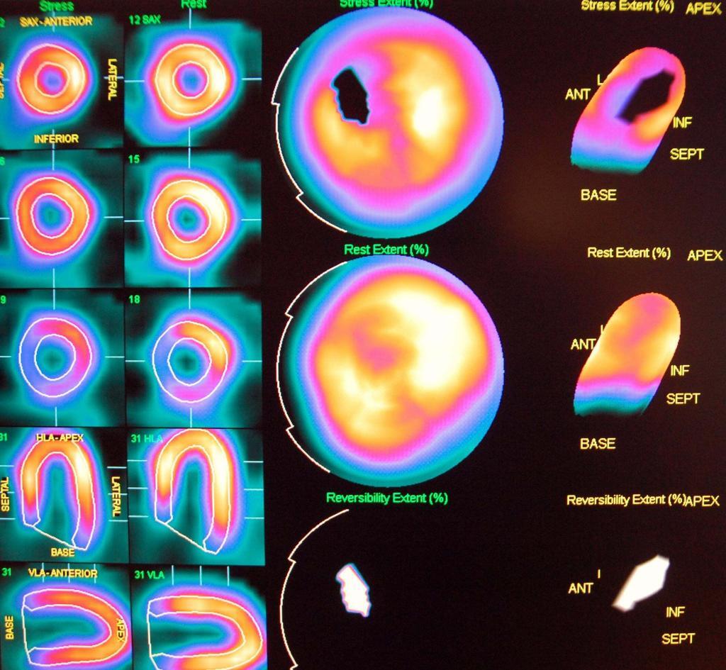
- SPECT-CT is used to assess myocardial perfusion, viability, and function. In addition, it can detect ischemic heart disease, evaluate the extent of myocardial infarction, and guide revascularisation procedures.
- The technique is employed to diagnose and manage neurological disorders like dementia, epilepsy, and Parkinson’s. In addition, SPECT-CT can provide valuable information on brain perfusion, neurotransmitter function, and receptor binding.
- SPECT-CT is used to diagnose bone and joint disorders, including infection, inflammation, and fractures. It is particularly useful in assessing prosthetic joint complications and spinal fusion.
Advantages of SPECT-CT
The integration of SPECT and CT offers several advantages over standalone imaging techniques:
- Combining functional and anatomical information helps clinicians accurately identify, localise, and characterise abnormalities.
- SPECT-CT can differentiate between benign and malignant lesions, leading to more accurate diagnoses and improved treatment planning.
- Using low-dose CT protocols minimises the radiation dose to patients while maintaining diagnostic quality.
home » SPECT-CT
 By
By