Summary: Urolithiasis, the formation of urinary tract stones, remains a prevalent condition affecting populations worldwide. The clinical presentation can vary from acute renal colic to chronic discomfort, making a timely and accurate diagnosis essential. Medical imaging has transformed the understanding and management of urolithiasis, providing clinicians with sophisticated tools to identify, classify, and monitor stones while guiding the development of appropriate treatment strategies. This article explores the evolution of imaging modalities, their relative advantages and limitations, and how emerging techniques are shaping the future of urolithiasis care.
Keywords: medical imaging in urolithiasis, renal colic diagnosis, ultrasound for kidney stones, CT in urinary stone disease, MRI urolithiasis imaging, future imaging technologies in urology
Introduction
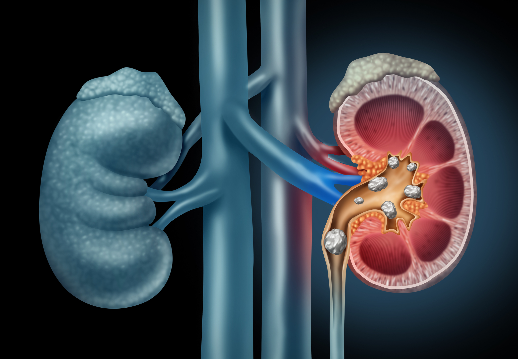
Urolithiasis, commonly referred to as urinary stone disease, is one of the most frequent urological disorders seen in both emergency and elective settings. The prevalence is increasing in many regions, partly due to dietary changes, sedentary lifestyles and genetic predispositions. The recurrence rate is also high, with many patients experiencing more than one episode over their lifetime. As symptoms can range from mild haematuria to severe pain, rapid diagnosis is essential to avoid complications such as obstruction, infection and renal impairment. Medical imaging plays a central role in identifying the presence, location and composition of stones, as well as informing treatment decisions.
Over the past decades, the spectrum of imaging techniques available to clinicians has expanded significantly. Each modality offers unique advantages, and the choice depends on clinical presentation, patient characteristics and resource availability. The shift towards precision imaging and the integration of advanced technologies continue to refine diagnostic accuracy while aiming to reduce radiation exposure and unnecessary interventions.
The Role of Ultrasound in Urolithiasis
Ultrasound remains the first-line investigation in many healthcare settings due to its wide availability, non-invasive nature and absence of ionising radiation. In patients presenting with suspected renal colic, ultrasound can detect hydronephrosis and identify stones within the kidney or proximal ureter. The modality is particularly valuable in vulnerable populations such as pregnant women and children, where radiation avoidance is paramount.
However, ultrasound has recognised limitations. Its sensitivity is operator-dependent, and small stones, particularly those in the distal ureter, may be missed. Acoustic shadowing can provide indirect evidence of stones, but definitive size measurement can be challenging. Nonetheless, ultrasound remains an essential screening tool and is frequently used in combination with other modalities for follow-up imaging.
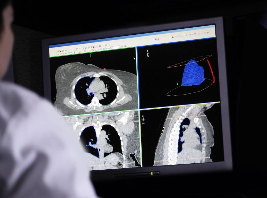
Computed Tomography as the Gold Standard
Non-contrast computed tomography (CT) has emerged as the gold standard for diagnosing urolithiasis. Its high sensitivity and specificity allow clinicians to detect even small stones with precision. CT provides detailed anatomical information, including stone size, density and location, which are crucial factors when determining the most appropriate management strategy.
The widespread adoption of CT has revolutionised the care of patients with suspected urinary stones, particularly in emergency settings where rapid and reliable diagnosis is required. The ability to identify alternative diagnoses when stones are absent also enhances its clinical utility. However, the associated radiation exposure remains a concern, particularly for younger patients and those with recurrent stone disease requiring multiple scans.
To address this, low-dose CT protocols have been developed. These reduce radiation exposure while maintaining diagnostic accuracy, making CT more sustainable for long-term use in recurrent stone formers. Advances in iterative reconstruction algorithms have further improved image quality at reduced dose levels, extending the safety profile of CT imaging.
Magnetic Resonance Imaging and Its Potential
Magnetic resonance imaging (MRI) is not routinely used in the assessment of urolithiasis due to its limited ability to visualise stones directly. Stones typically appear as signal voids, which can make detection less reliable than CT or ultrasound. Nevertheless, MRI has significant strengths in evaluating the urinary tract and detecting obstruction, oedema and secondary changes within the kidneys.
In specific patient populations, such as those who cannot undergo CT or where repeated radiation exposure is undesirable, MRI can provide valuable complementary information. Functional MRI techniques, including diffusion-weighted imaging and MR urography, are under investigation as potential tools for assessing urinary tract obstruction and renal function in the context of urolithiasis.
Although MRI is unlikely to replace CT as the primary diagnostic tool for stone disease, ongoing research into novel sequences and contrast techniques may expand its clinical relevance in the future.
Imaging in Paediatric and Pregnant Patients
Imaging strategies require particular consideration in children and pregnant women, where both diagnostic accuracy and patient safety are of paramount importance. In these groups, ultrasound is typically the preferred initial modality. In children, the absence of radiation exposure is particularly advantageous given their increased susceptibility to long-term radiation risks. In pregnancy, an ultrasound not only avoids radiation but also allows evaluation of the foetus when required.
When ultrasound findings are inconclusive, alternative imaging must be carefully considered. Low-dose CT may occasionally be necessary in children; however, MRI urography is emerging as a safer alternative in cases where the results are equivocal. During pregnancy, MRI offers a radiation-free alternative, although accessibility and expertise in interpretation can limit its use. Clinical decision-making in these populations requires balancing the need for accurate diagnosis against the potential risks associated with imaging.
Functional Imaging and Stone Composition Analysis
Beyond structural assessment, advances in imaging now allow the functional evaluation of renal performance and prediction of stone composition. Dual-energy CT (DECT) is an example of a technique that can differentiate stone types based on their attenuation profiles at different energy levels. This capability is clinically valuable because treatment strategies vary for stones of various compositions. For example, uric acid stones may respond to medical dissolution therapy, whereas calcium oxalate stones typically require interventional approaches.
Functional imaging also offers insights into renal perfusion and drainage patterns, aiding the management of complex cases. Nuclear medicine techniques, although less commonly used, provide quantitative assessments of renal function that can complement anatomical findings from CT or ultrasound. These developments represent a shift towards personalised medicine, where imaging not only detects stones but also guides tailored therapeutic strategies.
Emerging Technologies in Urolithiasis Imaging
The future of medical imaging in urolithiasis is closely linked to technological innovation. Advances in artificial intelligence (AI) and machine learning are being applied to imaging datasets to enhance stone detection, automate measurements and predict treatment outcomes. Algorithms capable of integrating radiological data with clinical parameters have the potential to reduce diagnostic errors and support real-time decision-making.
Photon-counting CT is another development gaining attention. By improving spatial resolution and contrast-to-noise ratio, this technology may enhance the characterisation of stones while further reducing radiation exposure. Similarly, novel MRI sequences are being investigated to improve stone visibility and overcome the limitations of conventional MRI in urolithiasis.
Point-of-care imaging devices are also emerging, particularly portable ultrasound systems that allow rapid bedside assessment. These tools may expand access to imaging in resource-limited settings and reduce delays in diagnosis and treatment.
Imaging in the Follow-up and Prevention of Recurrence
The role of imaging does not end with initial diagnosis and treatment. Follow-up imaging is essential to assess treatment outcomes, monitor residual fragments and evaluate for recurrence. Ultrasound is frequently used in this context due to its safety and repeatability, though CT remains valuable in cases where precise measurement of stone clearance is necessary.
Imaging findings can also inform preventive strategies. By identifying the stone composition and burden, clinicians can recommend dietary changes, pharmacological interventions, and lifestyle modifications aimed at reducing the risk of recurrence. Long-term imaging surveillance ensures that these measures are effective and provides early detection of new stones before symptoms arise.
Conclusion
Medical imaging has become an indispensable element in the management of urolithiasis. From the accessibility of ultrasound to the precision of CT and the evolving potential of MRI, each modality contributes to a comprehensive diagnostic pathway. Advances such as low-dose protocols, dual-energy techniques and AI integration continue to refine the field, making imaging safer and more informative than ever before.
For clinicians, the ability to select the most appropriate imaging strategy is crucial in delivering timely and effective care. For patients, these advances mean earlier detection, more tailored treatment and improved long-term outcomes. As technology progresses, the integration of anatomical, functional and computational imaging approaches will continue to transform the management of urinary stone disease.
Disclaimer
The content provided in Advances in Medical Imaging for the Diagnosis and Management of Urolithiasis is intended for informational and educational purposes only. It should not be considered a substitute for professional medical advice, diagnosis or treatment. Clinicians should rely on their professional training, clinical judgement and local guidelines when making decisions about patient care. Patients are advised to consult a qualified healthcare professional for any concerns regarding symptoms, diagnosis or treatment of urolithiasis or related conditions. Open MedScience does not accept responsibility for any loss, injury or damage arising from reliance on the information contained in this article.