Summary: Kilovoltage (kV) X-rays, commonly used in diagnostic imaging and superficial radiotherapy, represent a critical modality within medical imaging and treatment. This article outlines the fundamental physics behind kilovoltage X-rays, their distinct advantages, diverse clinical applications, potential limitations, and evolving technological advancements in the field.
Keywords: kilovoltage X-rays; diagnostic imaging; radiotherapy; superficial radiation; image quality; radiation safety.
Physics of X-ray Generation
X-rays have been instrumental in medicine since their discovery in 1895 by Wilhelm Röntgen. While modern medical imaging has undergone significant evolution, kilovoltage (kV) X-rays continue to be a cornerstone in both diagnostic and therapeutic medical applications. Kilovoltage X-rays typically operate between 20 and 150 kV, providing sufficient energy for clear imaging of soft tissues and superficial treatments. Understanding the physics, application scope, and clinical implications of their use ensures effective and safe patient care.
Fundamentals of Kilovoltage X-rays
Kilovoltage X-rays are produced by accelerating electrons in an X-ray tube towards a metal anode. Upon collision, kinetic energy is primarily converted into heat, with a small fraction (approximately 1%) emitted as X-rays. The voltage applied across the X-ray tube determines the kinetic energy of electrons, influencing the penetrating ability (quality) and intensity (quantity) of the emitted X-rays.
Lower kilovoltage settings (20-60 kV) produce softer X-rays, which are suitable for detailed imaging of softer structures, such as mammography. In contrast, higher kV ranges (60-150 kV) yield harder, more penetrating beams used in general diagnostic imaging, including chest and abdomen X-rays.
Beam Characteristics
Kilovoltage X-rays differ significantly from megavoltage X-rays (typically above 1 MV). The former have higher absorption in tissues, allowing clear imaging contrasts between structures with minor density differences, but are less penetrative, limiting their therapeutic use to superficial tissues. Moreover, kilovoltage beams exhibit a marked photoelectric effect, which enhances image contrast but increases patient radiation absorption.
Clinical Applications
Kilovoltage X-rays play an essential role in diagnostic radiology. Their ability to produce high-contrast images makes them suitable for imaging bones, soft tissues, and certain pathologies such as pulmonary diseases. Chest radiographs, one of the most common applications, utilise kV settings around 100-120 kV, balancing adequate penetration and optimal contrast between air-filled lungs and surrounding tissues.
Similarly, mammography employs low-energy kilovoltage X-rays (around 25-35 kV), specifically designed to visualise subtle tissue differences in breast tissue, facilitating the early detection of breast cancer.
Superficial Radiotherapy
Kilovoltage radiotherapy (also known as superficial or orthovoltage radiotherapy) is an effective treatment modality for superficial skin cancers and benign dermatological conditions. Its relatively low penetration depth is ideal for treating skin lesions, minimising the irradiation of deeper healthy tissues, thus reducing systemic side effects.
Typically, superficial X-ray therapy employs energies ranging from 50 to 150 kV, effectively delivering therapeutic doses to lesions at shallow depths (up to a few millimetres beneath the skin surface).
Advantages of Kilovoltage X-rays
Kilovoltage X-rays offer exceptional contrast resolution, which is significantly advantageous in diagnostic radiology. Lower energies enhance differential absorption between tissues, highlighting subtle pathological changes that may otherwise remain undetected with higher-energy modalities, such as CT or megavoltage radiotherapy.
Cost-effectiveness and Accessibility
Compared to advanced imaging techniques such as MRI and CT scans, kilovoltage X-ray systems are significantly cheaper to install, maintain, and operate. Their affordability and ease of use ensure wide accessibility, which is particularly valuable in resource-limited healthcare settings.
Reduced Radiation Exposure in Select Cases
Although kilovoltage X-rays often result in higher superficial absorption, an appropriate selection of kV settings can minimise radiation exposure while maintaining diagnostic image quality. For instance, careful modulation of energy reduces patient dose significantly in paediatric and mammographic imaging.
Limitations and Challenges
A key limitation of kilovoltage X-rays is their limited tissue penetration, restricting their therapeutic application to superficial layers. Conditions requiring deeper tissue penetration, such as internal tumours, necessitate the use of megavoltage radiation therapy or alternative modalities like electron beams or proton therapy.
Radiation Dose Concerns
Increased absorption in tissues enhances image contrast but simultaneously escalates radiation dose to superficial layers, raising potential concerns regarding radiation-induced skin damage, particularly in frequent, repetitive imaging scenarios or extended treatment regimens.
Image Quality Compromise with Higher Tissue Thickness
Kilovoltage X-rays are susceptible to reduced image quality and increased scatter radiation with thicker anatomical structures. Excessive scatter can obscure diagnostic detail, prompting reliance on alternative imaging techniques, such as CT or MRI, for complex anatomical assessments.
Safety and Radiation Protection
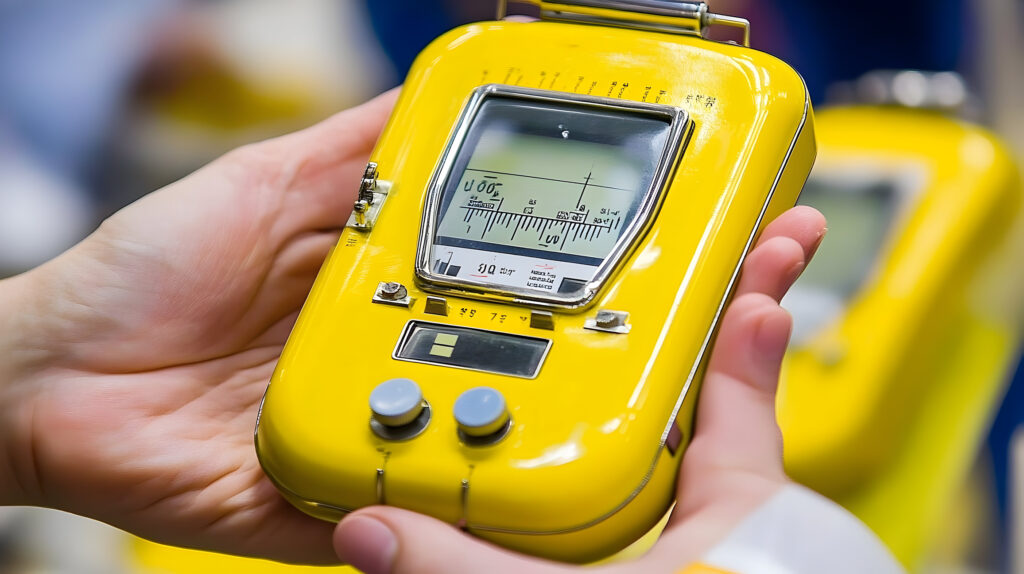
Ensuring patient and staff safety involves stringent adherence to radiation safety protocols, including employing lead shielding, limiting exposure time, optimising kV and mA settings, and regular equipment calibration. Additionally, radiographers undergo rigorous training to minimise unnecessary radiation exposure through proper positioning and technique.
Dose Optimisation
Optimising the radiation dose involves meticulous adjustments of kilovoltage, milliamperage, and exposure time. Implementing the ALARA (As Low As Reasonably Achievable) principle consistently reduces unnecessary radiation exposure, ensuring clinical effectiveness while safeguarding patient and occupational health.
Technological Advances
The transition from film-based to digital radiography has significantly enhanced kilovoltage X-ray imaging. Digital systems offer greater efficiency, enhanced diagnostic capabilities through post-processing techniques, and substantial reductions in patient radiation doses by minimising repeat exposures.
Adaptive X-ray Systems
Modern X-ray equipment incorporates automatic exposure control systems that dynamically adjust kV and mA based on patient thickness and tissue density, thereby improving image consistency, reducing operator error, and further optimising radiation dose.
Advanced Radiotherapy Techniques
Advances in superficial radiotherapy equipment now allow more precise dose delivery. Enhanced collimation techniques and real-time imaging guidance systems increase accuracy, minimising healthy tissue exposure and maximising therapeutic effectiveness.
Future Perspectives
The integration of AI into kilovoltage X-ray imaging and treatment planning promises substantial advancements. AI algorithms assist in optimising imaging parameters, predicting treatment outcomes, automating lesion detection, and reducing radiation dose, fostering more precise and personalised patient care.
Novel Imaging Detectors
Research continues into innovative detector technologies, such as photon-counting detectors, which promise significant improvements in image quality, lower patient doses, and advanced spectral imaging capabilities. These technologies could revolutionise kilovoltage imaging, enabling detailed material characterisation within tissues and enhanced diagnostic accuracy.
Conclusion
Kilovoltage X-rays remain integral to medical diagnostics and superficial radiotherapy, owing to their distinct characteristics, clinical versatility, and accessibility. Continued innovation in technology and rigorous adherence to radiation safety standards are essential to maximising clinical benefits while minimising potential risks. With ongoing advancements, kilovoltage X-ray imaging and treatment are expected to remain indispensable in modern healthcare, providing effective, safe, and widely accessible medical interventions for years to come.
Disclaimer
The information provided in “Kilovoltage X-rays: Fundamentals, Applications, and Clinical Implications” is intended solely for educational and informational purposes. It is not designed to replace professional medical advice, diagnosis, or treatment. Healthcare providers should rely on their clinical judgment and current medical standards when applying information contained herein to patient care.
While every effort has been made to ensure accuracy, Open Medscience does not assume responsibility for errors, omissions, or any actions taken based on the content presented. Readers are encouraged to consult relevant clinical guidelines, regulatory standards, and equipment manufacturers’ specifications for detailed guidance.
The use of kilovoltage X-rays involves inherent risks associated with radiation exposure. Proper training, adherence to radiation safety protocols, and ongoing technological updates are essential for mitigating these risks. Open Medscience disclaims liability for any harm, loss, or injury resulting from reliance upon or implementation of the information contained in this article.
You are here: home » diagnostic medical imaging blog »