Introduction: Why Accurate Vein Diagnosis Matters
The emergence of varicose veins is caused by a combination of increased blood volume, elimination of certain cues that influence blood flow (which reduces stagnation of blood flow in the veins) and increased hydrostatic pressure due in part to gravity, and impaired venous transport capacity.
Varicose veins have a more structural and functional nature, as they are not superficial to venous engagement.
Structurally, varicose veins are the result of elongation of the saphenous veins caused by increased venous pressure.
Medically, varicose veins are viewed as a complication of chronic venous insufficiency due to the significant impact they have on the patient’s quality of life.
Healthy veins flow to the heart and prevent blood from flowing backward through one-way valves. When these valves deteriorate, blood can begin to pool. This creates pressure and causes the veins to stretch and distort.
Even though surface veins are most visible, there can be problems even deeper in the venous system. Many patients develop venous reflux, which is the backward flow of blood due to bad valves. Many other patients experience normative circulatory issues that affect the deeper veins, which usually cannot be seen in a typical check-up.
This is the reason we must have the necessary equipment to analyze the condition of veins and the flow of blood.
As advanced vascular imaging becomes more widely available, patients experiencing symptoms such as leg heaviness, swelling, visible varicose veins, or unexplained discomfort can benefit from an early evaluation. Individuals seeking comprehensive assessment and modern diagnostic approaches may consider consulting a trusted vein clinic serving Mesa residents that utilizes advanced imaging technologies to identify underlying venous conditions and guide personalized treatment plans.
Problems of Historical Diagnostic Methods
For a long time, doctors had to be able to recognize vein disease with just a check-up due to a lack of innovations in imaging technologies. This was usually not a problem, but finding the specific cause of the disease was much more difficult.
There are many causes for symptoms like swelling, heaviness, and fatigue. These can be bone issues and even orthopedic and musculoskeletal ailments. Just like swelling, the presence of varicosities is not a sign of major circulation issues.
There are even patients that present severe discomfort and have no signs of vascular issues. This is one of the reasons for a more advanced diagnostic system.
The Development of Higher-Order Vascular Imaging
Medical imaging technology has significantly advanced healthcare in every specialty, and the same can be said for vascular medicine. Imaging technology now available to vascular specialists allows them to capture images of vascular structures in great detail and assess the flow of blood for consistency.
Modern imaging technology allows vascular specialists to view the structures of veins, assess the flow of blood, evaluate the integrity of the valves, and identify many of the abnormalities that are the causes of a patient’s signs and symptoms. This technology allows specialists to center their treatment plans on the evidence of the findings rather than their assumptions.
The transition from diagnosis by signs and symptoms to diagnosis by imaging has improved the outcomes of patients and increased the accuracy of vascular medicine.
Duplex Ultrasound: The Benchmark in Venous Imaging
Of all the available imaging tools for the assessment of veins, the duplex ultrasound, without a doubt, is the most clinically relevant. This non-invasive imaging modality integrates traditional ultrasound imaging and Doppler technology.
Ultrasound imaging provides the clinician with the ability to view a cross-section of the venous structure and the surrounding tissue and aids in the assessment of normal and variant anatomy of veins that may influence the treatment options.
The Doppler component provides the clinician with the capability to view the flow of blood and assess whether the flow is in a normal direction toward the heart or if the flow is in a retrograde direction, indicative of reflux.
One of the greatest advantages of the duplex ultrasound is the ability to assess venous reflux and determine the veins that are most involved in the abnormal flow of blood and, therefore, to enhance the target of treatment.
Duplex ultrasound, due to its speed, accuracy, and safety, is the preferred method for diagnosing varicose veins.
Mapping the Venous System for Patient-Centered Care
Each patient’s venous system is different and may show different severities as well as different veins that are affected.
With the use of advanced imaging, physicians are able to create thorough and detailed maps of the venous system. This is often referred to as vein mapping. This shows physicians where the veins are that are causing the most symptoms and how they are functioning.
Not only does vein mapping show where problematic varicose veins are, but displays inadequate venous reflux. Addressing reflux is necessary as failing to do so may cause symptoms to return.
Imaging is important throughout the entire diagnosis process as it is the first part of treatment customization.
Treatment Imaging
Improved diagnosis has a positive impact on treatment. Venous imaging aids physicians to select advanced treatment options that address the condition.
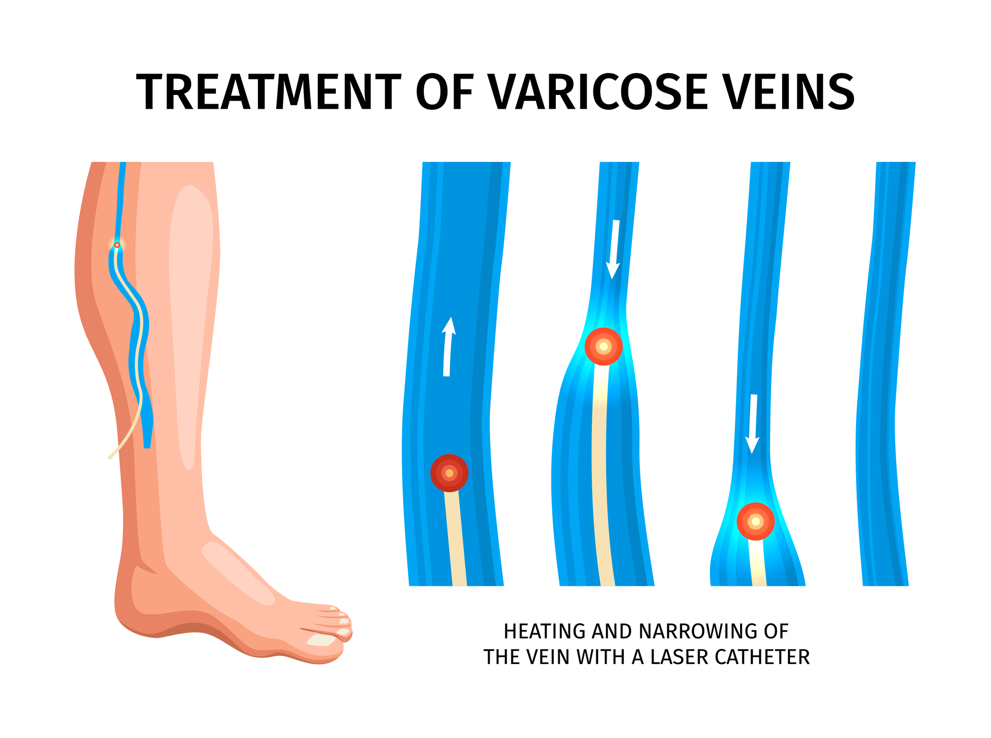
Modern treatment options that are less invasive and painful, such as endovenous laser therapy and sclerotherapy, rely on imaging. During treatment, ultrasound is used to view the veins to allow for better treatment.
Imaging aids the treatment process by increasing safety, improving target treatment, and decreasing pain. Better interventions and treatment options lead to an improved prognosis.
The combination of imaging in diagnosis and treatment has made vascular care a highly sophisticated specialty of medicine.
New Technologies in Vascular Imaging
Despite duplex ultrasound being the dominant modality in the diagnosis of venous disorders, advances in technology are expanding the frontiers of vascular imaging.
3D imaging is an example of advanced technology that provides enhanced resolution of venous anatomy and allows clinicians to visualize complex anatomy of an individual patient from different imaging angles.
To some extent, artificial intelligence has begun being introduced in the field of vascular diagnostics. Subtle imaging abnormalities may be detected by AI, as well as workflow automation and assistance in the formulation of a clinical hypothesis. For the time being, these adjuncts are a work in progress and are not a substitution for the clinician, but the technology has high potential for the future.
The invention of imaging devices with miniaturized ultrasound systems may make vascular imaging more convenient in numerous marketing channels within the healthcare industry, allowing clinicians to rapidly assess and image patients.
Innovations in diagnostic tools will increase the likelihood that diagnosis in the field of vascular imaging, in particular venous imaging, will continue to focus on the patient.
The Value of Prompt and Correct Diagnosis
The breakout use of advanced imaging has made it possible to identify venous disorders prior to the onset of complications. If vascular imaging is performed promptly, it becomes possible to prevent the exacerbation of symptoms and to avoid additional damage to the vascular system.
In addition to rapid intervention, the use of advanced imaging will reduce the likelihood that the patient will be required to undergo surgery. This is possible because with more accurate imaging clinicians may identify the etiology of the symptoms, which might be an explanation for conditions that may be confused with a venous disorder.
Having better diagnostic imaging leads to improvement in three areas that benefit patients: people feel more confident, they get more personalized care, and they understand their health better. Having better diagnostic imaging leads to better patient education and better patient communication.
When Should You Get a Comprehensive Vascular Evaluation?
You might want to get a comprehensive vascular evaluation if you have leg pain, swelling in your extremities, visible varicose veins, or if your legs feel heavy or fatigued.
Modern diagnostic imaging means advanced diagnostic testing. It means better care. Advanced diagnostic imaging is the best first step for any vascular care you may need. It means more accurate test results, improved patient care, and more successful treatment outcomes.
Imaging and modern diagnostic testing can easily identify and explain problems that affect circulation.
Conclusion: Modern Vein Care Imaging
Over the decades, the way that health care providers have been able to observe and diagnose varicose veins has improved significantly. What was once a difficult task that relied solely on patient and provider subjective reports has been improved by the great advancements and innovations in imaging technologies.
Advanced imaging, especially duplex ultrasound, is now at the heart of modern vascular care. By combining imaging solutions with ultrasound, health practitioners have the ability to make assessments of the anatomy, blood circulation, and the competency of the valves, and to apply this information with extreme diagnostic accuracy for the identification and management of venous disease.
With the continuous evolution and incorporation of novel technologies such as artificial intelligence and the capabilities of three-dimensional imaging and beyond, the future of precisely and personally diagnosing veins is bright. It portends for patients the benefits of diagnosis at an earlier stage and, therefore, a well-designed therapeutic intervention with the expectation of better outcomes.
Beyond the value as a diagnostic technique, modern imaging is a major transformative and advanced feature of contemporary vascular care that allows health practitioners to bridge the gap to solutions and provides patients with the best care in the fight for healthier veins and an improved wellbeing and quality of life.
Disclaimer: This article is provided for informational and educational purposes only and does not constitute medical advice, diagnosis, or treatment. Readers should consult a qualified healthcare professional regarding any symptoms, medical concerns, or treatment decisions. Open MedScience does not endorse any specific healthcare provider, clinic, or treatment mentioned in this article. Information may not reflect the latest medical research or clinical guidelines.


