Positron Emission Tomography
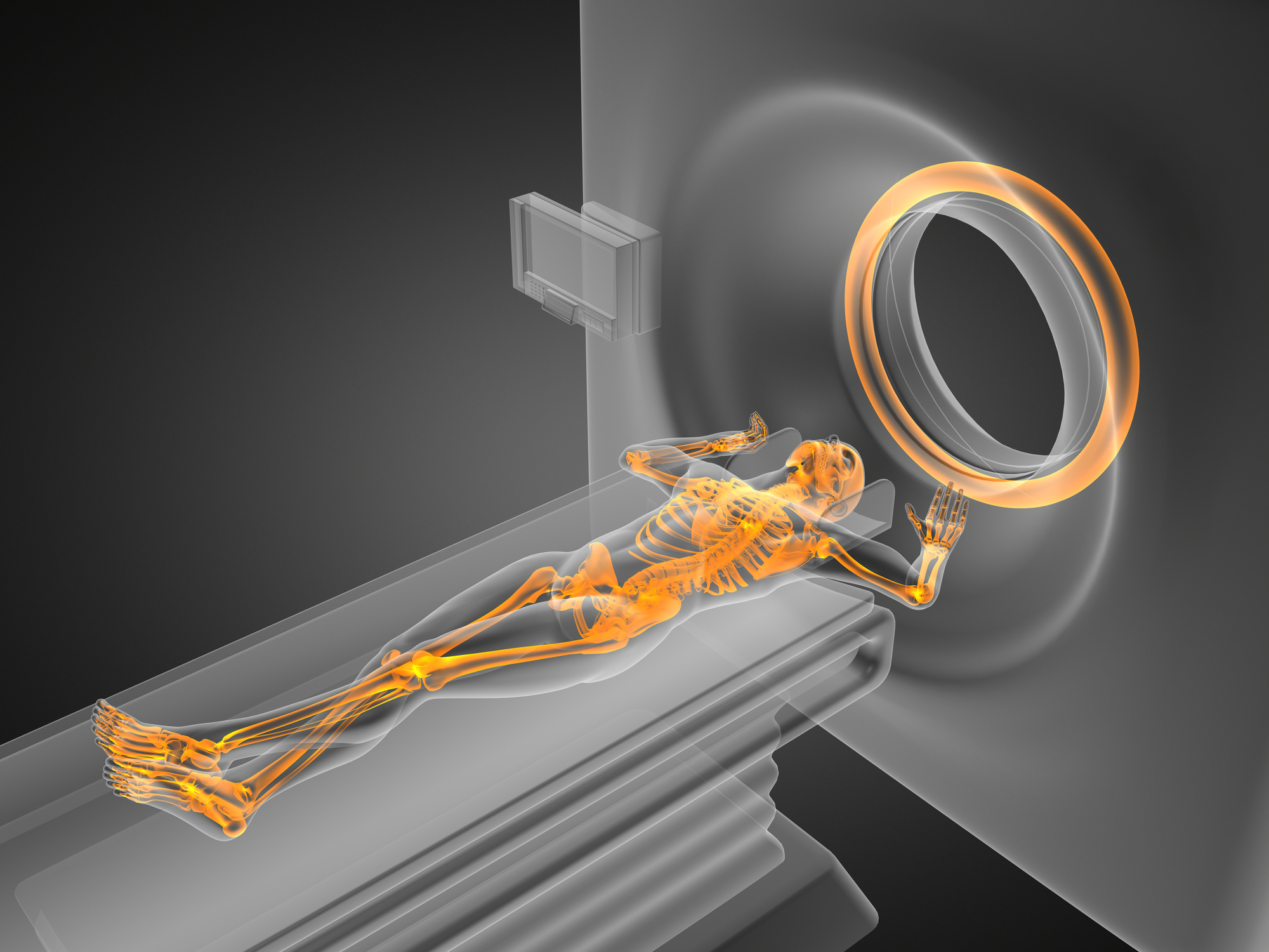
Positron Emission Tomography (PET) is an imaging modality used in neurology and oncology and, in some cases, used in conjunction with magnetic resonance imaging (MRI). PET defines the radiotherapeutic target volume of a tumour or the extent of disease before surgically removing a tissue structure or organ from the human body.
PET also assists in establishing a prognostic value about the patients’ overall cancer outcome, regardless of therapy, through molecular imaging. For example, the PET radiotracer fluorine-18-labelled fluoroethyltyrosine (18F-FET) has been shown to predict the prognosis of improved target delineation to assess treatment response.
A recent study using 18F-FET-PET and T1 weighted image MRI predicted the treatment effectiveness following glioblastoma patients’ chemoradiation therapy. The radiotracer fluorodopa F-18 (fluoro-deoxyphenylalanine 18F-DOPA) provides additional clinical information that histopathology may validate. Another target for brain tumour imaging is the translocator protein (TSPO).
This protein is overexpressed in glioblastoma patients, and clinical investigations have shown a link to the neuro-inflammatory component by observing a TSPO PET signal. Positron Emission Tomography is also used to evaluate head and neck cancers such as nasopharyngeal carcinomas, squamous cell cancer and salivary tumours.
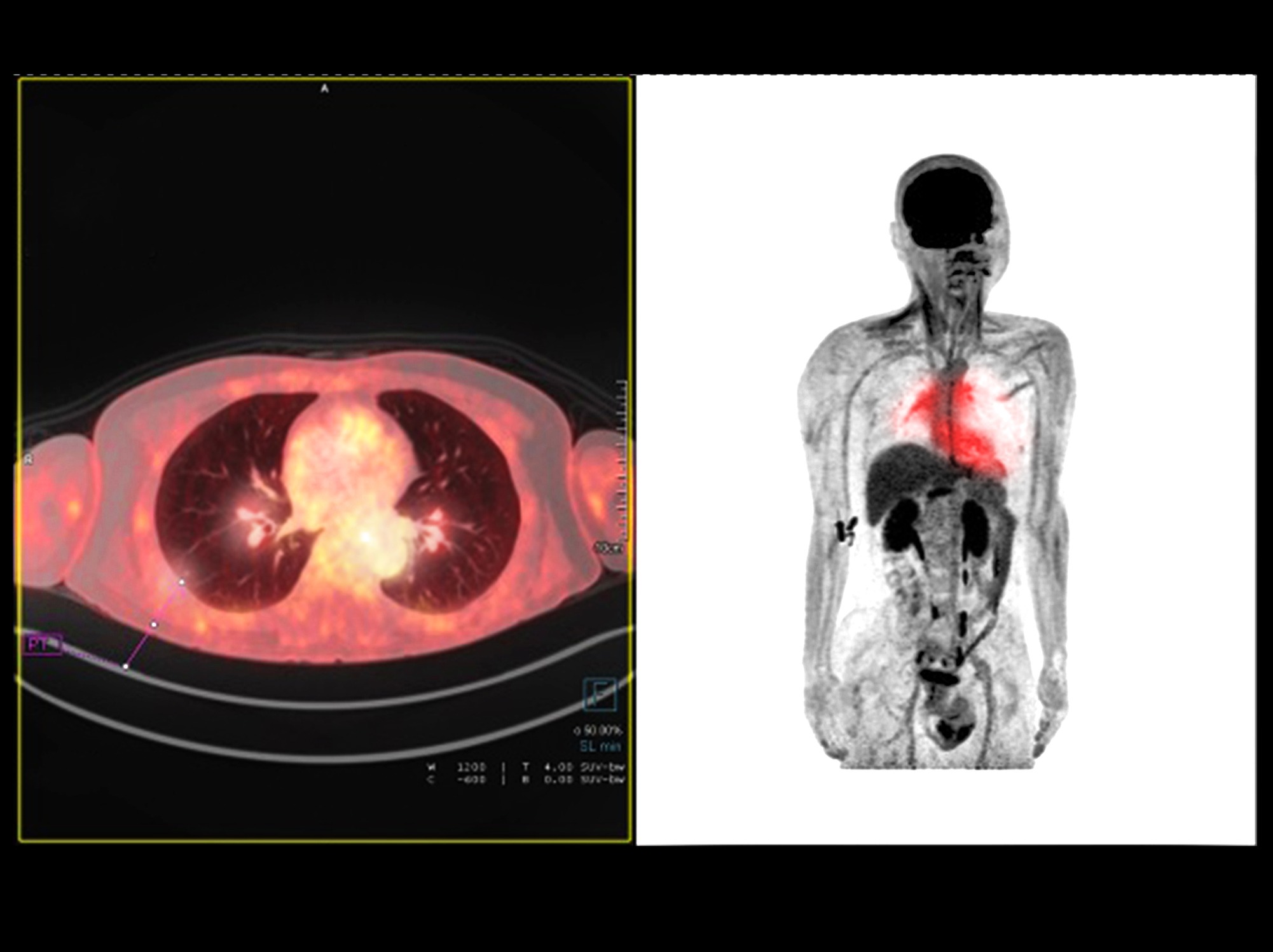
Furthermore, the hybrid PET and computed tomography (CT) scanner can provide insight into the status of nodal metastases of the tumour and tissue metabolism. PET/CT enables contouring for chemotherapy and radiotherapy in radiation treatment planning. Lung cancer occurs in 80-90% of non-small cell lung cancer (NSCLC) patients. PET imaging with 18F-FDG (fluorodeoxyglucose) is used to stage NSCLC patients.
In another diagnostic investigation, PET/CT PSMA (prostate-specific membrane antigen) ligands labelled with gallium-68 or fluorine-18 are used in prostate cancer screening programmes because they provide an excellent target-to-background ratio, leading to a better detection rate. PSMA is highly specific for prostatic tumoral tissue.
home » positron emission tomography
 By
By