
Laser vision correction changes the shape of the cornea so that light is focused more accurately inside the eye. Because the laser reshapes corneal tissue, the cornea’s existing shape, thickness, symmetry and structural stability all influence whether surgery is appropriate and how the procedure should be planned.
A routine prescription identifies nearsightedness, farsightedness and astigmatism, but two people with similar prescriptions may have very different corneal anatomy. Daniel M. Cotter, MD, from Eye Care & Vision Associates, treats corneal disease within a practice that offers LASIK in Buffalo. Before laser vision correction, corneal imaging helps clinicians assess the tissue’s shape, thickness and suitability for the proposed procedure. Prescription history and repeat measurements may also be needed to confirm that the cornea is stable [1,2].
Corneal topography is central to that assessment. It converts measurements of the corneal surface into colour-coded maps that reveal patterns a standard refraction cannot show. Modern planning may also include corneal tomography, epithelial thickness mapping, wavefront analysis and biomechanical measurements [1]. These technologies are related, but they do not provide identical information.
The map adds anatomical context to the prescription by showing how curvature and symmetry vary across the cornea.
What a corneal map shows that a standard eye test cannot
A standard eye examination can establish how clearly a person sees and which lens power provides the best correction. Keratometry may also estimate the curvature of the central cornea. These measurements are important, but they reduce a complex surface to a limited set of values.
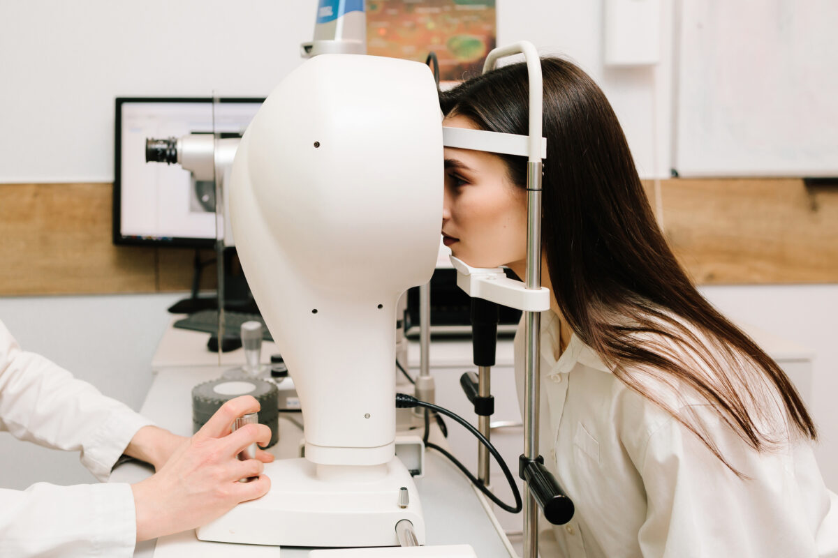
Corneal topography examines how curvature changes across a much wider area of the anterior corneal surface. Depending on the imaging system, reflected light patterns are analysed and translated into maps showing flatter and steeper regions. The resulting pattern can reveal the orientation of astigmatism, differences between the upper and lower cornea, localised steepening, decentration and other forms of asymmetry [1,2].
A prescription describes the optical error of the eye as a whole, but it does not show whether the corneal surface is uniformly shaped or where an irregularity originates. A patient may have what appears to be ordinary astigmatism during refraction, while topography shows that the curvature is uneven or displaced from the expected axis.
Topography should also be distinguished from tomography. Topography is particularly useful for describing anterior surface curvature. Tomographic systems reconstruct the cornea in three dimensions and can assess the front and back surfaces and the distribution of corneal thickness. Recent reviews emphasise that contemporary refractive-surgery screening benefits from combining these complementary forms of imaging rather than relying on a single central thickness measurement or anterior map [1,2].
Placido-based topography depends on reflections from the tear film, so dry eye, incomplete blinking, poor fixation or contact-lens-related distortion can reduce image quality [1,2]. Borderline scans may need to be repeated after the ocular surface has been treated or contact lenses have been discontinued, then interpreted alongside tomography, corneal thickness and the clinical examination.
Why subtle shape irregularities can affect treatment planning
Regular astigmatism usually produces a relatively orderly difference in curvature between two principal directions. Refraction and keratometry can usually characterise regular astigmatism, allowing it to be corrected with glasses or toric contact lenses.
Irregular astigmatism is less predictable. The steep and flat areas may not follow a simple, symmetrical pattern, and conventional lenses may not fully correct the resulting optical distortion. Irregular corneal optics may cause ghosting, glare, halos, reduced contrast or variable visual quality, even when standard-chart acuity remains relatively good [2,5].
One important reason for examining these patterns is keratoconus. In this condition, the cornea progressively thins and steepens, often asymmetrically, producing irregular astigmatism and reduced vision. Early or subclinical disease may not create obvious signs during slit-lamp examination, particularly when the patient still has good corrected visual acuity. Corneal topography and tomography can reveal characteristic changes before the condition becomes clinically apparent [3].
That information is particularly relevant before laser vision correction. LASIK and related procedures reshape the cornea by removing tissue. In an appropriately selected eye, the treatment is planned to preserve sufficient structural support. An eye with an underlying ectatic disorder, however, may have a greater risk of progressive postoperative weakening and distortion, known as corneal ectasia.
A systematic review estimated that reported ectasia in eyes without identifiable preoperative risk factors was uncommon, although the calculated rate differed among LASIK, photorefractive keratectomy and small-incision lenticule extraction. The authors also noted that incomplete detection of pre-existing keratoconus may account for some postoperative cases [4]. These figures should be interpreted cautiously because the review relied on published reports and estimated procedure volumes rather than a single prospective population.
Risk assessment considers the overall map alongside age, prescription, corneal thickness, expected tissue removal, the planned residual stromal bed and other clinical findings [4]. Epithelial thickness mapping and biomechanical measurements may add further information in selected cases [1].
Subtle asymmetry can therefore change the planning conversation even when it does not establish a diagnosis. It may lead to repeat imaging, additional testing, a period of observation or a decision that elective corneal surgery would introduce unnecessary risk.
How topography helps clinicians assess procedural suitability
Refractive surgery is often discussed as a choice between procedures, but the first question is whether corneal surgery is suitable at all.
A normal-looking prescription and stable vision are not sufficient on their own. The clinician also needs to consider corneal shape, thickness, tear-film quality, pupil characteristics, age, ocular health and the amount of correction required. Previous contact-lens wear, scarring, trauma or surgery may further affect the interpretation.
Topography can identify several reasons to pause. These include asymmetric steepening, irregular astigmatism, suspected keratoconus and contact-lens-induced warpage. Tomography may add evidence of abnormal posterior elevation or an unusual thickness profile. Together, these measurements can improve the detection of corneas that fall outside an expected pattern [1,2].
The result may alter which procedure is considered. LASIK involves creating a corneal flap before laser reshaping. PRK treats the surface without a flap, although it still removes corneal tissue. Other patients may be better suited to a lens-based procedure, continued use of glasses or contact lenses, or no elective surgery.
Choosing PRK rather than LASIK does not remove the need to investigate suspected ecstatic disease. Suspected or progressive keratoconus requires specialist assessment before elective corneal ablation is considered [3,4].
Imaging devices generate numerous indices, probability scores and colour displays, but borderline findings may have different significance in a young patient with changing astigmatism than in an older patient with stable measurements. The maps must therefore be considered alongside the examination and ocular history.
From screening data to a more personalised treatment plan
Once a patient has been judged suitable for laser vision correction, corneal imaging may contribute to the design of the treatment itself.
Conventional laser profiles primarily correct lower-order refractive errors such as myopia and regular astigmatism. Topography-guided treatment uses corneal-shape data to align the ablation with the measured surface pattern and may reduce selected higher-order aberrations in suitable patients [5,6].
This approach should not be confused with wavefront-guided or wavefront-optimised treatment. Wavefront analysis measures how light travels through the entire optical system of the eye, including the cornea and internal structures. Topography-guided treatment concentrates on corneal shape. Ray-tracing systems may integrate corneal, biometric and optical measurements into a more comprehensive model of the eye [1]. Each method has different assumptions, data requirements and potential advantages.
A meta-analysis comparing topography-guided and wavefront-optimised LASIK for myopia included 11 studies and 1,425 eyes. Both approaches produced effective and predictable results. The analysis found no significant difference in the proportion achieving 20/20 or better uncorrected distance vision, although topography-guided treatment showed advantages in some measures of refractive accuracy and higher-order aberrations [6]. The findings suggest potential benefits, but they do not establish that one profile is superior for every patient.
Planning can become more complicated when the astigmatism measured during refraction differs from the magnitude or axis shown on the topographic map. The surgeon must decide how much weight to give each dataset. A 2024 peer-reviewed clinical opinion article describes several strategies for reconciling topographic and refractive measurements, while noting that no single method resolves every discrepancy [5].
A personalised plan involves more than transferring a map directly into a laser. The surgeon must evaluate scan quality, repeatability, corneal thickness, expected tissue removal, ocular-surface health and the relationship between topographic and refractive measurements. Patient priorities also matter. Someone mainly concerned about daytime distance vision may have different expectations from a person who regularly drives at night or performs visually demanding work.
For readers considering refractive surgery in Western New York, Eye Care & Vision Associates offers LASIK and related eye-care services through four regional offices. A comprehensive evaluation can place the corneal maps alongside the prescription, ocular-surface findings and wider eye health rather than treating one scan as a stand-alone result [1,2].
Corneal topography may support an individualised treatment profile, prompt further investigation or point towards a different approach. Its value lies in showing how the corneal anatomy affects the options available.
References
[1] Fernandes VM, McGlone C, Fernandez KB, Rocha KM. Update on topography and tomography for refractive surgery. Curr Opin Ophthalmol. 2026 Jul 1;37(4):267-274. doi: 10.1097/ICU.0000000000001226.
[2] Fan R, Chan TC, Prakash G, Jhanji V. Applications of corneal topography and tomography: a review. Clin Exp Ophthalmol. 2018 Mar;46(2):133-146. doi: 10.1111/ceo.13136.
[3] Dina MS, Marinescu MC, Corbu CG, Constantin MM, Tataru CI, Tataru CP. Corneal Topography – a Review of Available Investigation Methods and Impact in the Diagnosis and Follow-Up of Keratoconus. Maedica (Bucur). 2025 Jun;20(2):374-382. doi: 10.26574/maedica.2025.20.2.374.
[4] Moshirfar M, Tukan AN, Bundogji N, Liu HY, McCabe SE, Ronquillo YC, Hoopes PC. Ectasia After Corneal Refractive Surgery: A Systematic Review. Ophthalmol Ther. 2021 Dec;10(4):753-776. doi: 10.1007/s40123-021-00383-w.
[5] İpek ŞC, Utine CA. Topography-guided excimer laser ablation in refractive surgery. Front Ophthalmol (Lausanne). 2024 Mar 7;4:1367258. doi: 10.3389/fopht.2024.1367258.
[6] Hu PC, Li L, Wu XH, Li YQ, Li KW. Visual differences in topography-guided versus wavefront-optimized LASIK in the treatment of myopia: a Meta-analysis. Int J Ophthalmol. 2021 Oct 18;14(10):1602-1609. doi: 10.18240/ijo.2021.10.19.
Disclaimer: This article is intended for educational and informational purposes only and should not be considered medical advice, diagnosis, or treatment. It does not replace consultation with a qualified ophthalmologist, optometrist, or other healthcare professional. Suitability for laser vision correction varies between individuals and requires a comprehensive eye examination, including appropriate corneal imaging and clinical assessment. Any references to specific clinicians, practices, procedures, or technologies are provided for informational purposes only and do not constitute an endorsement or guarantee of clinical outcomes. Readers should seek personalised medical advice before making decisions about their eye health or refractive surgery.
home » blog » optical imaging »