Neuroendocrine Tumour Imaging
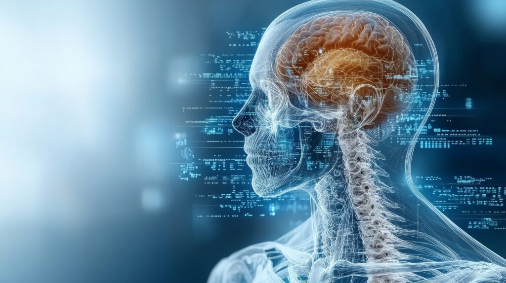
Neuroendocrine tumours (NETs) are a heterogeneous group of neoplasms arising from neuroendocrine system cells. These tumours can occur throughout the body but are most commonly found in the gastrointestinal tract, pancreas, and lungs. The diagnosis and management of NETs rely heavily on imaging, which is crucial for accurate tumour localisation, staging, and monitoring treatment response.
Recent advances in imaging technologies have significantly improved the detection and characterisation of NETs. Conventional imaging techniques such as computed tomography (CT) and magnetic resonance imaging (MRI) remain staples in the evaluation of these tumours. CT provides excellent anatomical detail, which is essential for surgical planning and detecting metastases. MRI is preferred for its superior contrast resolution, which is particularly beneficial for identifying liver metastases and for imaging pancreatic NETs.
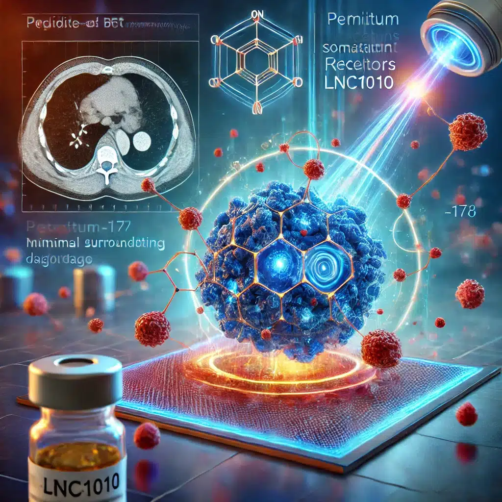
However, the cornerstone of NET imaging is nuclear medicine techniques, specifically positron emission tomography (PET) combined with CT (PET/CT). The most commonly used radiotracer, 68-Gallium-DOTATATE, binds to somatostatin receptors abundantly expressed on many NET cells’ surfaces. This method offers high sensitivity and specificity and enables the assessment of receptor status, which is crucial for planning receptor-targeted therapies.
Another promising advancement is the introduction of 18F-FDG PET/CT, which is particularly useful in imaging high-grade NETs that do not express somatostatin receptors. This technique assesses tumours’ metabolic activity, providing vital prognostic information and aiding in the differentiation of tumour grades.
As imaging technology evolves, the integration of artificial intelligence (AI) is poised to enhance the precision of NET diagnostics further. AI algorithms can help quantify tumour burden and predict treatment response, thereby personalising patient management. These technological advancements underscore the dynamic nature of neuroendocrine tumour imaging and its pivotal role in the comprehensive care of patients with NETs.
You are here:
home » Neuroendocrine Tumour Imaging

